Abstract
Water-filtered infrared-A (wIRA), as a special form of heat radiation with a high tissue penetration and a low thermal load to the skin surface, can improve the healing of acute and chronic wounds both by thermal and thermic as well as by non-thermal and non-thermic effects. wIRA increases tissue temperature (+2.7°C at a tissue depth of 2 cm), tissue oxygen partial pressure (+32% at a tissue depth of 2 cm) and tissue perfusion. These three factors are decisive for a sufficient supply of tissue with energy and oxygen and consequently also for wound healing and infection defense.
wIRA can considerably alleviate pain (without any exception during 230 irradiations) with substantially less need for analgesics (52–69% less in the groups with wIRA compared to the control groups). It also diminishes exudation and inflammation and can show positive immunomodulatory effects. The overall evaluation of the effect of irradiation as well as the wound healing and the cosmetic result (assessed on visual analogue scales) were markedly better in the group with wIRA compared to the control group. wIRA can advance wound healing (median reduction of wound size of 90% in severely burned children already after 9 days in the group with wIRA compared to 13 days in the control group; on average 18 versus 42 days until complete wound closure in chronic venous stasis ulcers) or improve an impaired wound healing (reaching wound closure and normalization of the thermographic image in otherwise recalcitrant chronic venous stasis ulcers) both in acute and in chronic wounds including infected wounds. After major abdominal surgery there was a trend in favor of the wIRA group to a lower rate of total wound infections (7% versus 15%) including late infections following discharge from hospital (0% versus 8%) and a trend towards a shorter postoperative hospital stay (9 versus 11 days).
Even the normal wound healing process can be improved.
The mentioned effects have been proven in six prospective studies, with most of the effects having an evidence level of Ia/Ib.
wIRA represents a valuable therapy option and can generally be recommended for use in the treatment of acute as well as of chronic wounds.
Keywords: water-filtered infrared-A (wIRA); infrared-A radiation; wound healing; thermal and non-thermal effects; thermic and non-thermic effects; energy supply; oxygen supply; tissue oxygen partial pressure; tissue temperature; tissue blood flow; reduction of pain; wound exudation; inflammation; immunomodulatory effects; acute wounds; chronic wounds; chronic venous stasis ulcers of the lower legs; problem wounds; wound infections; infection defense; contact-free method; absent expenditure of material; prospective, randomized, controlled, double-blind studies; visual analogue scales (VAS); quality of life; infrared thermography; thermographic image analysis
Abstract
Wassergefiltertes Infrarot A (wIRA) als spezielle Form der Wärmestrahlung mit hohem Eindringvermögen in das Gewebe bei geringer thermischer Oberflächenbelastung kann die Heilung akuter und chronischer Wunden sowohl über thermische und temperaturabhängige als auch über nicht-thermische und temperaturunabhängige Effekte verbessern. wIRA steigert Temperatur (+2,7°C in 2 cm Gewebetiefe) und Sauerstoffpartialdruck im Gewebe (+32% in 2 cm Gewebetiefe) sowie die Gewebedurchblutung. Diese drei Faktoren sind entscheidend für eine ausreichende Versorgung des Gewebes mit Energie und Sauerstoff und deshalb auch für Wundheilung und Infektionsabwehr.
wIRA vermag Schmerzen deutlich zu mindern (ausnahmslos bei 230 Bestrahlungen) mit bemerkenswert niedrigerem Analgetikabedarf (52–69% niedriger in den Gruppen mit wIRA verglichen mit den Kontrollgruppen) und eine erhöhte Wundsekretion und Entzündung herabzusetzen sowie positive immunmodulierende Effekte zu zeigen. Die Gesamtbeurteilung des Effekts der Bestrahlung wie auch die Wundheilung und das kosmetische Ergebnis (erhoben mittels visueller Analogskalen) waren in der Gruppe mit wIRA wesentlich besser verglichen mit der Kontrollgruppe. wIRA kann sowohl bei akuten als auch bei chronischen Wunden einschließlich infizierter Wunden die Wundheilung beschleunigen (Abnahme der Wundfläche im Median um 90% bei schwerbrandverletzten Kindern bereits nach 9 Tagen in der Gruppe mit wIRA verglichen mit 13 Tagen in der Kontrollgruppe; im Durchschnitt 18 versus 42 Tage bis zum kompletten Wundschluss bei chronischen venösen Unterschenkelulzera) oder bei stagnierender Wundheilung verbessern (mit Erreichen eines kompletten Wundschlusses und Normalisierung des thermographischen Bildes bei zuvor therapierefraktären chronischen venösen Unterschenkelulzera). Nach großen abdominalen Operationen zeigte sich ein Trend zugunsten der wIRA-Gruppe hin zu einer niedrigeren Rate von Wundinfektionen insgesamt (7% versus 15%) einschließlich später Infektionen nach der Entlassung aus dem Krankenhaus (0% versus 8%) und ein Trend hin zu einem kürzeren postoperativen Krankenhausaufenthalt (9 versus 11 Tage).
Selbst der normale Wundheilungsprozess kann verbessert werden.
Die erwähnten Effekte wurden in 6 prospektiven Studien belegt, die meisten mit einem Evidenzgrad von Ia/Ib.
wIRA stellt eine wertvolle Therapieoption dar und kann generell für die Therapie von akuten und chronischen Wunden empfohlen werden.
Introduction
The application of water-filtered infrared-A (wIRA) for the improvement of healing of acute and chronic wounds and the underlying principles are described more extensively than here in the three reviews [1], [2], [3], which belong together (in total 42 PDF pages). Please refer to these reviews for more details and references. Besides this, two further reviews concerning this subject [4], [5] and one review on a slightly broader subject [6] are available.
Working mechanisms of wIRA
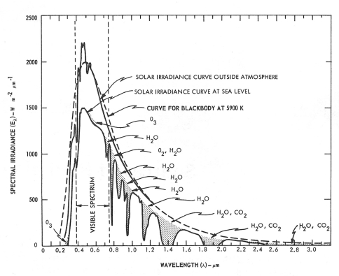
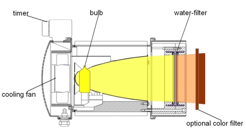
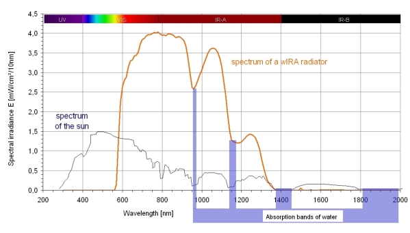
The experience of the pleasant heat of the sun in moderate climatic zones arises from the filtering of the heat radiation of the sun by water vapor in the Earth’s atmosphere [1], [4], [5], [6], [7], [8], see Figure 1 (Fig. 1). The filter effect of water decreases those parts of infrared radiation (most parts of infrared-B and -C and the absorption bands of water within infrared-A), which would otherwise – by reacting with water molecules in the skin – cause an undesired thermal load to the surface of the skin [1], [4], [5], [6], [7], [8]. Technically, water-filtered infrared-A (wIRA) is produced by special radiators, whose full spectrum of radiation of a halogen bulb is passed through a cuvette containing water, which absorbs or decreases the described undesired wavelengths of the infrared radiation [1], [9], see Figure 2 (Fig. 2). Within the infrared range, the remaining wIRA (within 780–1400 nm) mainly consists of radiation with good tissue penetration properties and therefore allows – compared to unfiltered heat radiation – a multiplication of the energy transfer into tissue without irritating the skin, similar to the sun’s heat radiation in moderate climatic zones. Typical wIRA radiators emit no ultraviolet (UV) radiation and almost no infrared-B and -C radiation and the amount of infrared-A radiation in relation to the amount of visible light (380–780 nm) is accentuated [1], [9], see Figure 3 (Fig. 3).
Figure 1. Spectral solar irradiance outside the atmosphere and on the surface of the Earth at sea level,
in both cases with the sun at the zenith and for a mean Earth-sun distance. Shaded areas indicate absorption before reaching the surface of the Earth at sea level due to the atmospheric constituents shown (from [1], [58], adapted from [59]).
For comparison of Figures 1 and 3: 1000 W · m–2 · µm–1 = 100 mW · cm–2 · µm–1 = 1 mW · cm–2 · (10 nm)–1
Figure 2. Cross-section of a water-filtered infrared-A radiator (Hydrosun, Müllheim, Germany) .
The whole incoherent non-polarized broadband radiation of a 3000 Kelvin halogen bulb is passed through a cuvette containing water, which absorbs or decreases the undesired wavelengths within the infrared region (most parts of infrared-B and -C and the absorption bands of water within infrared-A). The water is hermetically sealed within the cuvette. A fan provides air cooling of the cuvette to prevent the water from boiling. (from [1])
Figure 3. Comparison of the spectra of the sun on the surface of the Earth at sea level and of a water-filtered infrared-A radiator .
Spectral solar irradiance on the surface of the Earth at sea level (with the sun at the zenith and for a mean Earth-sun distance) as in Fig. 1 (from [1], adapted from [58]) and spectral irradiance of a water-filtered infrared-A radiator (Hydrosun® radiator 501 with 10 mm water cuvette and orange filter OG590) at approximately 210 mW/cm² (= 2.1 · 10³ W/m²) total irradiance (from [1], [4]).
The spectrum of the sun at sea level includes ultraviolet radiation (UV, <400 nm), visible light (VIS, 380–780 nm), and infrared radiation (IR, >780 nm). The spectrum of the water-filtered infrared-A radiator includes only visible light (VIS) and infrared radiation (IR); the visible part depends on the color filter used; the wIRA radiator does not emit ultraviolet radiation (UV).
Both spectra show the decreased irradiances of the absorption bands of water.
Within the spectra of infrared-A and water-filtered infrared-A, radiation effects in particular of the energy-rich wavelengths near to visible light – approximately 780–1000 nm (800–900 nm [10], [11], [12], 800 nm [13], 820 nm [14], [15], [16], 830 nm [17]) – have been described both in vitro and in vivo. These wavelengths seem to represent the clinically most important part of the infrared-A and wIRA range [1], [18].
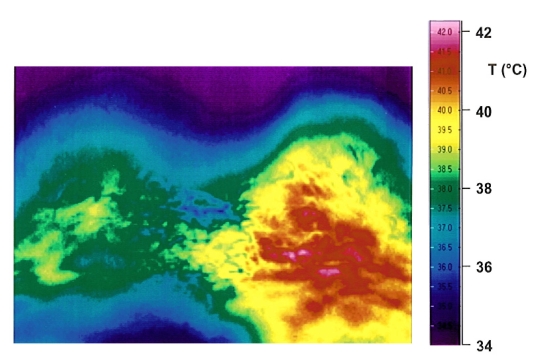
Water-filtered infrared-A as a special form of heat radiation with a high tissue penetration and with a low thermal load to the skin surface (see Figure 4 (Fig. 4)), acts both by thermal (related to heat energy transfer) and thermic (temperature dependent, with a relevant change of temperature) as well as by non-thermal (without a relevant transfer of heat energy) and non-thermic (not depending on temperature, without a relevant change of temperature) effects [1]. wIRA produces a therapeutically usable field of heat in the tissue and increases tissue temperature [19], [20], [21], [22], [23], [24], [25], [26], tissue oxygen partial pressure [19], and tissue perfusion [1], [24], [25], [26]. These three factors are vital for a sufficient supply of tissue with energy and oxygen.
Figure 4. Comparison of irradiation with water-filtered infrared-A and with conventional infrared .
Thermographical comparison of skin surface temperatures in the lumbar region 12 minutes after commencement of irradiation with water-filtered infrared-A (left) and conventional infrared (right) with the same irradiance: the skin surface temperature is higher in the case of irradiation with conventional infrared (presented in the thermography), while the temperature at 1 cm tissue depth is higher when irradiating with water-filtered infrared-A (from [1], [20]). Water-filtered infrared-A thus leads to a high tissue penetration combined with a low thermal load to the skin surface.
As wound healing and infection defense (e.g. granulocyte function including its antibacterial oxygen radical formation) depend decisively on a sufficient supply of tissue with energy and oxygen and since the centers of chronic wounds are often relatively hypothermic [1], [19], [23] (while e.g. both preoperative [27] and postoperative [19], [28] heat supply to the operation field can improve healing of acute wounds) and frequently have an oxygen partial pressure close to zero [1], [19], [23], [29], [30], [31], [32], [33], [34], [35], [36], [37], [38], one explanation for the good clinical effect of wIRA on wounds and wound infections could be the improvement of both the energy supply per time (increase of metabolic rate) and the oxygen supply [1]. In addition, wIRA has non-thermal and non-thermic effects, which are based on a direct stimulation of cells and cellular structures: Reactions of cells to infrared radiation – partly found even at very small irradiances – are e.g. target-oriented growth of surface extensions (plasmodia) [10], influence on cytochrome c oxidase [14], [39], [40], target-oriented growth of neurons [13], stimulation of wound repair [41], [42] as well as cell protective effects of infrared-A [43], [44], [45], [46] and water-filtered infrared-A (wIRA) [47], [48], [49].
wIRA can considerably alleviate pain (with remarkably less need for analgesics) and diminish an elevated wound exudation and inflammation and can show positive immunomodulatory effects. wIRA can advance wound healing or improve an impaired wound healing both in acute and in chronic wounds, including infected wounds. Even the normal wound healing process can be improved [1], [19].
wIRA is contact-free, easily applied, involves no discomfort to the patient or the use of expendable materials and is effective even in deeper-lying tissue regions. wIRA application, with appropriate therapeutic irradiances and doses, could be shown not only to be harmless for human skin [1], [4], [18], [47], [48], [50], but even to have protective effects in cells against damage caused by UV radiation [1], [4], [43], [44], [45], [46], [47], [48], [49]. Safety aspects of the clinical use of wIRA have been described extensively, especially in [1] and [18]. Particularly when [50] and the current review [51] are taken into consideration, the application of wIRA with adequate irradiances can be considered as being safe. The irradiation of the typically uncovered wound is carried out using a wIRA radiator, see Figure 5 (Fig. 5).
Figure 5. Example of an irradiation of a wound with a water-filtered infrared-A radiator .
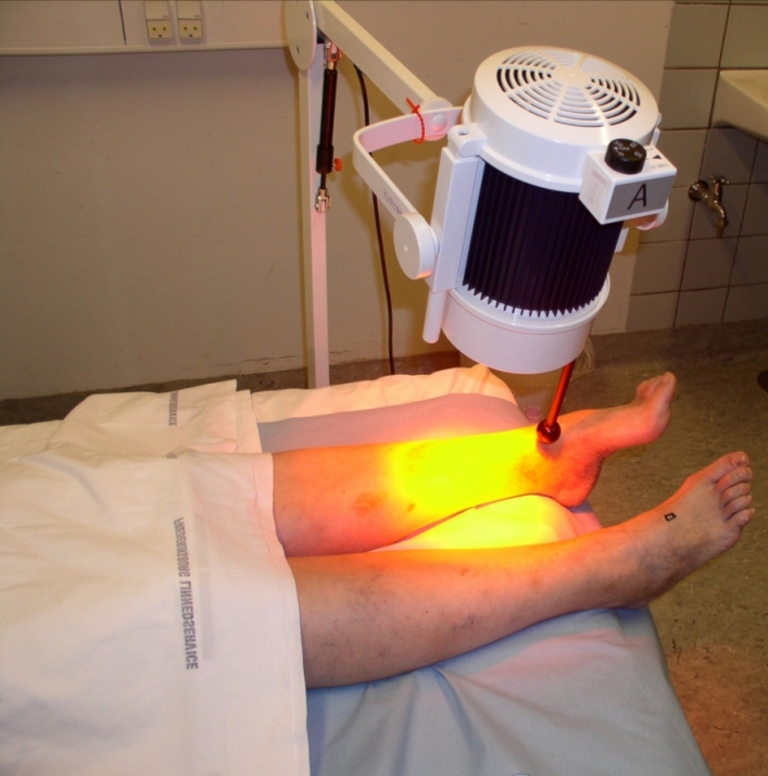
(published with kind approval of Prof. James Mercer, Tromsø/Norway) (from [1], [23])
Clinical effects of wIRA on wounds
Based on 6 clinical studies, the following has been proven with a level of evidence of Ia/Ib [1], [52]:
acute pain reduction during wIRA irradiation
reduction of the required dose of analgesics
faster reduction of wound area
better assessment of wound healing
better overall evaluation of the effects of irradiation (including pain, wound healing, cosmesis)
higher tissue oxygen partial pressure during wIRA
higher subcutaneous temperature during wIRA
better cosmesis
In addition, the following trends have been found:
lower rate of wound infections
shorter postoperative hospital stay
Additional clinical observations are:
reduction of inflammation
reduction of hypersecretion
Therapy of acute wounds with wIRA
wIRA for acute operation wounds (Study of the University Hospital Heidelberg, Department of Surgery)
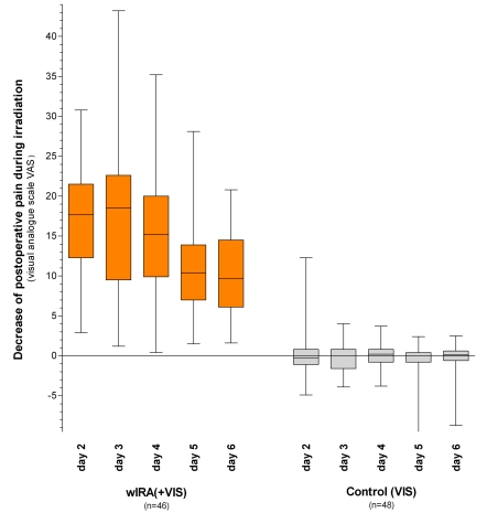
A prospective, randomized, controlled, double-blind study with 111 patients who had undergone major abdominal surgery at the University Hospital Heidelberg, Germany, and thereafter underwent 20 minutes irradiation 2 times per day (starting on the second postoperative day) showed a significant and relevant pain reduction combined with a markedly decreased dose of required analgesics in the group with wIRA and visible light VIS (wIRA(+VIS), approximately 75% wIRA, 25% VIS) compared to a control group with only VIS: during 230 single irradiations with wIRA(+VIS) pain decreased without any exception (median of decrease of pain on postoperative days 2–6 was 13.4 on a 100 mm visual analogue scale VAS 0–100), while pain remained unchanged in the control group (p<0.000001, see Figure 6 (Fig. 6)). The median of decrease of pain on the third postoperative day was 18.5 versus 0.0, the median difference between the groups was 18.4 (99% confidence interval 12.3/21.0), p<0.000001. (Semantic statistical remark in [2], [5].)
Figure 6. Decrease of postoperative pain during irradiation in the group with water-filtered infrared-A (wIRA) and visible light (VIS) and in the control group with only visible light (VIS) (Study Heidelberg) .
assessed with a visual analogue scale; given as minimum, percentiles of 25, median, percentiles of 75, and maximum (box and whiskers graph with the box representing the interquartile range), from [2], adapted from [19]).
During 230 single irradiations with wIRA(+VIS) the pain decreased without any exceptions, while pain remained unchanged in the control group (p<0.000001 for any single documented day as well as for all the days).
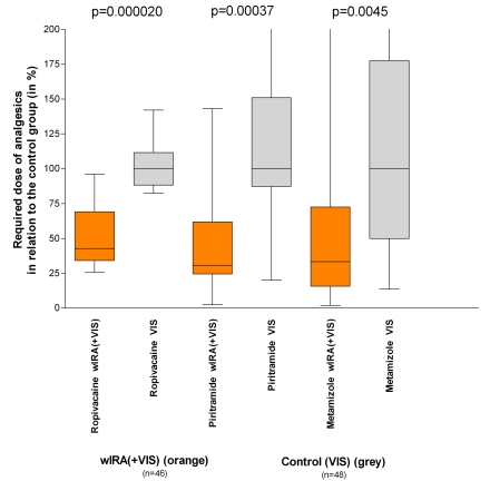
The required dose of analgesics was 52–69% lower (median differences) in the subgroups with wIRA(+VIS) compared to the control subgroups with only VIS (median 598 versus 1398 mL ropivacaine, p=0.000020, for peridural catheter analgesia; 31 versus 102 mg piritramide, p=0.00037, for patient-controlled analgesia; 3.4 versus 10.2 g metamizole, p=0.0045, for intravenous and oral analgesia, see Figure 7 (Fig. 7)).
Figure 7. Required dose of analgesics of the subgroups with water-filtered infrared-A (wIRA) and visible light (VIS) in relation to the control subgroups with only visible light (VIS) (medians of the control subgroups = 100) (Study Heidelberg) .
(given as minimum, percentiles of 25, median, percentiles of 75, and maximum (box and whiskers graph with the box representing the interquartile range), adapted from [2], data taken from [19]).
The required dose of analgesics was 52–69% lower (median differences) in the subgroups with wIRA(+VIS) compared to the control subgroups with only VIS.
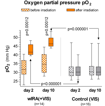
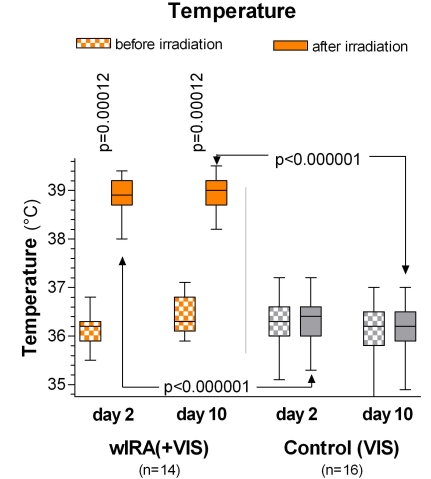
During irradiation with wIRA(+VIS) the subcutaneous oxygen partial pressure rose markedly by 32% and the subcutaneous temperature by 2.7°C (both measured at a tissue depth of 2 cm), whereas both remained unchanged in the control group. After irradiation, the median of the subcutaneous oxygen partial pressure was 41.6 (with wIRA) versus 30.2 mm Hg in the control group (median difference between the groups 11.9 mm Hg (+39%), 99% confidence interval 8.4/15.4 mm Hg (+28%/+51%), p<0.000001, see Figure 8 (Fig. 8)) and the median of the subcutaneous temperature was 38.9 versus 36.4°C (median difference between the groups 2.6°C, 99% confidence interval 2.1/2.9°C, p<0.000001, see Figure 9 (Fig. 9)). The baseline values (before irradiation) of the subcutaneous oxygen partial pressure rose from the second to the tenth postoperative day by 3.4 versus 0.3 mm Hg (median difference between the groups 3.1 mm Hg (+10%), 99% confidence interval 1.9/3.7 mm Hg, p=0.00051). The baseline values for the subcutaneous temperature rose by 0.4 versus –0.3°C (median difference 0.6°C, 95% confidence interval 0.2/0.8°C, p=0.0074) (effects which endured beyond the time period of the single irradiation).
Figure 8. Subcutaneous oxygen partial pressure at a tissue depth of 2 cm on the postoperative days 2 and 10 in the group with water-filtered infrared-A (wIRA) and visible light (VIS) and in the control group with only visible light (VIS) (Study Heidelberg) .
(given as minimum, percentiles of 25, median, percentiles of 75, and maximum (box and whiskers graph with the box representing the interquartile range); adapted from [2], [19]).
During irradiation with wIRA(+VIS), the subcutaneous oxygen partial pressure rose markedly by more than 30%, whereas it remained unchanged in the control group.
Figure 9. Subcutaneous temperature at a tissue depth of 2 cm on the postoperative days 2 and 10 in the group with water-filtered infrared-A (wIRA) and visible light (VIS) and in the control group with only visible light (VIS) (Study Heidelberg) .
(given as minimum, percentiles of 25, median, percentiles of 75, and maximum (box and whiskers graph with the box representing the interquartile range); adapted from [2], [19]).
During irradiation with wIRA(+VIS) the subcutaneous temperature rose markedly by approximately 2.7°C, whereas it remained unchanged in the control group.
The overall evaluation of the effect of irradiation, including wound healing, pain and cosmesis, assessed on a VAS (0-100 with 50 as the indifferent point of no effect) by the surgeon (median 79.0 versus 46.8, median difference 27.9, 99% confidence interval 17.2/37.3, p<0.000001) or the patient (79.0 versus 50.2, median difference 23.8, 99% confidence interval 9.5/34.1, p=0.000007) was considerably better in the group with wIRA compared to the control group. This was also true for single aspects: Wound healing assessed on a VAS by the surgeon (median 88.6 versus 78.5, p<0.000001) or the patient (median 85.8 versus 81.0, p=0.040, trend) and cosmetic result assessed on a VAS by the surgeon (median 84.5 versus 76.5, p=0.00027) or the patient (median 86.7 versus 73.6, p=0.00077).
In addition there was a trend towards a lower rate of total wound infections in favor of the wIRA group (3 of 46, 7%, versus 7 of 48, 15%, difference –8%, 95% confidence interval –20%/4%, p=0.21) including late infections following discharge. This was due to the different rate of late infections following discharge: 0 of 46 (0%) in the wIRA group and 4 of 48 (8%) in the control group (difference –8%, 95% confidence interval –18%/2%, p=0,12). There was also a trend towards a shorter postoperative hospital stay: 9 days in the wIRA group versus 11 days in the control group (median difference –2 days (–18%), 95% confidence interval –3/0 days, p=0.022).
The principal finding of this study was that postoperative irradiation with wIRA can improve even the normal wound healing process [2], [19].
wIRA for severely burned children (Study of the Children’s Hospital Park Schönfeld, Kassel, Department of Pediatric Surgery)
A prospective, randomized, controlled, double-blind study with 45 severely burned children was carried out at the Children’s Hospital Park Schönfeld, Kassel, Germany. 30 minutes irradiation was applied once a day (starting on the first day, with the day of burn being day 1). In the group with wIRA and visible light VIS (wIRA(+VIS), approximately 75% wIRA, 25% VIS) a markedly faster reduction of wound size was seen in comparison to a control group with only VIS. On the fifth day (after 4 days with irradiation), the decision was taken as to whether surgical debridement of necrotic tissue was necessary because of deeper (second degree, type b) burns (11 of 21 in the group with wIRA, 14 of 24 in the control group) or whether non-surgical treatment was possible (second degree, type a burns). The patients treated conservatively were kept within the study and irradiated until reepithelialisation was complete.
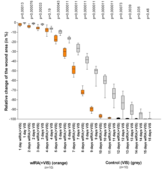
The patients in the group with wIRA showed a markedly faster reduction of wound area: a median reduction of wound size of 50% was reached already after 7 days compared to 9 days in the control group, a median reduction of wound size of 90% was already achieved after 9 days compared to 13 days in the control group, see Figure 10 (Fig. 10) and Figure 11 (Fig. 11). After 9 days, the median reduction in wound area was 89.2% versus 49.5%, the median difference between the groups was a 39.5% reduction of the wound area (99% confidence interval 34.4%/43.0%), p=0.000011. The median difference between the groups existed already after one day with p=0.00013 and after 2, 5, 6, 7, 8, 9, 10 and 11 days with p<0.0001. In addition, the group with wIRA showed superior results in terms of the overall surgical assessment of the wound and the assessment of effects of irradiation (the latter as a trend up to 3 months after the burn) compared to the control group [2].
Figure 10. Relative change of wound area in severely burned children as a function of duration of treatment (in days) in the group with water-filtered infrared-A (wIRA) and visible light (VIS) and in the control group with only visible light (VIS) (Study Kassel) .
(given as minimum, percentiles of 25, median, percentiles of 75, and maximum (box and whiskers graph with the box representing the interquartile range), adapted from [2]).
The figure presents the data from those 10+10 = 20 children (out of 21+24 = 45 children), who had second degree, type a burns (not second degree, type b burns) and were consequently treated non-surgically until complete cutaneous regeneration occurred including irradiation (starting on the day of the burn, until complete reepithelialization) with wIRA(+VIS) or with only VIS (control group).
Patients in the group with wIRA showed a markedly faster reduction of wound area compared to the control group: a median reduction of wound size of 50% was reached in the group with wIRA already after 7 days compared to 9 days in the control group, a median reduction of wound size of 90% was achieved in the group with wIRA already after 9 days compared to 13 days in the control group.
Figure 11. Example of a rapid improvement with wIRA in a severely burned child (Study Kassel) .

Left: 1 day after the burn, right: only 30 hours later than shown on the left side (from [2]).
wIRA for experimental wounds (Study of the University Medical Center Charité Berlin, Department of Dermatology)
In a prospective, randomized, controlled study with 12 volunteers at the University Medical Center Charité, Berlin, Germany, volunteers were inflicted with 4 experimental superficial wounds (5 mm diameter). In this acute wound model, wounds were generated by a suction cup technique, with the roof of the blister being removed with a scalpel and sterile forceps (day 1). 4 different treatments were used and investigated over 10 days: no therapy, wIRA(+VIS) only (approximately 75% wIRA, 25% VIS; 30 minutes irradiation once a day), only dexpanthenol (= D-panthenol) cream once a day, wIRA(+VIS) and dexpanthenol cream once a day. Healing of the small experimental wounds was, from a clinical point of view, excellent with all 4 treatments. Therefore there were only small differences between the treatments with slight advantages seen with the combination wIRA(+VIS) and dexpanthenol cream and with dexpanthenol cream alone as far as relative change of wound size and assessment of feeling of the wound area were concerned.
Laser scanning microscopy, however, together with a scoring system revealed differences between the 4 treatments concerning the formation of the stratum corneum (from first layer of corneocytes to full formation) especially on days 5–7: the fastest formation of the stratum corneum was seen in wounds treated with wIRA(+VIS) and dexpanthenol cream, second was wIRA(+VIS) alone, third dexpanthenol cream alone and lastly, untreated wounds. Bacterial counts of the wounds (taken every 2 days) showed that wIRA(+VIS) and the combination of wIRA(+VIS) with dexpanthenol cream were able to inhibit the colonisation with physiological skin flora up to day 5 when compared with the two other groups (untreated group and group with dexpanthenol cream alone). At any investigated time, the amount of colonisation under therapy with wIRA(+VIS) alone was lower (interpreted as being more suppressed) compared with the group with wIRA(+VIS) and dexpanthenol cream [2].
wIRA for wound seromas
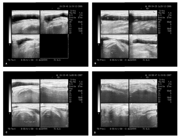
During rehabilitation after hip and knee endoprosthetic operations the resorption of wound seromas and wound hematomas was both clinically and sonographically faster and pain was reduced by irradiation with wIRA(+VIS) [2]. An additional example is presented in Figure 12 (Fig. 12).
Figure 12. Example of a successful treatment of recurrent wound seromas with wIRA .
A 64 year-old female patient had relapsing wound seromas and wound hematomas (without infection) after a hip operation (replacement of the acetabulum part of a 15 year-old endoprosthesis) even after an additional operation with the sole intention of stopping the wound seromas and after approximately 8 aspirations of seroma fluid (up to approximately 90 mL within one aspiration) within 2 months, and a third operation was seriously considered: Figure A shows the sonographic state. At that time, wIRA(+VIS) irradiation was commenced, beginning with 30 minutes twice per day and increasing up to 3 times one hour per day. Within a few days the seroma no longer increased as usual; after approximately one week a slight decrease of seroma size was noticed clinically (Figure B). Figure C shows reduced seroma size after 18 days and Figure D after 29 days. After approximately 2 months the seroma had resolved completely (both clinically and sonographically) without any aspiration of seroma fluid or operation since commencement of wIRA(+VIS) irradiation (sonographic pictures published with kind approval of Dr. Michael Paulus, Herzogenaurach, Germany) (from [2]).
wIRA for persistent postoperative pain
wIRA can be used successfully for persistent postoperative pain e.g. after thoracotomy [2].
Perspectives for wIRA for the improvement of healing of acute wounds
As far as perspectives for the use of wIRA are concerned, it seems clinically prudent to use wIRA both pre- and postoperatively, e.g. in abdominal and thoracic operations. wIRA can be used preoperatively (e.g. over 1–2 weeks) to precondition donor and recipient sites of skin flaps, transplants or partial-thickness skin grafts, and postoperatively to improve wound healing and to decrease pain, inflammation and infections at all mentioned sites. wIRA can be used to support routine pre- or intraoperative antibiotic administration or even a replacement of the latter with wIRA can be discussed under certain conditions [2].
Therapy of chronic wounds with wIRA
The central portion of chronic wounds is often hypoxic and relatively hypothermic, representing a deficient energy supply of the tissue, which impedes wound healing or even makes it impossible. wIRA increases temperature, oxygen partial pressure and perfusion of the tissue. These three factors are decisive for a sufficient supply of tissue with energy and oxygen and consequently for wound healing, especially in chronic wounds, and infection defense. wIRA can enable wound healing in non-healing chronic wounds [3].
wIRA for chronic venous stasis ulcers of the lower legs (Study in Basel)
In a prospective, randomized, controlled study of 40 patients with chronic venous stasis ulcers of the lower legs, irradiation with wIRA and visible light VIS 30 minutes three times per week over 6 weeks accelerated the wound healing process (on average 18 versus 42 days until complete wound closure, residual ulcer area after 42 days 0.4 cm² versus 2.8 cm²) and led to a reduction of the required dose of pain medication in comparison to the control group of patients treated with the same standard care (wound cleansing, wound dressing with antibacterial gauze, and compression therapy) without concomitant irradiation [3], [53].
wIRA for chronic venous stasis ulcers of the lower legs (Study of the University of Tromsø/Norway and the Hospital in Hillerød/Denmark)
Another prospective study of 10 patients with non-healing chronic venous stasis ulcers of the lower legs included extensive thermographic investigation. Therapy with wIRA(+VIS) resulted in a complete or almost complete wound healing in 7 patients and a marked reduction of the ulcer size in a further 2 of the 10 patients, a clear reduction of pain and required dose of pain medication (e.g. from 15 to 0 pain tablets per day), and a normalization of the thermographic image (before the beginning of the therapy, a hyperthermic rim of the ulcer together with a relative hypothermic ulcer base and a temperature difference of up to 4.5°C was typically seen).
In one patient the therapy of an ulcer of one leg was performed with the fully active radiator (wIRA(+VIS)), while the therapy of an ulcer of the other leg was carried out with a control group radiator (only VIS without wIRA), showing a clear difference in favor of the wIRA treatment.
All variables assessed with visual analogue scales – effect of the irradiation (assessed by patient and by clinical investigator), feeling of the wound area (assessed by patient), wound healing (assessed by clinical investigator), and cosmetic state (assessed by patient and by clinical investigator) – improved remarkably during the period of irradiation treatment, representing an increased quality of life.
Within the group of 6 patients with chronic venous stasis ulcers of the lower legs without any concomitant problems (i.e. without arterial insufficiency, without being a smoker and without lacking compression therapy) all 6 ulcers healed completely or almost completely (96–100% reduction of ulcer size) [3], [23].
The original publication [23] provides 10 appendices with detailed information about each patient und in addition two thermographic video sequences.
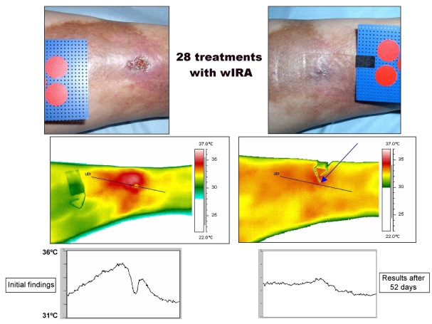
An example of the healing process in a chronic venous stasis ulcer of the lower leg under therapy with wIRA is presented in Figure 13 (Fig. 13).
Figure 13. Example of the healing process of a chronic venous stasis ulcer of the lower leg under therapy with wIRA (Study Tromsø/Hillerød) .
(28 times 30 minutes irradiation with water-filtered infrared-A (wIRA) and visible light (VIS) within 52 days = approximately 7 weeks) with normal view, thermographic image, and temperature profile across the ulcer, in each case to the left before therapy and to the right after completion of the course of therapy. The arrow in the thermographic image - taken after completion of the course of therapy - points to the place where the wound has been. Diameter of the red circles: 16 mm. (Study of the University of Tromsø/Norway and the Hospital in Hillerød/Denmark) (adapted from [3], [4], [23])
wIRA for chronic venous stasis ulcers of the lower legs (Study of the University of Freiburg, Department of Dermatology)
In a prospective, randomized, controlled, blinded study, 51 patients with non-healing chronic venous stasis ulcers of the lower legs were treated with compression therapy, wound cleansing, non-adhesive wound dressings and 30 minutes irradiation five times per week over 9 weeks. A preliminary analysis of this study has shown advanced wound healing, improved granulation and in the later phase of treatment a decrease of the bacterial burden in the group with wIRA(+VIS) compared to a control group with VIS only [3].
wIRA for chronic venous stasis ulcers of the lower legs (Example)
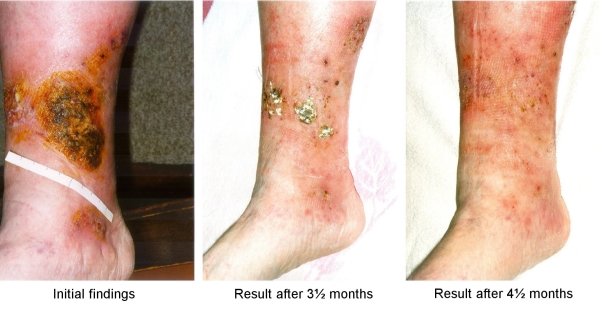
An additional example is presented in Figure 14 (Fig. 14).
Figure 14. Example of the healing process of a chronic venous stasis ulcer of the lower leg under therapy with wIRA .
88 year-old woman with an infected (lightly malodorous) crustaceous ulcer (of the right distal medial lower leg), which had persisted for 13 months and had increased despite conservative dermatological therapy including local antisepsis, systemic antibiotic, and non-adhesive wound dressing up to 10 cm in diameter. Chronic venous insufficiency with marked stasis-related edemas of the lower legs and extensive stasis dermatitis, diabetes mellitus type II (orally treated), slightly overweight, and decreased amount of daily motion. Under irradiation with wIRA(+VIS) 30 minutes once daily, compression therapy, local antisepsis, non-adhesive wound dressing and the possibility of ending the systemic antibiotic therapy, a complete wound closure was achieved within 4½ months:
initial findings, result after 3½ months, result after 4½ months (healed) (adapted from [3], [4])
Other wound-related indications of wIRA
Some case reports have demonstrated that wIRA can even be used for mixed arterial-venous ulcers or arterial ulcers, if an appropriately low irradiation intensity is chosen and if irradiation is monitored carefully [3].
wIRA can be used for decubital ulcers both as a preventive and as a therapeutic measure [3].
wIRA can also improve the resorption of topically applied substances [54], [55], [56] in wounds [3].
Endogenous PDT-like effect of wIRA
An irradiation with VIS and wIRA presumably acts with endogenous protoporphyrin IX (or protoporphyrin IX of bacteria) in a manner similar to a mild photodynamic therapy (endogenous PDT-like effect). This could lead to improved cell regeneration and wound healing and to antibacterial effects [3], [57].
Perspectives for wIRA for the improvement of healing of chronic wounds
In conclusion, these results indicate that wIRA can generally be recommended for use in the treatment of chronic wounds [3].
Résumé
wIRA can considerably alleviate pain, is capable of diminishing exudation and inflammation and can reduce infections. wIRA can advance wound healing or improve an impaired wound healing both in acute and in chronic wounds. Even the normal wound healing process can be improved.
wIRA can generally be recommended for use in the treatment of acute as well as of chronic wounds.
References
- 1.Hoffmann G. Principles and working mechanisms of water-filtered infrared-A (wIRA) in relation to wound healing [review] GMS Krankenhaushyg Interdiszip. 2007;2(2):Doc54. Available from: http://www.egms.de/en/journals/dgkh/2007-2/dgkh000087.shtml. [PMC free article] [PubMed] [Google Scholar]
- 2.Hartel M, Illing P, Mercer JB, Lademann J, Daeschlein G, Hoffmann G. Therapy of acute wounds with water-filtered infrared-A (wIRA) [review] GMS Krankenhaushyg Interdiszip. 2007;2(2):Doc53. Available from: http://www.egms.de/en/journals/dgkh/2007-2/dgkh000086.shtml. [PMC free article] [PubMed] [Google Scholar]
- 3.von Felbert V, Schumann H, Mercer JB, Strasser W, Daeschlein G, Hoffmann G. Therapy of chronic wounds with water-filtered infrared-A (wIRA) [review] [review]. GMS Krankenhaushyg Interdiszip. 2007;2(2):Doc52. (Ger). Available from: http://www.egms.de/en/journals/dgkh/2008-2/dgkh000085.shtml. [PMC free article] [PubMed] [Google Scholar]
- 4.Hoffmann G. Wassergefiltertes Infrarot A (wIRA) zur Verbesserung der Wundheilung [Übersichtsarbeit] [Water-filtered infrared A (wIRA) for the improvement of wound healing [review]]. GMS Krankenhaushyg Interdiszip. 2006;1(1):Doc20. (Ger). Available from: http://www.egms.de/en/journals/dgkh/2006-1/dgkh000020.shtml. [Google Scholar]
- 5.Hoffmann G. Wassergefiltertes Infrarot A (wIRA) zur Verbesserung der Wundheilung bei akuten und chronischen Wunden. [Water-filtered Infrared-A (wIRA) for the improvement of wound healing of acute and chronic wounds]. Wundmanagement. 2008;2:72–80. (Ger). Available from: http://publikationen.ub.uni-frankfurt.de/volltexte/2008/5429/ [Google Scholar]
- 6.Hoffmann G. Klinische Anwendungen von wassergefiltertem Infrarot A (wIRA) In: Kaase H, Serick F, editors. Sechstes Symposium "Licht und Gesundheit" [Sixth symposium "Light and health"]. Eine Sondertagung der Technischen Universität Berlin und der Deutschen Gesellschaft für Photobiologie mit der Deutschen Akademie für Photobiologie und Phototechnologie und der Deutschen Lichttechnischen Gesellschaft, Berlin, 13./14.03.2008. Berlin: 2008. pp. 130–146. (Ger). Available from: http://publikationen.ub.uni-frankfurt.de/volltexte/2008/5484/ [Google Scholar]
- 7.Fuchs SM, Fluhr JW, Bankova L, Tittelbach J, Hoffmann G, Elsner P. Photodynamic therapy (PDT) and waterfiltered infrared A (wIRA) in patients with recalcitrant common hand and foot warts. Ger Med Sci. 2004;2:Doc08. Available from: http://www.egms.de/en/gms/2004-2/000018.shtml. [PMC free article] [PubMed] [Google Scholar]
- 8.Cobarg CC. Physikalische Grundlagen der wassergefilterten Infrarot-A-Strahlung. In: Vaupel P, Krüger W, editors. Wärmetherapie mit wassergefilterter Infrarot-A-Strahlung. Grundlagen und Anwendungsmöglichkeiten. 2. Aufl. [Thermal therapy with water-filtered infrared-A radiation. The fundamentals and applications]. Stuttgart: Hippokrates; 1995. pp. 19–28. (Ger). [Google Scholar]
- 9.Rzeznik J. Die Technik zur loko-regionalen Wärmetherapie mit wassergefilterter Infrarot-A-Strahlung. In: Vaupel P, Krüger W, editors. Wärmetherapie mit wassergefilterter Infrarot-A-Strahlung. Grundlagen und Anwendungsmöglichkeiten. 2. Aufl. [Thermal therapy with water-filtered infrared-A radiation. The fundamentals and applications]. Stuttgart: Hippokrates; 1995. pp. 29–46. (Ger). [Google Scholar]
- 10.Albrecht-Buehler G. Surface extensions of 3T3 cells towards distant infrared light sources. J Cell Biol. 1991;114(3):493–502. doi: 10.1083/jcb.114.3.493. Available from: http://dx.doi.org/10.1083/jcb.114.3.493. [DOI] [PMC free article] [PubMed] [Google Scholar]
- 11.Albrecht-Buehler G. Cellular infrared detector appears to be contained in the centrosome. Cell Motil Cytoskeleton. 1994;27(3):262–271. doi: 10.1002/cm.970270307. Available from: http://dx.doi.org/10.1002/cm.970270307. [DOI] [PubMed] [Google Scholar]
- 12.Albrecht-Buehler G. A long-range attraction between aggregating 3T3 cells mediated by near-infrared light scattering. Proc Natl Acad Sci U S A. 2005;102(14):5050–5055. doi: 10.1073/pnas.0407763102. Available from: http://dx.doi.org/10.1073/pnas.0407763102. [DOI] [PMC free article] [PubMed] [Google Scholar]
- 13.Ehrlicher A, Betz T, Stuhrmann B, Koch D, Milner V, Raizen MG, Käs J. Guiding neuronal growth with light. Proc Natl Acad Sci USA. 2002;99(25):16024–16028. doi: 10.1073/pnas.252631899. Available from: http://dx.doi.org/10.1073/pnas.252631899. [DOI] [PMC free article] [PubMed] [Google Scholar]
- 14.Karu TI, Pyatibrat LV, Kalendo GS. Cell attachment to extracellular matrices is modulated by pulsed radiation at 820 nm and chemicals that modify the activity of enzymes in the plasma membrane. Lasers Surg Med. 2001;29(3):274–281. doi: 10.1002/lsm.1119. Available from: http://dx.doi.org/10.1002/lsm.1119. [DOI] [PubMed] [Google Scholar]
- 15.Karu TI, Pyatibrat LV, Kalendo GS. Donors of NO and pulsed radiation at lambda = 820 nm exert effects on cell attachment to extracellular matrices. Toxicol Lett. 2001;121(1):57–61. doi: 10.1016/S0378-4274(01)00315-0. Available from: http://dx.doi.org/10.1016/S0378-4274(01)00315-0. [DOI] [PubMed] [Google Scholar]
- 16.Karu TI, Pyatibrat LV, Kalendo GS. Cell attachment modulation by radiation from a pulsed light diode (lambda = 820 nm) and various chemicals. Lasers Surg Med. 2001;28(3):227–236. doi: 10.1002/lsm.1043. Available from: http://dx.doi.org/10.1002/lsm.1043. [DOI] [PubMed] [Google Scholar]
- 17.Chow RT, Heller GZ, Barnsley L. The effect of 300 mW, 830 nm laser on chronic neck pain: a double-blind, randomized, placebo-controlled study. Pain. 2006;124(1-2):201–210. doi: 10.1016/j.pain.2006.05.018. Available from: http://dx.doi.org/10.1016/j.pain.2006.05.018. [DOI] [PubMed] [Google Scholar]
- 18.Gebbers N, Hirt-Burri N, Scaletta C, Hoffmann G, Applegate LA. Water-filtered infrared-A radiation (wIRA) is not implicated in cellular degeneration of human skin. GMS Ger Med Sci. 2007;5:Doc08. Available from: http://www.egms.de/en/gms/2007-5/000044.shtml. [PMC free article] [PubMed] [Google Scholar]
- 19.Hartel M, Hoffmann G, Wente MN, Martignoni ME, Büchler MW, Friess H. Randomized clinical trial of the influence of local water-filtered infrared A irradiation on wound healing after abdominal surgery. Br J Surg. 2006;93(8):952–960. doi: 10.1002/bjs.5429. Available from: http://dx.doi.org/10.1002/bjs.5429. [DOI] [PubMed] [Google Scholar]
- 20.Vaupel P, Rzeznik J, Stofft E. Wassergefilterte Infrarot-A-Strahlung versus konventionelle Infrarotstrahlung: Temperaturprofile bei lokoregionaler Wärmetherapie. [Water-filtered infrared-A radiation versus conventional infrared-A radiation: temperature profiles upon loco-regional thermotherapy]. Phys Rehab Kur Med. 1995;5:77–81. doi: 10.1055/s-2008-1061959. (Ger). Available from: http://dx.doi.org/10.1055/s-2008-1061959. [DOI] [Google Scholar]
- 21.Stofft E, Vaupel P. Wassergefilterte Infrarot-A-Strahlung versus Fango-Paraffin-Packung: Temperaturprofile bei lokoregionaler Wärmetherapie. [Water-filtered infrared-A radiation versus fango-paraffin pack: temperature profiles upon loco-regional thermotherapy]. Phys Rehab Kur Med. 1996;6:7–11. doi: 10.1055/s-2008-1061893. (Ger). Available from: http://dx.doi.org/10.1055/s-2008-1061893. [DOI] [Google Scholar]
- 22.Vaupel P, Stofft E. Wassergefilterte Infrarot-A-Strahlung im Vergleich zu konventioneller Infrarotstrahlung oder Fango-Paraffin-Packungen: Temperaturprofile bei lokaler Wärmetherapie. In: Vaupel P, Krüger W, editors. Wärmetherapie mit wassergefilterter Infrarot-A-Strahlung. Grundlagen und Anwendungsmöglichkeiten. 2. Aufl. [Thermal therapy with water-filtered infrared-A radiation. The fundamentals and applications]. Stuttgart: Hippokrates; 1995. pp. 135–147. (Ger). [Google Scholar]
- 23.Mercer JB, Nielsen SP, Hoffmann G. Improvement of wound healing by water-filtered infrared-A (wIRA) in patients with chronic venous stasis ulcers of the lower legs including evaluation using infrared thermography. GMS Ger Med Sci. 2008;6:Doc11. Available from: http://www.egms.de/en/gms/2008-6/000056.shtml. [PMC free article] [PubMed] [Google Scholar]
- 24.Mercer JB, de Weerd L. The effect of water-filtered infrared-A (wIRA) irradiation on skin temperature and skin blood flow as evaluated by infrared thermography and scanning laser Doppler imaging. Thermology Int. 2005;15(3):89–94. [Google Scholar]
- 25.Pascoe DD, Mercer JB, de Weerd L. Physiology of thermal signals. In: Bronzino JD, editor. Biomedical Engineering Handbook. 3rd edition. Boca Raton (Florida/USA): Tailor and Francis Group, CRC press; 2006. pp. 21–1 . [Google Scholar]
- 26.Hellige G, Becker G, Hahn G. Temperaturverteilung und Eindringtiefe wassergefilterter Infrarot-A-Strahlung. In: Vaupel P, Krüger W, editors. Wärmetherapie mit wassergefilterter Infrarot-A-Strahlung. Grundlagen und Anwendungsmöglichkeiten. 2. Aufl. [Thermal therapy with water-filtered infrared-A radiation. The fundamentals and applications]. Stuttgart: Hippokrates; 1995. pp. 63–79. (Ger). [Google Scholar]
- 27.Melling AC, Ali B, Scott EM, Leaper DJ. Effects of preoperative warming on the incidence of wound infection after clean surgery: a randomised controlled trial. Lancet. 2001;358(9285):876–880. doi: 10.1016/S0140-6736(01)06071-8. Available from: http://dx.doi.org/10.1016/S0140-6736(01)06071-8. [DOI] [PubMed] [Google Scholar]
- 28.Plattner O, Akca O, Herbst F, Arkilic CF, Függer R, Barlan M, Kurz A, Hopf H, Werba A, Sessler DI. The influence of 2 surgical bandage systems on wound tissue oxygen tension. Arch Surg. 2000;135(7):818–822. doi: 10.1001/archsurg.135.7.818. Available from: http://dx.doi.org/10.1001/archsurg.135.7.818. [DOI] [PubMed] [Google Scholar]
- 29.Kivisaari J, Vihersaari T, Renvall S, Niinikoski J. Energy metabolism of experimental wounds at various oxygen environments. Ann Surg. 1975;181:823–828. doi: 10.1097/00000658-197506000-00011. Available from: http://dx.doi.org/10.1097/00000658-197506000-00011. [DOI] [PMC free article] [PubMed] [Google Scholar]
- 30.Kühne HH, Ullmann U, Kühne FW. New aspects on the pathophysiology of wound infection and wound healing - the problem of lowered oxygen pressure in the tissue. Infection. 1985;13(2):52–56. doi: 10.1007/BF01660413. Available from: http://dx.doi.org/10.1007/BF01660413. [DOI] [PubMed] [Google Scholar]
- 31.Niinikoski J, Gottrup F, Hunt TK. The role of oxygen in wound repair. In: Janssen H, Rooman R, Robertson JIS, editors. Wound healing. Petersfield: Wrightson Biomedical Publishing; 1991. pp. 165–174. [Google Scholar]
- 32.Hoffmann G. Improvement of wound healing in chronic ulcers by hyperbaric oxygenation and by waterfiltered ultrared A induced localized hyperthermia. Adv Exp Med Biol. 1994;345:181–188. doi: 10.1007/978-1-4615-2468-7_24. [DOI] [PubMed] [Google Scholar]
- 33.Buslau M, Hoffmann G. Hyperbaric oxygenation in the treatment of skin diseases [review] In: Fuchs J, Packer L, editors. Oxidative stress in dermatology. New York: Marcel Dekker; 1993. pp. 457–485. [Google Scholar]
- 34.Buslau M, Hoffmann G. Die hyperbare Oxygenation (HBO) - eine adjuvante Therapie akuter und chronischer Wundheilungsstörungen [Review] [Hyperbaric oxygenation - an adjuvant therapy of acute and chronic wound healing impairments]. Dermatol Monatsschr. 1993;179:39–54. (Ger). [Google Scholar]
- 35.Hoffmann G, Buslau M. Treatment of skin diseases by hyperbaric oxygenation. In: Cramer FS, editor. Proceedings of the Eleventh International Congress on Hyperbaric Medicine. Flaggstaff, USA: Best Publishing Company; 1995. pp. 20-1, 153–9. [Google Scholar]
- 36.Wright J. Hyperbaric oxygen therapy for wound healing. World Wide Wounds. 2001 Available from: http://www.worldwidewounds.com/2001/april/Wright/|HyperbaricOxygen.html. [Google Scholar]
- 37.Knighton DR, Silver IA, Hunt TK. Regulation of wound-healing angiogenesis - effect of oxygen gradients and inspired oxygen concentration. Surgery. 1981;90:262–270. [PubMed] [Google Scholar]
- 38.Jünger M, Hahn M, Klyscz T, Steins A. (Progr. Appl. Microc.; Vol. 23).Role of microangiopathy in the development of venous leg ulcers. 1999;23:180–193. [Google Scholar]
- 39.Karu TI. Primary and secondary mechanisms of action of visible to near-IR radiation on cells [review] J Photochem Photobiol B. 1999;49(1):1–17. doi: 10.1016/S1011-1344(98)00219-X. Available from: http://dx.doi.org/10.1016/S1011-1344(98)00219-X. [DOI] [PubMed] [Google Scholar]
- 40.Karu TI. Low-power laser effects. In: Waynant RW, editor. Lasers in medicine. Boca Raton: CRC Press; 2002. pp. 171–209. [Google Scholar]
- 41.Danno K, Mori N, Toda K, Kobayashi T, Utani A. Near-infrared irradiation stimulates cutaneous wound repair: laboratory experiments on possible mechanisms. Photodermatol Photoimmunol Photomed. 2001;17(6):261–265. doi: 10.1034/j.1600-0781.2001.170603.x. [DOI] [PubMed] [Google Scholar]
- 42.Horwitz LR, Burke TJ, Carnegie D. Augmentation of wound healing using monochromatic infrared energy. Exploration of a new technology for wound management. Adv Wound Care. 1999;12(1):35–40. [PubMed] [Google Scholar]
- 43.Menezes S, Coulomb B, Lebreton C, Dubertret L. Non-coherent near infrared radiation protects normal human dermal fibroblasts from solar ultraviolet toxicity. J Invest Dermatol. 1998;111(4):629–633. doi: 10.1046/j.1523-1747.1998.00338.x. Available from: http://dx.doi.org/10.1046/j.1523-1747.1998.00338.x. [DOI] [PubMed] [Google Scholar]
- 44.Frank S, Menezes S, Lebreton-De Coster C, Oster M, Dubertret L, Coulomb B. Infrared radiation induces the p53 signaling pathway: role in infrared prevention of ultraviolet B toxicity. Exp Dermatol. 2006;15(2):130–137. doi: 10.1111/j.1600-0625.2005.00397.x. Available from: http://dx.doi.org/10.1111/j.1600-0625.2005.00397.x. [DOI] [PubMed] [Google Scholar]
- 45.Frank S, Oliver L, Lebreton-De Coster C, Moreau C, Lecabellec MT, Michel L, Vallette FM, Dubertret L, Coulomb B. Infrared radiation affects the mitochondrial pathway of apoptosis in human fibroblasts. J Invest Dermatol. 2004;123(5):823–831. doi: 10.1111/j.0022-202X.2004.23472.x. Available from: http://dx.doi.org/10.1111/j.0022-202X.2004.23472.x. [DOI] [PubMed] [Google Scholar]
- 46.Danno K, Horio T, Imamura S. Infrared radiation suppresses ultraviolet B-induced sunburn-cell formation. Arch Dermatol Res. 1992;284(2):92–94. doi: 10.1007/BF00373376. Available from: http://dx.doi.org/10.1007/BF00373376. [DOI] [PubMed] [Google Scholar]
- 47.Applegate LA, Scaletta C, Panizzon R, Frenk E, Hohlfeld P, Schwarzkopf S. Induction of the putative protective protein ferritin by infrared radiation: implications in skin repair. Int J Mol Med. 2000;5(3):247–251. doi: 10.3892/ijmm.5.3.247. [DOI] [PubMed] [Google Scholar]
- 48.Burri N, Gebbers N, Applegate LA. Chronic infrared-A radiation repair: Implications in cellular senescence and extracellular matrix. In: Pandalai SG, editor. Recent Research Developments in Photochemistry & Photobiology. Vol. 7. Trivandrum: Transworld Research Network; 2004. pp. 219–231. [Google Scholar]
- 49.Hoffmann G, Meffert H. Apparent contradiction between negative effects of UV radiation and positive effects of sun exposure. GMS Ger Med Sci. 2005;3:Doc01. Available from: http://www.egms.de/en/gms/2005-3/000019.shtml. [PMC free article] [PubMed] [Google Scholar]
- 50.Jung T, Höhn A, Piazena H, Grune T. Effects of water-filtered infrared A irradiation on human fibroblasts. Free Radic Biol Med. 2009 Oct 21; doi: 10.1016/j.freeradbiomed.2009.10.036. Available from: http://dx.doi.org/10.1016/j.freeradbiomed.2009.10.036. [DOI] [PubMed] [Google Scholar]
- 51.Piazena H, Kelleher DK. Effects of infrared-A irradiation on skin: discrepancies in published data highlight the need for an exact consideration of physical and photobiological laws and appropriate experimental settings. Photochem Photobiol. 2009 doi: 10.1111/j.1751-1097.2010.00729.x. [DOI] [PubMed] [Google Scholar]
- 52.Hoffmann G. Wassergefiltertes Infrarot A (wIRA) in der Wundbehandlung: Interview; 3 Fragen an Prof. Dr. med. Gerd Hoffmann. [Water-filtered infrared-A (wIRA) in the treatment of wounds: interview; 3 questions to Prof. Gerd Hoffmann, MD]. Heilberufe. 2009;61(7):14. (Ger). Available from: http://publikationen.ub.uni-frankfurt.de/volltexte/2009/6802/ [Google Scholar]
- 53.Biland L, Barras J. Die wassergefilterte Infrarot-A-Hyperthermie zur Behandlung venöser Ulcera. [Water-filtered infrared-A induced hyperthermia used as therapy of venous ulcers]. Hefte Wundbehand. 2001;5:41. (Ger). [Google Scholar]
- 54.Haupenthal H. In vitro- und in vivo-Untersuchungen zur temperaturgesteuerten Arzneistoff-Liberation und Permeation [Thesis] [In vitro and in vivo investigations of temperature dependent drug liberation and permeation]. Mainz: Johannes Gutenberg-Universität; 1997. [Google Scholar]
- 55.Bankova L, Heinemann C, Fluhr JW, Hoffmann G, Elsner P. Improvement of penetration of a topical corticoid by waterfiltered infrared A (wIRA). 1st Joint Meeting 14th International Congress for Bioengineering and the Skin & 8th Congress of the International Society for Skin Imaging; 2003 May 21-24; Hamburg. 2003. p. P96. [Google Scholar]
- 56.Otberg N, Grone D, Meyer L, Schanzer S, Hoffmann G, Ackermann H, Sterry W, Lademann J. Water-filtered infrared-A (wIRA) can act as a penetration enhancer for topically applied substances. GMS Ger Med Sci. 2008;6:Doc08. Available from: http://www.egms.de/en/gms/2008-6/000053.shtml. [PMC free article] [PubMed] [Google Scholar]
- 57.Hoffmann G. Wassergefiltertes Infrarot A (wIRA) In: Kramer A, Assadian O, editors. Wallhäußers Praxis der Sterilisation, Desinfektion, Antiseptik und Konservierung. Qualitätssicherung der Hygiene in Industrie, Pharmazie und Medizin. [Wallhäußer's practice of sterilization, disinfection, antisepsis and conservation. Quality assurance of the hygiene in industry, pharmacy, and medicine]. Stuttgart: Thieme; 2008. pp. 899–900. (Ger). Available from: http://publikationen.ub.uni-frankfurt.de/volltexte/2009/6135/ [Google Scholar]
- 58.Carter DR, editor. Lancester, PA, USA: Burle Industries; 2008. Electro-Optics Handbook [company publication] pp. Section 6, 61–80. Available from: http://www.burle.com/cgi-bin/byteserver.pl/pdf/Electro_Optics.pdf. [Google Scholar]
- 59.Valley SL, editor. Air Force Cambridge Research Laboratories, Office of Aerospace Research, U.S. Air Force; 1965. Handbook of geophysics and space environments. Also published: New York, N.Y.: McGraw-Hill Book Co.; 1965. [Google Scholar]


