Abstract
Background
Selective inhibition of the BCR-ABL tyrosine kinase by RNA interference has been demonstrated in leukemic cells. We, therefore, evaluated specific BCR-ABL small interfering RNA silencing in BCR-ABL-positive cell lines, including those resistant to imatinib and particularly those with the T315I mutation.
Design and Methods
The factor-independent 32Dp210 BCR-ABL oligoclonal cell lines and human imatinib-resistant BCR-ABL-positive cells from patients with leukemic disorders were investigated. The effects of BCR-ABL small interfering RNA or the combination of BCR-ABL small interfering RNA with imatinib and nilotinib were compared with those of the ABL inhibitors imatinib and nilotinib.
Results
Co-administration of BCR-ABL small interfering RNA with imatinib or nilotinib dramatically reduced BCR-ABL expression in wild-type and mutated BCR-ABL cells and increased the lethal capacity. BCR-ABL small interfering RNA significantly induced apoptosis and inhibited proliferation in wild-type (P<0.0001) and mutated cells (H396P, T315I, P<0.0001) versus controls. Co-treatment with BCR-ABL small interfering RNA and imatinib or nilotinib resulted in increased inhibition of proliferation and induction of apoptosis in T315I cells as compared to imatinib or nilotinib alone (P<0.0001). Furthermore, the combination of BCR-ABL small interfering RNA with imatinib or nilotinib significantly (P<0.01) reversed multidrug resistance-1 gene-dependent resistance of mutated cells. In T315I cells BCR-ABL small interfering RNA with nilotinib had powerful effects on cell cycle distribution.
Conclusions
Our data suggest that silencing by BCR-ABL small interfering RNA combined with imatinib or nilotinib may be associated with an additive antileukemic activity against tyrosine kinase inhibitor-sensitive and resistant BCR-ABL cells, and might be an alternative approach to overcome BCR-ABL mutations.
Keywords: small interfering RNA, CML, BCR-ABL
Introduction
Chronic myeloid leukemia (CML) is a relatively well differentiated myeloproliferative disorder originating from transformed hematopoietic stem cells. CML is characterized by the Philadelphia chromosome, which results in the expression of the BCR-ABL fusion gene, and is derived from the fusion of the cellular breakpoint cluster region (BCR) gene and the Abelson murine leukemia oncogene (ABL).1,2 Treatment with imatinib mesylate, a selective inhibitor of ABL, the chimeric BCR-ABL fusion protein, platelet-derived growth factor receptor (PDGF-R) α and β, and stem cell factor receptor (c-KIT), has shown remarkable clinical activity with minimal side effects in BCR-ABL+, C-KIT+ or PDGFR+ malignancies and resulted in complete cytogenetic remission in a majority of CML patients in chronic phase, but in fewer patients with accelerated and blast phase disease.3,4 However, despite the impressive efficacy of imatinib, resistance to this drug can emerge and is a significant clinical issue. In the phase III International Randomized Interferon versus STI571 (IRIS) trial, an estimated 5% of patients with newly diagnosed chronic phase-CML failed to achieve a complete hematologic response at 3 months, 16% failed to achieve a major cytogenetic response with imatinib at 12 months, and 24% failed to achieve a complete cytogenetic response at 18 months. The estimated relapse rate of patients in this trial was 17% and 7% of patients developed disease progression.5 Although allografting is still considered to be a curative option in CML, it is associated with significant mortality and morbidity, thus the number of transplants performed for this disease has dropped dramatically since imatinib became available.6 To overcome resistance, strategies such as dose escalation, combination with conventional drugs (cytarabine, interferon), alternative BCR-ABL inhibitors, and BCR-ABL protein down-regulating agents have been adopted.7
Nilotinib (ANM-107, Tasigna®, Novartis Pharmaceuticals Corp.) is a high-affinity aminopyrimidine-based ATP-competitive inhibitor that decreases proliferation and viability of wild-type BCR-ABL and imatinib-resistant BCR-ABL mutant-expressing cells in vitro by selectively inhibiting BCR-ABL autophosphorylation. Nilotinib is more potent than imatinib as an inhibitor of BCR-ABL in a wide range of CML-derived and transfected cell lines.8–10 In 2007 the U.S. Food and Drug Administration granted accelerated approval for the use of nilotinib in the treatment of chronic and accelerated phase Philadelphia chromosome-positive CML in adult patients resistant or intolerant to prior therapy that included imatinib.11 Gene targeting of BCR-ABL fusion transcripts is an ideal way to selectively kill those cells, leaving normal cells unaffected.
RNA interference is an evolutionarily conserved cellular mechanism that mediates sequence-specific post-transcriptional gene silencing initiated by double-stranded RNA. Small interfering RNA (siRNA) are the mediators of mRNA degradation in the process of RNA interference. Synthetic siRNA are able to mediate cleavage of the target RNA, as shown by Elbashir et al.12 Scherr et al. published a study showing that siRNA directed against BCR-ABL can specifically inhibit BCR-ABL expression in Philadelphia chromosome-positive cell lines and cells from CML patients.13 Furthermore, Wohlbold et al. showed that siRNA treatment might sensitize cells to imatinib contributing to its therapeutic potential.14 We demonstrated that combined transfection with Wilms’ tumor 1 gene (WT1) siRNA and BCR-ABL siRNA in Philadelphia chromosome-positive cell lines and cells of CML patients enhanced inhibition of cell proliferation and induction of apoptosis compared to transfection with BCR-ABL siRNA or WT1 siRNA alone.15 In addition, we showed that BCR-ABL siRNA had anti-proliferative and pro-apoptotic effects on Philadelphia chromosome-positive AML cells in vitro.16 Importantly, we demonstrated the feasibility of targeted, non-viral delivery of BCR-ABL siRNA as a therapeutic approach in a female CML patient with imatinib-resistant bone marrow and extramedullary relapse after allogeneic hematopoietic stem cell transplantation.17
In this study, we investigated the activity of the two protein tyrosine kinase inhibitors imatinib and nilotinib. We compared these agents to BCR-ABL siRNA in several murine bcr-abl-positive cell lines which differ in their sensitivity to imatinib or nilotinib and in human imatinib-resistant BCR-ABL-positive cells from patients with acute leukemias.
Design and Methods
Cell culture
The factor-independent 32Dp210 BCR-ABL oligoclonal cell line was generated by transfection of parental cells with the retroviral vector Migp210, Migp210-Thr315Ile, or Migp210-His396Pro, as previously described.18,19 All transfected 32Dp210 cell lines were a generous gift from Dr. H. van der Kuip (Stuttgart, Germany) and Prof. Dr. J. Duyster (Munich, Germany). The cells were grown in RPMI 1640 medium (Invitrogen, Heidelberg, Germany) supplemented with 10% fetal bovine serum (FBS) complemented with glutamine as described. All cells were maintained in a humidified 37°C incubator with 5% CO2.
Leukemia cells from bone marrow of patients were also used. All patients gave informed written consent to the donation and use of their cells.
Drug dosing and administration
Imatinib (IM; Gliveec®) and nilotinib (Tasigna®) were kindly provided by Novartis Pharma AG (Novartis, Germany). A stock solution of imatinib (10 mg/mL) was prepared by dissolving the compound in dimethylsulfoxide (DMSO): H2O (1:1) and was then stored at −20°C. Imatinib was used at a concentration of 1 μM for 32Dp210-wt and 32Dp210-His396Pro cells and at 1 or 3 μM for 32Dp210Thr315Ile cells. A stock solution of nilotinib (10 mg/mL) was prepared by dissolving the compound in DMSO:H2O (1:1) and this too was stored at −20°C. Nilotinib was used at a concentration of 1 μM for 32Dp210wt and 32Dp210-His396Pro cells and 1 or 3 μM for 32Dp210-Thr315Ile cells. For the tests using a combination of BCR-ABL siRNA with imatinib or nilotinib, cells were treated with imatinib or nilotinib and 54 pM siRNA for 24–48 h.
Transfection of small interfering RNA
Sequences of siRNA directed against the BCR-ABL transcript were published by Scherr et al.13 The siRNA was purchased from Qiagen-Xeragon (Hilden, Germany) with the sense 5′-GCAGAG UUCAAAAGCCCUUdTdT and antisense 5′-AAGGGCUUU UGAACUCUGCdTdT and a scrambled siRNA sequence was used as a control (sense 5′-UUGUACGGCAUC AGCGUUAdTdT and antisense 5′-UUACGUGAUGCCGUA CAAdTdT). In vitro transfections with 0.8 μg (54 pM) siRNA were performed in 24-well plates using the DOTAP® liposomal transfection reagent (1×10⁵ cells/well) (Roche Applied Science, Indianapolis, USA) following the manufacturer’s protocol. Transfection of siRNA was performed at least five times in each experiment. Each experiment was repeated at least twice. As a control we used two or more non-silencing siRNA (mismatched or scrambled siRNA) from Qiagen.
RNA isolation and real-time reverse transcriptase polymerase chain reaction
RNA was isolated using the RNeasy Mini Kit (Qiagen, Hilden, Germany), according to the manufacturer’s instructions. Real time reverse transcription-polymerase chain reaction (RT-PCR) was performed as previously described for BCR-ABL and glyceraldehyde-3-phosphate dehydrogenase (GAPDH).20 For BCR-ABL and 6-glucosephosphate dehydrogenase we used a commercial Light-Cycler®-t(9;22) Quantification Kit (Roche Molecular Diagnostics, Mannheim, Germany) following the manufacturer’s protocol. For MDR1 RT-PCR we used the following primers and hybridization probes: primers 5′-ggA AgC CAA TgC CTA TgA CTT TA-3′ and 5′gAA CCA Ctg CTT CgC TTT CTg-3′, hybridization probes 5′-6FAM-TgA AAC TgC CTC ATA AAT TTg ACA CCC Tgg-TAMRA. For quantification, RNA transcript expression was normalized by determining the ratio between expression levels of targets and GAPDH.
Growth inhibition assay
Cells were cultured in 96-well plates at a density of 5000 cells/well and left to recover. The quantity of viable cells was estimated by a colorimetric assay using 3-(4,5-dimethylthiazol-2-yl)-2,5-diphenyltetrazolium bromide (MTT). MTT (10 μL of 5 mg/mL solution, Sigma Chemical Co., Germany) was added to each well of the titration plate and incubated for 4 h at 37°C. The cells were then treated with DMSO (40 μL/well) and incubated for 60 min at 37°C. The absorbance of each well was determined in an enzyme linked immunosorbent assay (ELISA) plate reader using an activation wavelength of 570 nm and a reference wavelength of 630 nm. The percentage of viable cells was determined by comparison to untreated control cells.
Terminal transferase dUTP nick-end labeling assay
Apoptotic cells were determined using the in situ cell Death Detection kit from Roche Molecular Diagnostics (Mannheim, Germany) following the manufacturer’s instructions. The apoptotic cells (brown staining) were counted under a microscope. The apoptotic index was defined by the percentage of brown (dark) cells among the total number of cells in each sample. Five fields with 100 cells in each field were randomly counted for each sample. At least three samples with 15 single analyses in total were counted.
Cell proliferation assay
Cell proliferation was determined by 5-bromo-2-deoxyuridine (BrdU) incorporation. Twenty-four hours (and for up to 48 h) after transfection of siRNA, the 1×10⁵ cells were split into four-well chamber slides and incubated with culture medium containing BrdU for 4 h. BrdU staining was performed using the Roche Kit (Mannheim, Germany) following the manufacturer’s instructions. Proliferation was determined from the percentage of brown stained cells among the total number of cells in each sample with the analyses done in the same way as for apoptotic cells.
Cell cycle analysis
Cells were fixed in ice-cold ethanol (70% v/v) and stained with a solution of propidium iodide (PI) (20 μg/mL PI; 100 μg/mL RNase; 0.1% Triton X-100; and in 870 μg/mL trisodium citrate; Sigma Chemical, Germany) The DNA content was determined using a FACScan flow cytometer (Coulter, Krefeld, Germany). Cell-cycle distribution was analyzed using MultiCycle for Windows (Phoenix Flow System, San Diego, USA). Red fluorescence (585 ± 42 nm) was evaluated on a linear scale, and pulse width analysis was used to exclude cell doublets and aggregates from the analysis. Cells with a DNA content between 2N and 4N were designated as being in the G1, S, or G2/M phase of the cell cycle. The number of cells in each compartment of the cell cycle was expressed as a percentage of the total number of cells present. In sequential combination experiments, cells were pretreated with BCR-ABL siRNA or mismatched BCR-ABL siRNA for 24–48 h, washed twice with phosphate-buffered saline to remove the siRNA, or without BCR-ABL siRNA and analyzed for cell cycle distribution.
Flow cytometry
Between 1.0×10⁵ to 5.0×10⁵ cells were labeled with antibody for multi-color flow cytometry using phycoerythrin- or peridin chlorophyll-conjugated monoclonal antibodies directed against CD45, anti-phosphotyrosine protein (p-Tyr, PY99) and 7-amino-actinomycin D (7-AAD). For intracellular anti-phosphotyrosine protein (p-Tyr, PY99) and anti-phospho-Crkl (p-Tyr207) staining, cells were fixed with 2% paraformaldehyde for 15 min and permeabilized with 0.1% Nonidet P40 for 4 min before intracellular staining. Anti-phosphor-Crkl (p-Tyr207) was added for 30 min at room temperature in the dark. After incubation with the primary antibody, cells were washed twice with buffer and incubated with phycoerythrin cynanin 5-labeled goat anti-rabbit IgG secondary antibody. Cells were again washed and resuspended in 500 μL of phosphate-buffered saline for flow cytometry analysis. All antibodies were obtained from Beckmann-Coulter (Krefeld, Germany) or Santa Cruz Biotechnology Inc. (Heidelberg, Germany). Non-specific binding was corrected with isotype-matched controls. Flow cytometric data were acquired using a four-color Epics XL AF 14075 flow cytometer with Expo 32 ADC software (Coulter, Krefeld, Germany).
Statistics
Differences in data from the various groups were tested by the two-tailed unpaired t test or Mann-Whitney U-test using the SPSS 14 program (SPSS Inc, Chicago, USA).
Results
Efficiency of transfection in 32Dp210 BCR-ABL cell lines and in primary leukemic cells of different BCR-ABL-positive acute leukemic patients
Using fluorescence-marked, non-silencing siRNA (Qiagen) we evaluated the transfection rate in 32Dp210 BCR-ABL oligoclonal cells and in primary BCR-ABL positive leukemic cells. Twenty-four hours after transfection the number of fluorescence-marked cells was quantified using a fluorescence microscope. For this, 100 cells were counted in quintuplicate. We found a mean ± standard deviation (SD) transfection rate of 74.4±7.6% in 32Dp210 BCR-ABL cells and a mean transfection rate of 51.8±11.8% in primary BCR-ABL positive leukemic cells.
Inhibition of cell growth in 32Dp210 BCR-ABL cell lines and leukemic cells of BCR-ABL- positive acute leukemia patients
To assess whether treatment with imatinib, nilotinib, BCR-ABL siRNA or the combination of these drugs with BCR-ABL siRNA affects the viability of BCR-ABL-expressing leukemic cells, these were transfected with BCR-ABL siRNA, scrambled siRNA or without siRNA with constant doses (54 pM) of siRNA or incubated with imatinib or nilotinib for 24–48 h, harvested, and then cell viability analyzed by an MTT assay. As shown in Figure 1A, imatinib, nilotinib, BCR-ABL siRNA and the combination of imatinib or nilotinib with BCR-ABL siRNA significantly decreased the cell viability in 32Dp210-wt cells. Imatinib, nilotinib and the combination with siRNA had the strongest effects on reducing the viability of 32Dp210 BCR-ABL cells to 24.6–27.4% (mean) of control levels. siRNA reduced the viability of these cells to 48.6%. The combination of siRNA with imatinib or nilotinib reduced cell viability to a similar extent as the treatment with a single tyrosine kinase inhibitor (imatinib or nilotinib) in 32Dp210-wt cells. The mismatched control BCR-ABL siRNA, even when used at a dose up to 108 pM, did not inhibit cell viability since 32Dp cells are not sensitive to the scrambled siRNA (data not shown).
Figure 1.
Cell growth rate was measured 24–48 h after treatment with imatinib, nilotinib and transfection with BCR-ABL siRNA or cotreatment of BCR-ABL siRNA with imatinib or nilotinib in tyrosine kinase inhibitor-sensitive (A) and –resistant BCR-ABL oligoclonal cell lines (B: H396P, C: T315I) by the MTT assay. H396P mutated cells are resistant to imatinib. T315I mutated cells are resistant to imatinib and nilotinib. Mean and SD are presented. The percentage of viable cells was determined by comparison with untreated tyrosine kinase inhibitor or mismatched siRNA transfected cells.
Next, we studied whether the inhibition of BCR-ABL translation by siRNA is sufficient to overcome resistance to imatinib or nilotinib, as illustrated in Figure 1B and 1C. In 32Dp210-His396Pro cells, which render the respective cells 5-fold less sensitive to imatinib, we observed a marked reduction of cell viability with nilotinib and the combination of nilotinib with BCR-ABL siRNA, to 46.3% and 31.3% of control values, respectively. siRNA treatment alone resulted in a moderate reduction of viability of the transfected cells to about 65%. The combination of imatinib and BCR-ABL siRNA could reduce cell viability to 42.7% that of control cells. The combination of imatinib with siRNA was more effective than BCR-ABL siRNA or nilotinib alone. A much greater inhibition of 32Dp210-His396Pro cells was found with the combination of nilotinib and BCR-ABL siRNA than with the combination of imatinib and BCR-ABL siRNA (P<0.02), as shown in Figure 1B. In the 32Dp cells with the Thr315Ile mutation, which are completely resistant to imatinib and nilotinib, only BCR-ABL siRNA and the two combinations of siRNA with either imatinib or nilotinib caused a significant reduction of cell viability to 50.3% and 63.5%, respectively. siRNA alone led to a moderate reduction in the viability of the transfected cells to about 64%. The combination of BCR-ABL siRNA with imatinib or nilotinib resulted in a powerful reduction compared with that achieved by siRNA alone (P<0.05) or resistant 32DP control cells (P<0.02). Nilotinib with BCR-ABL siRNA had the strongest effects, reducing the viability of 32Dp210-Thr315Ile cells to 50.3%, but this was not significantly different from the reduction achieved with the combination of imatinib and siRNA (P<0.062), as shown in Figure 1C.
In order to assess the biological significance of these findings, we subsequently studied the effects of BCR-ABL siRNA on cell growth. Cells from patients with imatinib-resistant leukemias were treated with 54 pM BCR-ABL siRNA. To exclude the possibility that effects were not specific to BCR-ABL siRNA, we performed a control experiment using a scrambled BCR-ABL siRNA up to a maximal dose of 108 pM for 48 h, followed by harvest and analysis of cell viability, as presented in the Online Supplementary Table S1.
Taken together, these data show that BCR-ABL siRNA inhibits the growth of both imatinib-resistant 32Dp cells and cells from patients with imatinib-resistant hematologic malignancies.
BCR-ABL gene expression measured by real time reverse-transcriptase polymerase chain reaction analysis
We found a marked reduction of BCR-ABL expression in 32Dp210-wt cells, which was quantified by correlation to the housekeeping gene GAPDH by real-time RT-PCR (ratio of BCR-ABL/GAPDH). The levels of BCR-ABL mRNA were reduced to between 54.7% and 31.9% (mean) after treatment with imatinib, nilotinib or BCR-ABL siRNA transfection in comparison with controls (set at 100%), as shown in Online Supplementary Figure S1A. The combination of a tyrosine kinase inhibitor with BCR-ABL siRNA produced a further reduction of BCR-ABL mRNA to 36.2% when using the siRNA with imatinib and to 15.8% when using siRNA combined with nilotinib. The combination of BCR-ABL siRNA with nilotinib was, indeed, significantly more effective than siRNA combined with imatinib (P<0.01).
In 32Dp210-His396Pro cells we found a significant reduction of BCR-ABL mRNA levels to 40.3% of control levels after nilotinib treatment and to 46.4% after transfection with the BCR-ABL siRNA. The combination of either imatinib or nilotinib with BCR-ABL siRNA reduced the BCR-ABL mRNA to levels ranging from 29.1% to 31.2% in 32Dp210-His396Pro cells (Online Supplementary Figure S1B). Again, the combination of nilotinib with BCR-ABL siRNA caused a significantly greater reduction, to 29%, compared with that achieved by imatinib combined with siRNA (P<0.05).
In 32Dp210-Thr315Ile cells, a significant reduction of BCR-ABL mRNA to 60.4% was seen after transfection with siRNA. Combining the BCR-ABL siRNA with imatinib resulted in a reduction to 46.6% compared with a reduction to 40.9% when siRNA was co-administered with nilotinib (P<0.05 for imatinib with siRNA compared to nilotinib with siRNA) (Online Supplementary Figure S1C). To assess the biological significance of these findings, we subsequently studied the effects of BCR-ABL siRNA on BCR-ABL mRNA levels of the several patients, as presented in the Online Supplementary Table S1. Taken together, these data show that BCR-ABL siRNA inhibits the expression of BCR-ABL mRNA in both imatinib-resistant 32Dp cells and in cells from patients with imatinib-resistant hematologic malignancies.
Phosphotyrosine protein and phospho-Crkl expression measured by flow cytometry
To verify the above described findings, we next performed flow cytometric studies on the effects on phosphotyrosine protein (p-Tyr) and phosphor-Ckrl (p-Crkl) expression, accepted as an excellent method for assessing BCR-ABL status, in the tyrosine kinase inhibitor resistant 32Dp210Thr315Ile cells. We found a significant reduction of p-Tyr expression to 69% or 66% after high doses (up to 3 μM) of imatinib or nilotinib (P<0.01). We did not, however, detect any reduction in p-Crkl expression in 32Dp210-Thr315Ile cells after treatment with high doses of imatinib or nilotinib (Online Supplementary Figure S2). The administration of BCR-ABL siRNA reduced the level of p-Tyr to 48.2% of that in untreated control cells (P<0.0001) and the level of p-Crkl to 45.9% (P<0.001). The combination of nilotinib with BCR-ABL siRNA also achieved significant reduction of the tyrosyl-phosphoproteins, to 35.9% for p-Tyr (P<0.05) and to 5.4% for p-Crkl (P<0.001), as compared to BCR-ABL siRNA alone. Comparing the combination of imatinib and BCR-ABL siRNA (reduction to 11.8%) with that of nilotinib and BCR-ABL siRNA (reduction to 5.4%), the latter had greater efficacy in reducing p-Crkl expression in 32Dp210-Thr315Ile cells (P<0.02). Taken together, these data suggest that the combination of nilotinib and BCR-ABL siRNA results in enhanced inhibition of tyrosyl-phosphoproteins.
Cell proliferation measured by 5-bromo-2-deoxyuridine
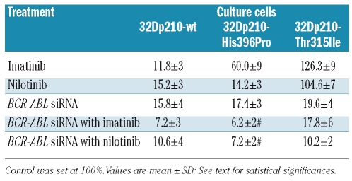
We determined whether treatment with imatinib, nilotinib, BCR-ABL siRNA or the combination of siRNA with either imatinib or nilotinib affects cell proliferation. Cell proliferation was determined by a 5-bromo-2-deoxyuridine (BrdU) incorporation assay following the manufacturer’s instructions. The control was set at 100%. In 32Dp210-wt cells, the administration of imatinib, nilotinib or siRNA alone significantly decreased cell proliferation. The greatest reduction, to 11.8% (mean), occurred with imatinib, whereas the administration of nilotinib and BCR-ABL siRNA caused reductions to 15.2% and 15.8%, respectively (P<0.0001). The combination of siRNA with either imatinib or nilotinib further reduced proliferation to 7.2% and 10.6% (P<0.0001), respectively. In 32Dp210-His396Pro cells, the administration of imatinib led to a lesser reduction in proliferation, as expected, to only 60.0% (P<0.001), whereas nilotinib and siRNA led to reduction in proliferation to 14.2% and 17.4%, respectively (P<0.0001). Despite the limited effect of imatinib alone, the combination of imatinib with siRNA led to the most impressive reduction to 6.2% (P<0.0001), whereas the combination of siRNA with nilotinib caused a reduction to 7.2% (P<0.0001). In 32Dp210-Thr315Ile cells only BCR-ABL siRNA (P<0.0001) and combinations of siRNA with imatinib and nilotinib caused significant reductions of cell proliferation to 19.6% and 10.2%, respectively (P<0.0001). The combination of nilotinib with BCR-ABL siRNA reduced the proliferation rate of the resistant 32Dp210-Thr315Ile cells to 10.2% (P<0.01) of the proliferation rate of these cells after treatment with imatinib combined with siRNA (Table 1).
Table 1.
Proliferation rate, measured by BrdU incorporation after treatment with imatinib, nilotinib and transfection with BCR-ABL siRNA or co-treatment of BCR-ABL siRNA with imatinib or nilotinib in tyrosine kinase inhibitor-sensitive and –resistant BCR-ABL oligoclonal cell lines.
Effect on cell apoptosis, determined by terminal transferase dUTP nick-end labeling assay
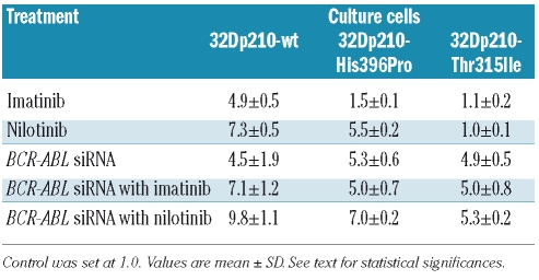
We further investigated the effect of imatinib, nilotinib, or BCR-ABL siRNA alone or in combination with either imatinib or nilotinib on cell apoptosis (Table 2). Numbers of apoptotic cells were determined using an in situ cell death kit according to the manufacturer’s instructions. The increase in apoptosis was compared to a control level set at one. Apoptosis of 32Dp210-wt cells was increased from 4.5 to 9.8-fold by the various treatment strategies: imatinib and siRNA, when administered alone, induced 4.9-fold and 4.5-fold increases in apoptosis, whereas their combination increased apoptosis by 7.1-fold (P<0.0001) as compared to control levels. Effects were greater with nilotinib which, alone, induced a 7.3-fold increase and in combination with the siRNA resulted in a 9.8-fold increase in apoptosis (P<0.0001).
Table 2.
Apoptosis rate, measured by terminal transferase dUTP nick-end labeling assay, after treatment with imatinib, nilotinib and transfection with BCR-ABL siRNA or co-treatment with BCR-ABL siRNA and imatinib or nilotinib in tyrosine kinase inhibitor-sensitive and –resistant BCR-ABL oligoclonal cell lines.
We also examined the apoptotic effect of the drugs administered to 32Dp210-His396Pro cells, which are 5-fold less sensitive to imatinib, and to the fully resistant 32Dp210-Thr315Ile cells. With regards to 32Dp210-His396Pro cells, the administration of imatinib alone failed to induce significant apoptosis, whereas either nilotinib or siRNA alone or the combination of siRNA with either imatinib or nilotinib induced significant, 5.0 to 7.0-fold increases in apoptosis (P<0.0001). Similarly, in 32Dp210-Thr315Ile cells only the administration of siRNA alone or in combination with imatinib or nilotinib induced significant, 4.9-to 5.3-fold increases in apoptosis (P<0.0001 for all), whereas the administration of imatinib or nilotinib alone failed to induce significant apoptosis.
The combination of nilotinib with BCR-ABL siRNA induced the greatest increase in apoptosis (7.0-fold, P<0.01) compared to all other treatments in the less sensitive 32Dp210-His396Pro cells. In the 32Dp210-Thr315Ile cells, the increase in apoptosis was limited to approximately 5-fold (Table 2).
Effects of imatinib, nilotinib, short interfering RNA or their combinations on cell cycle
We next investigated the influence of imatinib, nilotinib, BCR-ABL siRNA or the combination of siRNA with either imatinib or nilotinib on the cell cycle, i.e., the relative distribution of cells in the G1, S, G2 and sub-G1 phases. In 32Dp210-wt cells, the highest percentage of cells in the apoptotic sub-G1 phase was found following treatment with nilotinib alone (36.8%) or in combination with siRNA (41.9%). The sub-G1 values with imatinib alone, siRNA alone and the combination of imatinib and siRNA were significantly lower (range, 6.9–14.4%). Interestingly, nilotinib and the combination of siRNA with nilotinib led to a reduction of cells in S-phase (decreases to 66.7% and 72.9%) and a moderate reduction of cells in G0/G1-phase to 10% and 18%, respectively. The percentage of cells in G2/M-phase ranged from 2.5% to 5%. With imatinib alone, siRNA alone and the combination of siRNA with imatinib, the percentages of cells in the G0/G1 and G2/M phases changed moderately, whereas cells in S-phase decreased to 20.8% after treatment with imatinib or the combination of siRNA with imatinib.
In 32Dp210-His396Pro cells, which are 5-fold less sensitive to imatinib, the effect of imatinib alone or the combination of imatinib and siRNA on the apoptotic sub-G1 values were, as expected, less. Furthermore, the effect of siRNA on 32Dp210-His396Pro cells was lower than that on wild-type cells. Again, nilotinib alone and in combination with siRNA was more effective, inducing the highest numbers of cells in the sub-G1 phase (17.8% and 28.3%, respectively), and reducing those in S-phase (to 52.1% and 52.7%, respectively). We observed moderate changes in the distribution of cells in the G0/G1 and G2/M phases after treatment with nilotinib alone or in combination with siRNA. Imatinib alone, siRNA alone and the combination of siRNA with imatinib had moderate effects on cell cycle distribution.
Much smaller effects were seen in the completely imatinib-resistant 32D cell line with the Thr315Ile mutation: cells were induced into the apoptotic sub-G1 phase after transfection with siRNA or the combination of siRNA with imatinib or nilotinib (5.6% for siRNA, 8.0% for siRNA with imatinib, and 12.7% for siRNA with nilotinib, compared to the controls, imatinib or nilotinib). Interestingly, the greatest reduction of the S-phase was achieved with BCR-ABL siRNA alone and in combination with nilotinib (42.5% and 42.1%, respectively, compared to controls). The distribution of cells in all other phases of the cell cycle was changed moderately in 32Dp210-Thr315Ile cells (Online Supplementary Figure S3).
Effects on multiple drug resistance, assessed by real-time polymerase chain reaction
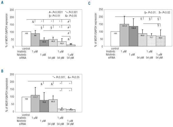
It has been reported that the MDR1 gene and its product, Pgp protein, are major obstacles to more successful therapy for CML. We, therefore, analyzed MDR1 gene expression relative to expression levels of the housekeeping gene GAPDH (Figure 2). The control was set at 100%. In 32Dp210-wt cells, imatinib decreased MDR1 levels slightly (86%, mean), whereas nilotinib and siRNA had greater effects (reductions to 42.5% and 46.5%, respectively). The lowest MDR1 levels were observed with the combinations of siRNA and imatinib (32.2%), and siRNA with nilotinib (9.9%).
Figure 2.
Effect of treatment with imatinib, nilotinib and transfection with BCR-ABL siRNA or co-treatment of BCR-ABL siRNA with imatinib or nilotinib in tyrosine kinase inhibitor-sensitive (A) and –resistant BCR-ABL oligoclonal cell lines (B: H396P, C: T315I) on MDR1 gene expression. MDR1 gene expression was detected by real time RT-PCR and normalized to GAPDH expression. Control was set at 100%.
In 32Dp210-His396Pro cells, we found significant effects of nilotinib (78.6%) and siRNA (82.5%), although treatment with the combination of siRNA with either imatinib or nilotinib caused significantly greater reductions in MRD1 levels (to 20.7% and 17.4% of control levels, respectively). This effect was also pronounced in 32Dp210-Thr315Ile cells, with co-administration of siRNA with either imatinib or nilotinib producing the lowest MRD1 levels (72.7% to 69.6%). Treatment with BCR-ABL siRNA alone resulted in a mean reduction of MRD1 level to 86.6%, whereas after treatment with either imatinib or nilotinib the MRD1 levels increased to 151.3% and 136.6%, respectively. Interestingly, the most significant reduction of MRD1 levels in 32Dp210-Thr315Ile cells was achieved with nilotinib combined with BCR-ABL siRNA (69.6%), which was significantly greater than that achieved by BCR-ABL siRNA alone (P<0.01). Taken together, these data show that combining BCR-ABL siRNA with nilotinib can strongly influence the expression of MDR1 in both imatinib-sensitive and imatinib-resistant 32Dp210 cells.
Discussion
Given that siRNA can now be delivered effectively to mammalian cells, selective interventions in leukemic cell gene regulation might be feasible for the treatment of leukemia.13,21 There are increasing numbers of reports of in vitro and in vivo selective silencing of the BCR-ABL fusion gene by RNA interference.15–17,22 Importantly, this construct was found to induce target-specific BCR-ABL mRNA cleavage without affecting the expression of BCR or ABL mRNA.14 Moreover, the first study on BCR-ABL siRNA showed that BCR-ABL silencing was accompanied by strong induction of apoptotic cell death.22 The rate of induced apoptosis was as high as that induced by 1 μM imatinib. Other investigators have confirmed these effects of BCR-ABL siRNA on CML cells.14,23,24
In the current study, we found that siRNA directed against the BCR-ABL gene significantly inhibited BCR-ABL gene expression in factor-independent 32Dp210 BCR-ABL oligoclonal cell lines, including those resistant to imatinib (particularly those with the T315I mutation). The response to increasing doses of BCR-ABL siRNA reached a plateau at 60% in the resistant 32Dp210-Thr315Ile cells, and the maximum BCR-ABL gene reduction in the 32Dp210 cells was two-fold as compared to the controls. As expected, we found differential reductions of BCR-ABL gene expression in response to two tyrosine kinase inhibitors (imatinib and nilotinib) in 32Dp210 cell lines. The results of BCR-ABL gene expression reduction and inhibition of cell proliferative capacity were concordant. O’Hare et al. compared the in vitro activity of BCR-ABL inhibitors in clinically relevant imatinib-resistant ABL domain mutants, and showed higher efficacy of nilotinib in many imatinib-resistant cell lines, with the exception of the T315I mutant.25 In a phase II study, nilotinib produced hematologic and cytogenetic responses in imatinib-resistant and -intolerant patients with CML in accelerated phase with the exception of those expressing the T315I mutation.26 In this study, we recorded large reductions in the growth of 32Dp210 cells (72.6% for imatinib, 74% for nilotinib, and 51.4% for siRNA) and smaller, but still significant reductions of as much as 35% for BCR-ABL siRNA in imatinib-resistant 32Dp210-His396Pro cells and imatinib-resistant 32Dp210-Thr315Ile cells. Consistent with the findings of Wohlbold et al.,27 who showed that BCR-ABL can be completely down-regulated by RNA interference in all common p210 and p190 BCR-ABL variants, we found reduced cell growth rates and BCR-ABL expression in primary leukemic cells from BCR-ABL-positive patients with acute lymphoblastic and myeloid leukemia and CML after transfection with BCR-ABL siRNA. Furthermore, we detected relevant differences in siRNA delivery to primary cells and to 32Dp cell lines. These results might, to some extent, reflect differences in the effectiveness of transfection with BCR-ABL siRNA in primary cells and 32Dp cell lines, which we found to vary from 52% and 75%. Furthermore, the expression of BCR-ABL protein is 5- to 6-fold higher in 32Dp210 cell lines than in primary cells.14 It should be emphasized that the efficacy of siRNA-mediated gene silencing is affected by a combination of factors (variation in transfection efficiency, siRNA sequences, properties of the target mRNA, and others). Moreover, Hu et al.28 suggested that low-abundant transcripts are less susceptible to siRNA-mediated degradation than medium- and high-abundant transcripts.
Surprisingly, we found that co-administration of BCR-ABL siRNA with imatinib or nilotinib resulted in enhanced inhibition of cell growth and BCR-ABL gene expression in 32Dp210 cells, including cells with the T315I mutation, which are highly resistant to imatinib and nilotinib. This might be the result of effective modulation by a breakpoint-specific siRNA. Consistent with our findings it was previously reported that transfection with BCR-ABL-specific siRNA increased the sensitivity of both imatinib-sensitive and imatinib-resistant CML cell lines to imatinib.14 Furthermore, Baker et al. showed that the IC50 of imatinib was 3-fold lower in K562 cells transfected with synthetic or recombinant-generated BCR-ABL siRNA enabling imatinib to efficiently inhibit the high levels of BCR-ABL protein found in these cells.29
Complex signal transduction pathways are also involved in controlling the proteolysis of potential BCR-ABL effectors that may be responsible for some features of BCR-ABL-transformed cells such as deregulated cellular proliferation and apoptosis control through effects on multiple intracellular signaling pathways, including the Ras, phosphatidylinositol 3-kinase (PI3K), JAK-STAT, and NF-κB pathways.30 For example, Perrotti et al. found that the levels of TLS/FUS, an RNA/DNA-binding protein involved in transcriptional and post-transcriptional regulation of gene expression, are markedly increased in growth factor-dependent hematopoietic cell lines ectopically expressing BCR-ABL and in CML blast crisis bone marrow cells.31 In addition to regulating gene expression at the transcriptional level, it is increasingly clear that BCR-ABL is also involved in post-transcriptional regulation via shuttling of heterogeneous nuclear ribonucleoproteins (hnRNP). These proteins play an important role in the control of pre-mRNA processing, nuclear mRNA export, and translation.32
Notably, the inhibitory effect on intracellular p-Tyr, measured by flow cytometry, declined significantly in the resistant 32Dp210-Thr315Ile cells after treatment with high doses of imatinib or nilotinib or transfection with BCR-ABL siRNA or the co-administration of siRNA with imatinib or nilotinib in comparison with that of mismatched siRNA or non-manipulated controls. Moreover, the inhibitory effect on p-Crkl was significantly correlated with siRNA treatment and with co-treatment with BCR-ABL siRNA and imatinib or nilotinib (P<0.0001). The tyrosine kinase activity of BCR-ABL constitutively induces the phosphorylation of a large panel of proteins, activating multiple signaling pathways responsible for protection against apoptosis, stimulation of growth factor-independent proliferation, and alterations of cellular adhesion. P-Crkl, a BCR-ABL adaptor protein, serves as a surrogate for BCR-ABL kinase activity in vivo.33 Our data on p-Crkl and p-Tyr support the efficacy of BCR-ABL siRNA transfection in vitro and validate our above described findings on cell growth and BCR-ABL expression in mutated 32Dp210 cells. Furthermore, we found that combining BCR-ABL siRNA transfection with imatinib or nilotinib had additive effects on induction of apoptosis and inhibition of proliferation of 32Dp210 cell lines, including mutated 32Dp210-His396Pro cells and mutated 32Dp210-Thr315Ile cells. The combination reduced the proliferation rate by about 2–2.5 fold compared to the use of BCR-ABL siRNA alone (P<0.01). This suggests that BCR-ABL siRNA and tyrosine kinase inhibitors affect BCR-ABL-positive cells by modulating or transforming genes via different pathways promising additive selective antitumor activity.
Ong et al. recently reported that mammalian target of rapamycin (mTOR)-dependent pathways are constitutively activated in BCR-ABL-transformed cells.34,35 These studies raised the possibility that regulation of mTOR may be a mechanism by which BCR-ABL promotes cell growth and survival of leukemic cells, but the mTOR effectors mediating such responses are not fully understood. Tyrosine kinase inhibitors, such as imatinib and nilotinib, target and block engagement of the mTOR pathway in BCR-ABL-expressing cells. Such effects of BCR-ABL kinase inhibitors are observed in cells transformed by imatinib-resistant BCR-ABL kinase mutants, establishing that the mTOR/p70 S6K pathway plays a critical role in promoting the leukemogenic effects of BCR-ABL.36 Recently, Ohba et al. demonstrated, by microarray analysis of K562 cells, that there is cross-talk between siRNA interference and BCR-ABL oncogeny and found that the expression of approximately 250 genes was changed. RNA interference of BCR-ABL was accompanied by decreased expression of various proto-oncogenes, growth factors, factors related to kinase activity, and factors related directly to cell proliferation. Several factors responsible for the development of apoptosis increased, including STAT-induced STAT inhibitors and apoptosis-related RNA binding proteins.37 Furthermore, Zhou et al. demonstrated that Adelson helper integration site 1 (Ahi-1/AHI-1) is a novel BCR-ABL interacting protein that is also physically associated with JAK2. This AHI-1-BCR-ABL-JAK2 complex seems to modulate BCR-ABL-transforming activity and the response/resistance of CML stem/progenitor cells to tyrosine kinase inhibitors through an interleukin-3-dependent BCR-ABL and JAK2-STAT5 pathway.38
In experiments focused on cell cycle analysis, the cell cycle distribution and apoptotic responses induced by BCR-ABL siRNA, tyrosine kinase inhibitors or co-treatment with BCR-ABL siRNA and tyrosine kinase inhibitors seemed to differ. We found that the effects of transfection with BCR-ABL siRNA depend on cell cycle arrest in the early S phase rather than on the disruption of the G2 checkpoint and on an increased apoptotic distribution with DNA fragmentation(sub-G0/G1 phase). By adding imatinib or nilotinib to BCR-ABL siRNA treatment these effects increased approximately 1.3-fold (P<0.05). This strongly suggests that dual inhibition of BCR-ABL by BCR-ABL siRNA combined with nilotinib additively enhances 32Dp210 cytotoxicity and apoptosis. In K562 cells, imatinib enhanced the inhibitory effect of radiation on proliferation of K562 cells and decreased the G2 phase content due to an efficient arrest in the S phase.39 Mazzacurati et al. found that imatinib also induces growth arrest by activating the Chk2-Cdc25A-Cdk2 axis, a pathway complementary to p53 in the activation of the G1/S cell cycle checkpoint, involved in cell fate decisions such as regulation of cell cycle progression, DNA repair and survival rather than apoptosis.40 Other investigators suggested that alternate pathways in addition to apoptosis may lead to the inhibition of cell growth mediated by tyrosine kinase inhibitors.41,42
There are several pharmacological mechanisms by which CML cells develop resistance to tyrosine kinase inhibitors, such as reduced drug-uptake and increased drug-efflux-related surface molecules including MDR-1, which reportedly mediates the efflux of imatinib. Such mechanisms may also lead to the resistance of other tyrosine kinase inhibitors, such as nilotinib.43–45 Specifically, a predominant acquisition of MDR1-dependent resistance is found in the majority of patients with advanced CML and other leukemias, as well as in patients during relapse.46,47 These pharmacokinetic mechanisms of resistance to tyrosine kinase inhibitors may occur in addition or may induce the well-described pharmacodynamic mechanisms involving the BCR-ABL gene in patients with CML.48 It should be stressed that our real-time RT-PCR data show a highly significant reduction of more than 13% in MDR1 gene expression (P<0.02) after transfection with BCR-ABL siRNA in mutated 32Dp210Thr315Ile cells. The combination of BCR-ABL siRNA with imatinib or nilotinib resulted in an additional reduction of MDR1 gene expression of 27% or 30% (P<0.01). In addition to impaired drug transport, aberrant activation of signal transduction proteins, including nuclear factor-κB, has been implicated in treatment failure in hematologic malignancies including CML.49,50 Our study demonstrates that BCR-ABL siRNA reliably and effectively induces apoptosis and inhibition of proliferation in factor-independent 32Dp210 BCR-ABL oligoclonal cell lines, including those resistant to imatinib and particularly those with the T315I mutation. Moreover, additive effects of BCR-ABL siRNA with tyrosine kinase inhibitors, such as imatinib or nilotinib, result in anti-proliferative and enhanced pro-apoptotic effects in 32Dp210 cells. In addition, the combination of BCR-ABL siRNA with imatinib or nilotinib significantly reversed MDR1-dependent multidrug resistance of mutated 32Dp210 cells. Modulation of breakpoint-specific siRNA by agents such as tyrosine kinase inhibitors might open the door to new important alternative or complementary approaches for the treatment of BCR-ABL-expressing hematologic disorders.
Acknowledgments
The authors thank Silke Gottwald, Melanie Kroll, Ines Riepenhoff and Christiane Schary for their excellent technical performance of the PCR analyses and siRNA experiments and Martina Franke and Ursula Hill for the flow cytometric analyses.
Footnotes
The online version of this article has a Supplementary Appendix.
Funding: this work was supported in parts by grants from Novartis Pharma AG, Germany.
Authorship and Disclosures
MK designed, performed, and analyzed the research and wrote the manuscript; LK contributed patients’ samples and analyzed data; DWB and AHE participated in coordination of the study, and obtained funding for the study.
MK, LK and DWB have no financial support to disclose; AHE received grant support and travel reimbursement from Novartis Pharma AG, Germany.
References
- 1.Goldman JM, Melo JV. Chronic myeloid leukemia – advances in biology and new approaches to treatment. N Engl J Med. 2003;349(15):1451–61. doi: 10.1056/NEJMra020777. [DOI] [PubMed] [Google Scholar]
- 2.Melo JV. The diversity of BCR-ABL fusion proteins and their relationship to leukemia phenotype. Blood. 1996;88(7):2375–84. [PubMed] [Google Scholar]
- 3.Jabbour E, Cortes JE, Ghanem H, O’Brien S, Kantarjian HM. Targeted therapy in chronic myeloid leukemia. Expert Rev Anti-cancer Ther. 2008;8(1):99–110. doi: 10.1586/14737140.8.1.99. [DOI] [PubMed] [Google Scholar]
- 4.De Bree F, Sorbera L, Fernandez R, Castancer J. Imatinib mesilate. Drugs Fut. 2001;26(6):545–52. [Google Scholar]
- 5.O’Brien SG, Guilhot F, Larson RA, Gathmann I, Baccarani M, Cervantes F, et al. IRIS Investigators. Imatinib compard with interferon and low-dose cytarabine for newly diagnosed chronic-phase chronic myeloid leukemia. N Engl J Med. 2003;348 (11):994–1004. doi: 10.1056/NEJMoa022457. [DOI] [PubMed] [Google Scholar]
- 6.Gratwohl A, Baldomero H, Horisberger B, Schmid C, Passweg J, Urbano-Ispizua A. Current trends in hematopoietic stem cell transplantation in Europe. Blood. 2002;100(7):2374–86. doi: 10.1182/blood-2002-03-0675. [DOI] [PubMed] [Google Scholar]
- 7.Dean M, Fojo T, Bates S. Tumour stem cells and drug resistance. Nat Rev Cancer. 2005;5(4):275–84. doi: 10.1038/nrc1590. [DOI] [PubMed] [Google Scholar]
- 8.Golemovic M, Verstovsek S, Giles F, Cortes J, Manshouri T, Manley PW, et al. AMN107, a novel aminopyrimidine inhibitor of Bcr-Abl, has in vitro activity against imatinib-resistant chronic myeloid leukaemia. Clin Cancer Res. 2005;11(13):4941–7. doi: 10.1158/1078-0432.CCR-04-2601. [DOI] [PubMed] [Google Scholar]
- 9.Weisberg E, Manley PW, Breitenstein W, Brüggen J, Cowan-Jacob SW, Ray A, et al. Characterization of AMN107, a selective inhibitor of native and mutant Bcr-Abl. Cancer Cell. 2005;7(2):129–41. doi: 10.1016/j.ccr.2005.01.007. [DOI] [PubMed] [Google Scholar]
- 10.Weisberg E, Manley P, Mestan J, Cowan-Jacob S, Ray A, Griffin JD. AMN107 (nilotinib): a novel and selective inhibitor of BCR-ABL. Br J Cancer. 2006;94(12):1765–9. doi: 10.1038/sj.bjc.6603170. [DOI] [PMC free article] [PubMed] [Google Scholar]
- 11.Hazarika M, Jiang X, Liu Q, Lee SL, Ramchandani R, Garnett C, et al. Tasigna for chronic and accelerated phase Philadelphia chromosome--positive chronic myelogenous leukemia resistant to or intolerant of imatinib. Clin Cancer Res. 2008;14(17):5325–31. doi: 10.1158/1078-0432.CCR-08-0308. [DOI] [PubMed] [Google Scholar]
- 12.Elbashir SM, Harborth J, Lendeckel W, Yalcin A, Weber K, Tuschl T. Duplexes of 21- nucleotide RNAs mediate RNA interference in cultured mammalian cells. Nature. 2001;411(6836):494–8. doi: 10.1038/35078107. [DOI] [PubMed] [Google Scholar]
- 13.Scherr M, Battmer K, Winkler T, Heidenreich O, Ganser A, Eder M. Specific inhibition of BCR-ABL gene expression by small interfering RNA. Blood. 2003;101(4):1566–9. doi: 10.1182/blood-2002-06-1685. [DOI] [PubMed] [Google Scholar]
- 14.Wohlbold L, Van Der Kuip H, Miething C, Vornlocher HP, Knabbe C, Duyster J, Aulitzky WE. Inhibition of bcr-abl gene expression by small interfering RNA sensitizes for imatinib mesylate (ST571) Blood. 2003;102(6):2236–9. doi: 10.1182/blood-2002-12-3899. [DOI] [PubMed] [Google Scholar]
- 15.Elmaagacli AH, Koldehoff M, Peceny R, Klein-Hitpass L, Ottinger H, Beelen DW, Opalka B. WT1 and BCR-ABL specific small interfering RNA have additive effects in the induction of apoptosis in leukemic cells. Haematologica. 2005;90(3):326–34. [PubMed] [Google Scholar]
- 16.Koldehoff M, Steckel NK, Beelen DW, Elmaagacli AH. Synthetic small interfering RNAs reduce bcr-abl gene expression in leukaemic cells of de novo Philadelphia (+) acute myeloid leukemia. Clin Exp Med. 2006;6(1):45–7. doi: 10.1007/s10238-006-0093-8. [DOI] [PubMed] [Google Scholar]
- 17.Koldehoff M, Steckel NK, Beelen DW, Elmaagacli AH. Therapeutic application of small interfering RNA directed against bcr-abl transcripts to a patient with imatinib-resistant chronic myeloid leukaemia. Clin Exp Med. 2007;7(2):47–55. doi: 10.1007/s10238-007-0125-z. [DOI] [PubMed] [Google Scholar]
- 18.Pear WS, Miller JP, Xu L, Pui JC, Soffer B, Quackenbush RC, et al. Efficient and rapid induction of a chronic myelogenous leukemia-like myeloproliferative disease in mice receiving P210 bcr/abl-transduced bone marrow. Blood. 1998;92(10):3780–92. [PubMed] [Google Scholar]
- 19.Bai RY, Ouyang T, Miething C, Morris SW, Peschel C, Duyster J. Nucleophosmin-anaplastic lymphoma kinase associated with anaplastic large-cell lymphoma activates the phosphatidylinositol 3-kinase/Akt antiapoptotic signaling pathway. Blood. 2000;96(13):4319–27. [PubMed] [Google Scholar]
- 20.Elmaagacli AH, Freist A, Hahn M, Opalka B, Seeber S, Schaefer SW, Beelen DW. Estimating the relapse stage in chronic myeloid leukaemia patients after allogeneic stem cell transplantation by the amount of BCR-ABL fusion transcripts detected using a new real-time polymerase chain reaction method. Br J Haematol. 2001;113(4):1072–5. doi: 10.1046/j.1365-2141.2001.02858.x. [DOI] [PubMed] [Google Scholar]
- 21.Novina CD, Sharp PA. The RNAi revolution. Nature. 2004;430(6996):161–4. doi: 10.1038/430161a. [DOI] [PubMed] [Google Scholar]
- 22.Wilda M, Fuchs U, Wössmann W, Borkhardt A. Killing of leukemic cells with a BCR/ABL fusion gene by RNA interference. Oncogene. 2002;21(37):5716–24. doi: 10.1038/sj.onc.1205653. [DOI] [PubMed] [Google Scholar]
- 23.Merkerova M, Klamova H, Brdicka R, Bruchova H. Targeting of gene expression by siRNA in CML primary cells. Mol Biol Rep. 2007;34(1):27–33. doi: 10.1007/s11033-006-9006-x. [DOI] [PubMed] [Google Scholar]
- 24.Withey JM, Marley SB, Kaeda J, Harvey AJ, Crompton MR, Gordon MY. Targeting primary human leukaemia cells with RNA interference: Bcr-Abl targeting inhibits myeloid progenitor self-renewal in chronic myeloid leukaemia cells. Br J Haematol. 2005;129(3):377–80. doi: 10.1111/j.1365-2141.2005.05468.x. [DOI] [PubMed] [Google Scholar]
- 25.O’Hare T, Walters DK, Stoffregen EP, Jia T, Manley PW, Mestan J, et al. In vitro activity of Bcr-Abl inhibitors AMN107 and BMS-354825 against clinically relevant imatinib-resistant Abl kinase domain mutants. Cancer Res. 2005;65(11):4500–5. doi: 10.1158/0008-5472.CAN-05-0259. [DOI] [PubMed] [Google Scholar]
- 26.le Coutre P, Ottmann OG, Giles F, Kim DW, Cortes J, Gattermann N, et al. Nilotinib (formerly AMN107), a highly selective BCR-ABL tyrosine kinase inhibitor, is active in patients with imatinib-resistant or -intolerant accelerated-phase chronic myelogenous leukemia. Blood. 2008;111(4):1834–9. doi: 10.1182/blood-2007-04-083196. [DOI] [PubMed] [Google Scholar]
- 27.Wohlbold L, van der Kuip H, Moehring A, Granot G, Oren M, Vornlocher HP, Aulitzky WE. All common p210 and p190 Bcr-abl variants can be targeted by RNA interference. Leukemia. 2005;19(2):290–2. doi: 10.1038/sj.leu.2403603. [DOI] [PubMed] [Google Scholar]
- 28.Hu X, Hipolito S, Lynn R, Abraham V, Ramos S, Wong-Staal F. Relative gene-silencing efficiencies of small interfering RNAs targeting sense and antisense transcripts from the same genetic locus. Nucleic Acids Res. 2004;32(15):4609–17. doi: 10.1093/nar/gkh790. [DOI] [PMC free article] [PubMed] [Google Scholar]
- 29.Baker BE, Kestler DP, Ichiki AT. Effects of siRNAs in combination with Gleevec on K-562 cell proliferation and Bcr-Abl expression. J Biomed Sci. 2006;13(4):499–507. doi: 10.1007/s11373-006-9080-z. [DOI] [PubMed] [Google Scholar]
- 30.Druker BJ, Tamura S, Buchdunger E, Ohno S, Segal GM, Fanning S, et al. Effects of a selective inhibitor of the Abl tyrosine kinase on the growth of Bcr-Abl positive cells. Nat Med. 1996;2(5):561–6. doi: 10.1038/nm0596-561. [DOI] [PubMed] [Google Scholar]
- 31.Perrotti D, Iervolino A, Cesi V, Cirinná M, Lombardini S, Grassilli E, et al. BCR-ABL prevents c-jun-mediated and proteasome-dependent FUS (TLS) proteolysis through a protein kinase CbetaII-dependent pathway. Mol Cell Biol. 2000;20(16):6159–69. doi: 10.1128/mcb.20.16.6159-6169.2000. [DOI] [PMC free article] [PubMed] [Google Scholar]
- 32.Perrotti D, Calabretta B. Post-transcriptional mechanisms in BCR/ABL leukemogenesis: role of shuttling RNA-binding proteins. Oncogene. 2002;21(56):8577–83. doi: 10.1038/sj.onc.1206085. [DOI] [PubMed] [Google Scholar]
- 33.Oda T, Heaney C, Hagopian JR, Okuda K, Griffin JD, Druker BJ. CRKL is the major tyrosine-phosphorylated protein in neutrophils from patients with chronic myelogenous leukemia. J Biol Chem. 1994;269(37):22925–8. [PubMed] [Google Scholar]
- 34.Ly C, Arechiga AF, Melo JV, Walsh CM, Ong ST. Bcr-Abl kinase modulates the translation regulators ribosomal protein S6 and 4E-BP1 in chronic myelogenous leukemia cells via the mammalian target of rapamycin. Cancer Res. 2003;63(18):5716–22. [PubMed] [Google Scholar]
- 35.Parmar S, Smith J, Sassano A, Uddin S, Katsoulidis E, Majchrzak B, et al. Differential regulation of the p70 S6 kinase pathway by interferon alpha (IFNα) and imatinib mesylate (STI571) in chronic myelogenous leukemia cells. Blood. 2005;106(7):2436–43. doi: 10.1182/blood-2004-10-4003. [DOI] [PMC free article] [PubMed] [Google Scholar]
- 36.Carayol N, Katsoulidis E, Sassano A, Altman JK, Druker BJ, Platanias LC. Suppression of programmed cell death 4 (PDCD4) protein expression by BCR-ABL-regulated engagement of the mTOR/p70 S6 kinase pathway. J Biol Chem. 2008;283(13):8601–10. doi: 10.1074/jbc.M707934200. [DOI] [PMC free article] [PubMed] [Google Scholar]
- 37.Ohba H, Zhelev Z, Bakalova R, Ewis A, Omori T, Ishikawa M, et al. Inhibition of bcr-abl and/or c-abl gene expression by small interfering, double-stranded RNAs: cross-talk with cell proliferation factors and other oncogenes. Cancer. 2004;101(6):1390–403. doi: 10.1002/cncr.20468. [DOI] [PubMed] [Google Scholar]
- 38.Zhou LL, Zhao Y, Ringrose A, DeGeer D, Kennah E, Lin AE, et al. AHI-1 interacts with BCR-ABL and modulates BCR-ABL transforming activity and imatinib response of CML stem/progenitor cells. J Exp Med. 2008;205(11):2657–71. doi: 10.1084/jem.20072316. [DOI] [PMC free article] [PubMed] [Google Scholar]
- 39.Huguet F, Giocanti N, Hennequin C, Croisy M, Touboul E, Favaudon V. Growth inhibition by STI571 in combination with radiation in human chronic myelogenous leukemia K562 cells. Mol Cancer Ther. 2008;7(2):398–406. doi: 10.1158/1535-7163.MCT-07-2023. [DOI] [PubMed] [Google Scholar]
- 40.Mazzacurati L, Pattacini L, Brusa G, Mancini M, Benvenuti M, Barbieri E, et al. Chk2 drives late G1/early S phase arrest of clonal myeloid progenitors expressing the p210 BCR-ABL tyrosine kinase in response to STI571. Hematol J. 2004;5(2):168–77. doi: 10.1038/sj.thj.6200365. [DOI] [PubMed] [Google Scholar]
- 41.Verstovsek S, Golemovic M, Kantarjian H, Manshouri T, Estrov Z, Manley P, et al. AMN107, a novel aminopyrimidine inhibitor of p190 Bcr-Abl activation and of in vitro proliferation of Philadelphia-positive acute lymphoblastic leukemia cells. Cancer. 2005;104(6):1230–6. doi: 10.1002/cncr.21299. [DOI] [PubMed] [Google Scholar]
- 42.Belloc F, Moreau-Gaudry F, Uhalde M, Cazalis L, Jeanneteau M, Lacombe F, et al. Imatinib and nilotinib induce apoptosis of chronic myeloid leukemia cells through a Bim-dependant pathway modulated by cytokines. Cancer Biol Ther. 2007;6(6):912–9. doi: 10.4161/cbt.6.6.4101. [DOI] [PubMed] [Google Scholar]
- 43.Borst P, Elferink RO. Mammalian ABC transporters in health and disease. Annu Rev Biochem. 2002;71:537–92. doi: 10.1146/annurev.biochem.71.102301.093055. [DOI] [PubMed] [Google Scholar]
- 44.Jiang X, Zhao Y, Smith C, Gasparetto M, Turhan A, Eaves A, Eaves C. Chronic myeloid leukemia stem cells possess multiple unique features of resistance to BCR-ABL targeted therapies. Leukemia. 2007;21(5):926–35. doi: 10.1038/sj.leu.2404609. [DOI] [PubMed] [Google Scholar]
- 45.Borst P, Zelcer N, van Helvoort A. ABC transporters in lipid transport. Biochim Biophys Acta. 2000;1486(1):128–44. doi: 10.1016/s1388-1981(00)00053-6. [DOI] [PubMed] [Google Scholar]
- 46.Zhou S, Schuetz JD, Bunting KD, Colapietro AM, Sampath J, Morris JJ, et al. The ABC transporter Bcrp1/ABCG2 is expressed in a wide variety of stem cells and is a molecular determinant of the side-population phenotype. Nat Med. 2001;7(9):1028–34. doi: 10.1038/nm0901-1028. [DOI] [PubMed] [Google Scholar]
- 47.Jordanides NE, Jorgensen HG, Holyoake TL, Mountford JC. Functional ABCG2 is overexpressed on primary CML CD34+ cells and is inhibited by imatinib mesylate. Blood. 2006;108(4):1370–3. doi: 10.1182/blood-2006-02-003145. [DOI] [PubMed] [Google Scholar]
- 48.Thomas J, Wang L, Clark RE, Pirmohamed M. Active transport of imatinib into and out of cells: implications for drug resistance. Blood. 2004;104(12):3739–45. doi: 10.1182/blood-2003-12-4276. [DOI] [PubMed] [Google Scholar]
- 49.Breedveld P, Pluim D, Cipriani G, Wielinga P, van Tellingen O, Schinkel AH, Schellens JH. The effect of Bcrp1 (Abcg2) on the in vivo pharmacokinetics and brain penetration of imatinib mesylate (Gleevec): implications for the use of breast cancer resistance protein and P-glycoprotein inhibitors to enable the brain penetration of imatinib in patients. Cancer Res. 2005;65(7):2577–82. doi: 10.1158/0008-5472.CAN-04-2416. [DOI] [PubMed] [Google Scholar]
- 50.Ozvegy-Laczka C, Hegedus T, Várady G, Ujhelly O, Schuetz JD, Váradi A, et al. High-affinity interaction of tyrosine kinase inhibitors with the ABCG2 multidrug transporter. Mol Pharmacol. 2004;65(6):1485–95. doi: 10.1124/mol.65.6.1485. [DOI] [PubMed] [Google Scholar]
- 51.Shukla S, Sauna ZE, Ambudkar SV. Evidence for the interaction of imatinib at the transport-substrate site(s) of the multidrug-resistance-linked ABC drug transporters ABCB1 (P-glycoprotein) and ABCG2. Leukemia. 2008;22(2):445–7. doi: 10.1038/sj.leu.2404897. [DOI] [PubMed] [Google Scholar]
- 52.Brendel C, Scharenberg C, Dohse M, Robey RW, Bates SE, Shukla S, et al. Imatinib mesylate and nilotinib (AMN107) exhibit high-affinity interaction with ABCG2 on primitive hematopoietic stem cells. Leukemia. 2007;21(6):1267–75. doi: 10.1038/sj.leu.2404638. [DOI] [PubMed] [Google Scholar]
- 53.Mahon FX, Hayette S, Lagarde V, Belloc F, Turcq B, Nicolini F, et al. Evidence that resistance to nilotinib may be due to BCR-ABL, Pgp, or Src kinase overexpression. Cancer Res. 2008;68(23):9809–16. doi: 10.1158/0008-5472.CAN-08-1008. [DOI] [PubMed] [Google Scholar]
- 54.Hegeds C, Ozvegy-Laczka C, Apáti A, Magócsi M, Német K, Orfi L, et al. Interaction of nilotinib, dasatinib and bosutinib with ABCB1 and ABCG2: implications for altered anti-cancer effects and pharmacological properties. Br J Pharmacol. 2009;158(4):1153–64. doi: 10.1111/j.1476-5381.2009.00383.x. [DOI] [PMC free article] [PubMed] [Google Scholar]
- 55.Dulucq S, Bouchet S, Turcq B, Lippert E, Etienne G, Reiffers J, et al. Multidrug resistance gene (MDR1) polymorphisms are associated with major molecular responses to standard-dose imatinib in chronic myeloid leukemia. Blood. 2008;112(5):2024–7. doi: 10.1182/blood-2008-03-147744. [DOI] [PubMed] [Google Scholar]
- 56.Galimberti S, Cervetti G, Guerrini F, Testi R, Pacini S, Fazzi R, et al. Quantitative molecular monitoring of BCR-ABL and MDR1 transcripts in patients with chronic myeloid leukemia during imatinib treatment. Cancer Genet Cytogenet. 2005;162(1):57–62. doi: 10.1016/j.cancergencyto.2005.01.015. [DOI] [PubMed] [Google Scholar]
- 57.Valent P. Imatinib-resistant chronic myeloid leukemia (CML): current concepts on pathogenesis and new emerging pharmacologic approaches. Biologics. 2007;1(4):433–48. [PMC free article] [PubMed] [Google Scholar]
- 58.Tiwari AK, Sodani K, Wang SR, Kuang YH, Ashby CR, Jr, Chen X, et al. Nilotinib (AMN107, Tasigna) reverses multidrug resistance by inhibiting the activity of the ABCB1/Pgp and ABCG2/BCRP/MXR transporters. Biochem Pharmacol. 2009;78(2):153–61. doi: 10.1016/j.bcp.2009.04.002. [DOI] [PubMed] [Google Scholar]
- 59.Grosjean-Raillard J, Tailler M, Adès L, Perfettini JL, Fabre C, Braun T, et al. ATM mediates constitutive NF-κB activation in high-risk myelodysplastic syndrome and acute myeloid leukemia. Oncogene. 2008;28(8):1099–109. doi: 10.1038/onc.2008.457. [DOI] [PubMed] [Google Scholar]
- 60.Monteghirfo S, Tosetti F, Ambrosini C, Stigliani S, Pozzi S, Frassoni F, et al. Antileukemia effects of xanthohumol in Bcr/Abl-transformed cells involve nuclear factor-kappaB and p53 modulation. Mol Cancer Ther. 2008;7(9):2692–702. doi: 10.1158/1535-7163.MCT-08-0132. [DOI] [PubMed] [Google Scholar]


