Abstract
The detection and compensatory response to the accumulation of unfolded proteins in the endoplasmic reticulum (ER), termed the Unfolded Protein Response (UPR), represents a conserved cellular homeostatic mechanism with important roles in normal development and in the pathogenesis of disease1. The IRE1-XBP1/Hac1p pathway is a major branch of the UPR that has been conserved from yeast to human2,3,4,5,6. XBP-1 is required for the differentiation of the highly secretory plasma cells of the mammalian adaptive immune system7,8, but recent work also points to reciprocal interactions between the UPR and other aspects of immunity and inflammation9,10,11. We have been studying innate immunity in the nematode Caenorhabditis elegans, having established a key role for a conserved PMK-1 p38 mitogen-activated protein kinase (MAPK) pathway in mediating resistance to microbial pathogens12. Here, we show that during C. elegans development, XBP-1 has an essential role in protecting the host during activation of innate immunity. Activation of the PMK-1-mediated response to infection with Pseudomonas aeruginosa induces the XBP-1-dependent UPR. Whereas a loss-of-function xbp-1 mutant develops normally in the presence of relatively non-pathogenic bacteria, infection of the xbp-1 mutant with P. aeruginosa leads to disruption of ER morphology and larval lethality. Unexpectedly, the larval lethality phenotype on pathogenic P. aeruginosa is suppressed by loss of PMK-1-mediated immunity. Furthermore, hyperactivation of PMK-1 causes larval lethality in the xbp-1 mutant even in the absence of pathogenic bacteria. Our data establish innate immunity as a physiologically relevant inducer of ER stress during C. elegans development and suggest that an ancient, conserved role for XBP-1 may be to protect the host organism from the detrimental effects of mounting an innate immune response to microbes.
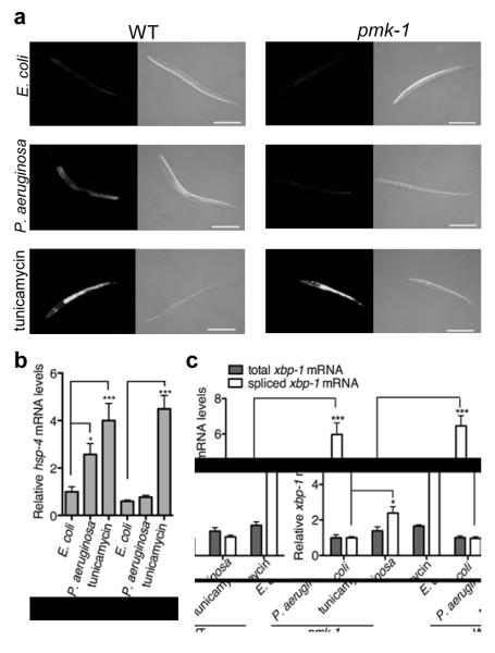
We began our analysis of the role of the UPR in C. elegans immunity by asking if the UPR is activated upon exposure of C. elegans larvae to P. aeruginosa strain PA14, a human opportunistic pathogen that can also infect and kill C. elegans13. First, we assessed expression of the transgenic transcriptional reporter Phsp-4∷GFP(zcIs4) (the C. elegans hsp-4 gene encodes a homolog of mammalian BiP/GRP78), which reflects activation of the IRE-1-XBP-1 branch of the UPR5. After exposure of C. elegans larvae to P. aeruginosa, GFP expression was induced in the intestine, the site of infection (Fig. 1a). We confirmed the pathogen-induced activation of the endogenous hsp-4 gene by quantitative RT-PCR (qRT-PCR) (Fig. 1b).
Figure 1. PMK-1 p38 MAPK-dependent activation of the IRE-1-XBP-1-dependent UPR by P. aeruginosa infection.
(a) Fluorescence and DIC microscopy of WT and mutant larvae carrying the Phsp-4∷GFP(zcIs4) transgene 24 h after egg lay on indicated treatments (Scale bar = 100μm). (b) qRT-PCR of endogenous hsp-4 mRNA levels. (c) qRT-PCR analysis of total and spliced xbp-1 mRNA levels. Values represent fold change relative to WT on OP50 ± s.e.m. (n = 4 independent experiments, *P < 0.05 and ***P < 0.001, two-way ANOVA with Bonferroni post test).
The mechanism by which IRE-1 activates XBP-1 in response to ER stress is conserved from yeast to mammals2,4,5,6. Upon accumulation of unfolded proteins in the ER, the integral ER-membrane protein IRE-1 activates XBP-1 by alternative splicing of the xbp-1 mRNA, thereby changing the reading frame. The activated “spliced form” of XBP-1 regulates expression of genes involved in ER homeostasis, such as those encoding chaperones. Using qRT-PCR to quantify the IRE-1-mediated splicing of xbp-1 mRNA after exposure to P. aeruginosa, we found there was a marked induction of IRE-1-spliced xbp-1 transcript within 4 h of exposure to pathogen (Fig. 1c).
We hypothesized that the rapid activation of the IRE-1-XBP-1 pathway after P. aeruginosa exposure might in fact be caused by induction of the host immune response. A conserved p38 MAPK pathway mediates innate immunity in organisms ranging from C. elegans to humans12,14. We have previously shown that loss-of-function of the p38 MAPK ortholog PMK-1 results in markedly enhanced susceptibility to killing of C. elegans by P. aeruginosa. In the pmk-1(km25) mutant, we found no increase in IRE-1-spliced XBP-1 mRNA and no induction of the Phsp-4∷GFP reporter in response to P. aeruginosa (Fig. 1). RNAi of sek-1, encoding a MAPKK required for PMK-1 activation12, also abrogated induction of Phsp-4∷GFP expression in response to P. aeruginosa infection (Supplementary Fig. 1a).
Consistent with the induction of XBP-1 splicing arising as a consquence of a transcriptional response to infection, we found that a mutant, atf-7(qd22 qd130), carrying a mutation in a conserved transcription factor that abrogates the pathogen-induced expression of genes regulated by the PMK-1 pathway15, also blocked induction of the Phsp-4∷GFP reporter in response to P. aeruginosa PA14 (Supplementary Fig. 1b). Activation of the IRE-1-XBP-1 pathway in response to tunicamycin (an N-glycosylation inhibitor), however, occurred in the pmk-1 mutant as in the wild-type strain N2 (WT), as reported by Bischof et al. in their study of PMK-1-dependent activation of the IRE-1-XBP-1 pathway in the C. elegans response to a pore-forming toxin from Bacillus thurigiensis16. Whereas our data show that activation of the IRE-1-XBP-1 pathway by P. aeruginosa infection is dependent on the PMK-1 pathway, we did not observe a reciprocal dependence of activation of the PMK-1 pathway on XBP-1 (Supplementary Fig. 2).
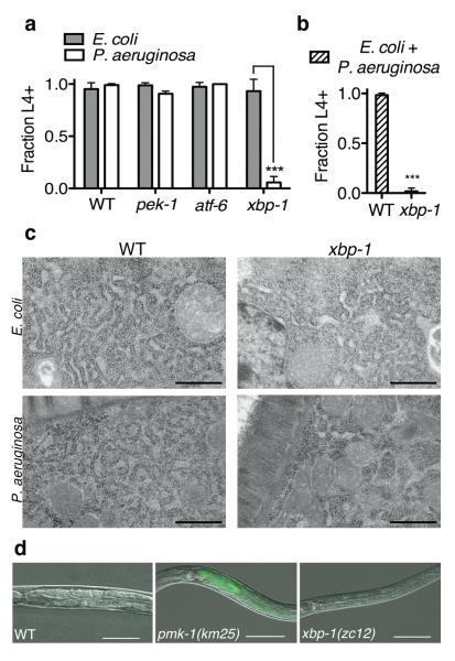
We next asked if XBP-1 is required for resistance to P. aeruginosa, focusing on its role during larval development. Synchronized eggs of WT and mutants deficient in each of the three branches of the UPR were propagated on plates with P. aeruginosa PA14 as the only food source and development was monitored over time. Nearly all the eggs of the WT strain developed to at least the fourth larval stage (L4) in this assay, comparable to their growth and development on the relatively nonpathogenic bacterial strain and standard laboratory food source Escherichia coli OP50 (Fig. 2a). In contrast, the xbp-1(zc12) mutant on P. aeruginosa exhibited severely attenuated larval development and growth, as measured by the rate of progression between molts, size of larvae, and ability of larvae to reach the L4 stage by 72 h (Fig. 2a), eventually resulting in larval lethality. Loss-of-function mutations in the each of the other two major branches of the UPR, atf-6(ok551) and pek-1(ok275)4,17, did not affect larval development in the presence of P. aeruginosa (Fig. 2a). These data implicate a specific and essential role for the IRE-1-XBP-1 branch of the UPR in C. elegans development in the presence of P. aeruginosa.
Figure 2. XBP-1 is required for C. elegans development and survival on P. aeruginosa.
(a) Development of indicated mutants to the L4 larval stage or older after 3 d at 25° C on either P. aeruginosa PA14 or E. coli OP50, or (b) mixture of PA14 and E. coli HB101. Plotted is mean ± s.d. (n = 3-4 plates, ***P<0.001, two-way ANOVA with Bonferroni post test in (a), and n = 4 plates, *** p<0.001 two tailed t-test in (b)). Results in (a) and (b) are representative of 3 independent experiments. (c) Transmission electron microscopy of L3 larvae at 60,000x magnification (Scale bar = 500 nm). (d) Fluorescence and DIC microscopy overlay of larvae 40 h after egg-lay on P. aeruginosa PA14 expressing GFP.
Strain Construction
The xbp-1 developmental phenotype was alleviated in the presence of the gacA mutant of P. aeruginosa PA14 (Supplementary Fig. 3a), which has diminished pathogenicity in C. elegans and mammals13,18. On a lawn of wild-type P. aeruginosa PA14 mixed with relatively non-pathogenic E. coli, the xbp-1 mutant again exhibited the severely attenuated developmental phenotype that was observed in the presence of P. aeruginosa alone, suggestive that the pathogenicity of P. aeruginosa, not a nutritional deficiency, caused the xbp-1 developmental phenotype (Fig. 2b and Supplementary Fig. 3b). Behavioural avoidance of pathogenic bacteria19,20 can influence survival21,22,23, but we found that the development of WT was unaffected by experimental conditions in which the P. aeruginosa could not be avoided, demonstrating that the inability of the xbp-1 mutant to develop on P. aeruginosa was not due to a defect in behavioural avoidance (C. E. R. and D. H. K., unpublished data). In addition, the attenuated development and death of the xbp-1 mutant on P. aeruginosa was also unaffected by the blocking of cell necrosis (Supplementary Table 1).
WT and xbp-1 mutant larvae exhibit characteristic ribosome-studded tubular structures of the ER by transmission electron microscopy when propagated on relatively non-pathogenic E. coli OP50 (Fig. 2c). The ER architecture of WT worms was not markedly perturbed upon exposure to P. aeruginosa (Fig. 2c). In contrast, xbp-1(zc12) larvae propagated on P. aeruginosa PA14 revealed disruption in ER morphology of xbp-1 mutant worms upon pathogen exposure (Fig. 2c), with localized regions of dilated ER lumen, comparable to that induced by tunicamycin treatment of xbp-1(zc12) worms (Supplementary Fig. 4). Such changes in ER morphology are consistent with ER stress and perturbation24, and strongly suggest that a disruption in ER homeostasis is induced by PA14 infection in the xbp-1 mutant.
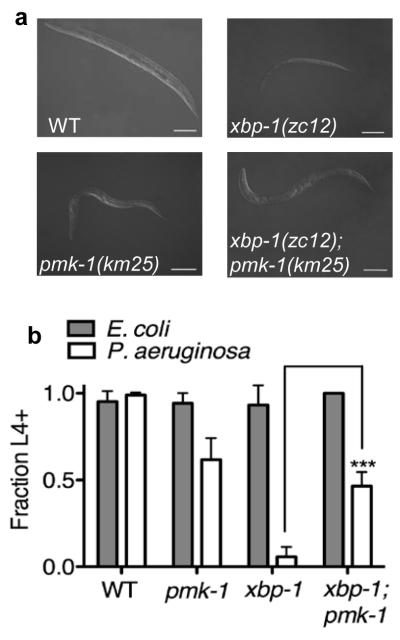
We considered whether the developmental lethality of the xbp-1 mutant might be due to the toxicity of PA14 arising from accelerated infection due to diminished resistance to P. aeruginosa in the xbp-1 mutant. Because activation of the PMK-1 pathway results in the splicing of XBP-1 upon P. aeruginosa exposure, we first asked whether XBP-1 confers a survival benefit against P. aeruginosa by facilitating resistance to intestinal infection. Diminished activation of PMK-1-dependent innate immunity is accompanied by an increased rate of P. aeruginosa accumulation in the intestine12(Fig. 2d). If XBP-1 functioned downstream of PMK-1 to promote the immune response to P. aeruginosa, we would anticipate that xbp-1 deficiency would lead to an increased rate of P. aeruginosa accumulation relative to WT. Interestingly, in spite of the severely attenuated development and survival of the xbp-1 mutant larvae on P. aeruginosa, the kinetics of accumulation of a PA14-derived strain of P. aeruginosa expressing GFP in the xbp-1(zc12) mutant were comparable to that observed for WT (Fig. 2d). Although the pmk-1(km25) mutant exhibited an increased rate of accumulation of P. aeruginosa (Fig. 2d), larval development and survival was markedly greater than that observed for the xbp-1 mutant (Figs. 3a and 3b, and Supplementary Fig. 5). These data decouple enhanced susceptibility to P. aeruginosa infection from the attenuated larval development phenotype of the xbp-1 mutant and suggest that while activation of XBP-1 by P. aeruginosa infection is PMK-1-dependent, the primary protective role of XBP-1 is not to facilitate the killing of P. aeruginosa.
Figure 3. Suppression of the P. aeruginosa-induced larval lethality phenotype of xbp-1(zc12) by pmk-1(km25).
(a) DIC microscopy of representative worms of each genotype 3 d after egg-lay on P. aeruginosa PA14 (Scale bar = 100 μm). (b) Development of indicated mutants to the L4 larval stage or older after 3 d at 25° C on either P. aeruginosa PA14 or E. coli OP50. Plotted is the mean ± s.d. (n = 3-4 plates, ***P < 0.001, two-way ANOVA with Bonferroni post test). Representative results of 3 independent experiments.
In view of these data, we considered whether the XBP-1-dependent UPR functions in this setting to protect against the ER stress induced by the immune response itself. If this were the case, then diminishing the immune response might actually improve the survival of the xbp-1 mutant on P. aeruginosa. To test this hypothesis, we examined the larval development of a xbp-1(zc12);pmk-1(km25) double mutant. Strikingly, the xbp-1;pmk-1 double mutant showed markedly increased development and survival relative to the xbp-1 mutant, to a degree that was comparable to the development and survival of the pmk-1 mutant (Figs. 3a and 3b). RNAi of sek-1, and RNAi of atf-7 in the xbp-1(zc12) mutant each alleviated the attenuated development phenotype in the presence of P. aeruginosa (Supplementary Figs. 6a and b). These data indicate that activation of the PMK-1-dependent transcriptional innate immune response to pathogen is indeed detrimental to survival during development in the absence of the IRE-1-XBP-1 pathway, and that the principal mechanism by which XBP-1 promotes development and survival during infection with P. aeruginosa is by protecting against the innate immune response.
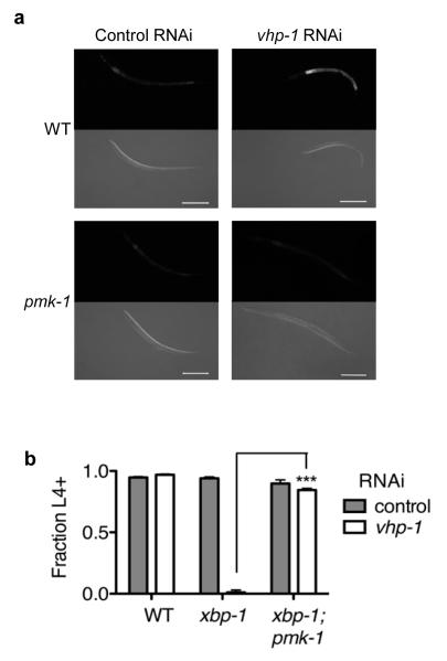
To separate further the effect of toxicity caused by pathogenic P. aeruginosa from that caused specifically by immune activation on the xbp-1 developmental phenotype, we next asked if the xbp-1 mutant would be compromised in the setting of hyperactivation of the PMK-1-mediated immune response in the absence of P. aeruginosa. RNAi against vhp-1, which encodes a MAPK phosphatase that negatively regulates PMK-1, results in hyperactivation of PMK-125. We observed that RNAi of vhp-1 resulted in sustained induction of the Phsp-4∷GFP(zcIs4) reporter with the relatively non-pathogenic, standard laboratory food source E. coli OP50 as the only bacteria present in the assay (Fig. 4a). RNAi-mediated knockdown of vhp-1 had little effect on development of WT (Fig. 4b). In contrast, RNAi of vhp-1 severely impaired development of the xbp-1(zc12) mutant in the absence of pathogenic bacteria (Fig. 4b) in a manner similar to that observed for the xbp-1 mutant when propagated on P. aeruginosa (Fig. 2a). Both the induction of hsp-4 and the larval developmental phenotype of the xbp-1 mutant resulting from RNAi of vhp-1 were suppressed by pmk-1 (Figs. 4a and 4b), demonstrating that the observed effects of RNAi of vhp-1 were mediated by the PMK-1-dependent immune response.
Figure 4. XBP-1 is required for development and survival during pathogen-independent constitutive activation of PMK-1.
(a) Fluorescence and DIC microscopy of WT and xbp-1 larvae carrying the Phsp-4∷GFP(zcIs4) transgene, which have been treated with control (empty vector) or vhp-1 RNAi, 24 h after egg lay. (b) Development to the L4 larval stage or older after 3 d at 25° C of WT, xbp-1, and xbp-1;pmk-1 mutants that have been subjected to RNAi of vhp-1 and propagated on E. coli OP50.
Plotted is mean ± s.e.m (n = 3 independent experiments, ***P < 0.001, two-way ANOVA with Bonferroni post test).
Our data suggest that the XBP-1-mediated UPR plays an essential role during C. elegans larval development by protecting against the activation of innate immunity. Transcriptional profiling of the C. elegans response to infection with P. aeruginosa26,27 has revealed the induction of at least 300 genes26, nearly half of which are predicted to be trafficked through the ER. Although this innate immune response promotes pathogen resistance, retarding the intestinal accumulation of P. aeruginosa and substantially increasing the fraction of the population that survives through larval development (as illustrated by the survival of WT relative to the pmk-1(km25) mutant (Fig. 3 and Supplementary Fig. 5)), our data suggest that innate immunity also constitutes a physiologically relevant source of ER stress that necessitates the compensatory activity of the UPR in C. elegans larval growth and development. An ancient role for the UPR may be to facilitate the development and survival of metazoan cell types that are exposed to the environment and mount a defensive, secretory response to microbial pathogens and abiotic toxins4,28. Our data not only support this hypothesis, but more specifically suggest that in fact the essential role of XBP-1 lies not in the neutralization of these environmental insults, but instead in conferring protection against the potentially lethal ER stress that is induced by this response. Our findings support the idea that the microbiota of multicellular organisms may represent the most commonly encountered and physiologically relevant inducer of such ER stress. Consistent with this hypothesis is the Paneth cell death observed in XBP1 intestinal knockout mice10. Evolutionary conservation of the function of XBP-1 that we have defined in C. elegans would implicate the UPR in protection against ER toxicity caused by activation of immune and inflammatory pathways in mammals, where the UPR may have a critical role in the pathogenesis of infectious and inflammatory autoimmune diseases.
Methods Summary
C. elegans strains were maintained as described29. Strains used in this study were N2 Bristol WT strain, SJ4005 Phsp-4∷GFP(zcIs4), ZD305 pmk-1(km25);Phsp-4∷GFP(zcIs4), ZD441 atf-7(qd22 qd130); Phsp-4∷GFP(zcIs4), ZD361 xbp-1(zc12), RB545 pek-1(ok275), RB772 atf-6(ok551), ZD363 xbp-1(zc12);pmk-1(km25), ZD416 xbp-1(tm2457), ZD417 xbp-1(tm2457);pmk-1(km25), ZD418 xbp-1(tm2482) and ZD419 xbp-1(tm2482);pmk-1(km25). Strain construction is detailed in Supplementary Methods. All P. aeruginosa (strain PA14) plates were prepared as described13, exceptions are detailed in Supplementary Methods. For tunicamycin treatments, tunicamycin was added to E. coli plates at 5 μg/ml final concentration. Images were acquired with an Axioimager Z1 microscope using worms anesthetized in 0.1% sodium azide. Transmission electron microscopy was performed as described in Supplementary Methods. For RNA analysis, L1 larvae were synchronized by hypochlorite treatment, washed onto OP50 plates and grown at 20 C for 23 h on OP50, then washed in M9 onto treatment plates. After 4 h, worms were washed off plates and frozen in liquid nitrogen. RNA and qRT-PCR methods are detailed in Supplementary Methods. For development assays, E. coli OP50 plates were prepared in parallel with PA14 plates. Strains were egg laid in parallel onto 4 plates each of PA14 and OP50 (at least 80 eggs for each strain and treatment), and fraction of worms growing to at least the L4 larval stage (“L4+”) between the plates was averaged. The L4 stage of development was chosen due to ease of scoring vulval development. RNAi by feeding of bacteria30 was performed as described by propagating stains on E. coli HT115 carrying the RNAi clones as indicated at 20° C, and adult progeny were used to lay eggs in experiments, which were performed at 25° C. Details regarding RNAi clones are in Supplementary Methods. Statistical analysis was performed using GraphPad Prism (GraphPad Software, Inc.).
Supplementary Material
Acknowledgments
Electron microscopy was performed by M. McKee in the Microscopy Core of the Center for Systems Biology/Program in Membrane Biology at Massachusetts General Hospital (supported by NIH grants DK43351 and DK57521). We thank E. Hartwieg and G. Voeltz for discussions regarding the interpretation of electron microscopy images. We thank T. Stiernagle and the Caenorhabditis Genetics Center (supported by the NIH), and S. Mitani and the National Bioresource Project of Japan for strains. T.K. was supported by summer research fellowships from the Howard Hughes Medical Institute. This work was supported by NIH grant R01-GM084477, a Career Award in the Biomedical Sciences from the Burroughs Wellcome Fund, and an Ellison Medical Foundation New Scholar Award (to D.H.K.).
Footnotes
Supplementary Information is linked to the online version of this paper atwww.nature.com/nature.
Author Information Reprints and permissions information is available at npg.nature.com/reprintsandpermissions. The authors have no competing financial interests.
References
- 1.Schröder M, Kaufman RJ. The mammalian Unfolded Protein Response. Annu. Rev. Biochem. 2005;74:739–789. doi: 10.1146/annurev.biochem.73.011303.074134. [DOI] [PubMed] [Google Scholar]
- 2.Cox JS, Walter P. A novel mechanism for regulating activity of a transcription factor that controls the Unfolded Protein Response. Cell. 1996;87:391–404. doi: 10.1016/s0092-8674(00)81360-4. [DOI] [PubMed] [Google Scholar]
- 3.Mori K, Kawahara T, Yoshida H, Yanagi H, Yura T. Signalling from Endoplasmic Reticulum to nucleus: transcription factor with a basic-leucine zipper motif is required for the Unfolded Protein Response pathway. Genes Cells. 1996;1:803–817. doi: 10.1046/j.1365-2443.1996.d01-274.x. [DOI] [PubMed] [Google Scholar]
- 4.Shen X, et al. Complementary signaling pathways regulate the Unfolded Protein Response and are required for C. elegans development. Cell. 2001;107:893–903. doi: 10.1016/s0092-8674(01)00612-2. [DOI] [PubMed] [Google Scholar]
- 5.Calfon M, et al. IRE1 couples endoplasmic reticulum load to secretory capacity by processing the xbp-1 mRNA. Nature. 2002;415:92–96. doi: 10.1038/415092a. [DOI] [PubMed] [Google Scholar]
- 6.Yoshida H, Matsui T, Yamamoto A, Okada T, Mori K. XBP1 mRNA is induced by ATF6 and spliced by IRE1 in response to ER stress to produce a highly active transcription factor. Cell. 2001;107:881–891. doi: 10.1016/s0092-8674(01)00611-0. [DOI] [PubMed] [Google Scholar]
- 7.Reimold AM, et al. Plasma cell differentiation requires the transcription factor XBP-1. Nature. 2001;412:300–307. doi: 10.1038/35085509. [DOI] [PubMed] [Google Scholar]
- 8.Iwakoshi NN, et al. Plasma cell differentiation and the Unfolded Protein Response intersect at the transcription factor XBP-1. Nature Immunol. 2003;4:321–329. doi: 10.1038/ni907. [DOI] [PubMed] [Google Scholar]
- 9.Todd DJ, Lee A-H, Glimcher LH. The endoplasmic eeticulum stress response in immunity and autoimmunity. Nat. Rev. Immunol. 2008;8:663–674. doi: 10.1038/nri2359. [DOI] [PubMed] [Google Scholar]
- 10.Zhang K, et al. Endoplasmic reticulum stress activates cleavage of CREBH to induce a systemic inflammatory response. Cell. 2006;124:587–599. doi: 10.1016/j.cell.2005.11.040. [DOI] [PubMed] [Google Scholar]
- 11.Kaser A, et al. XBP1 links ER stress to intestinal inflammation and confers genetic risk for human inflammatory bowel disease. Cell. 2008;134:743–756. doi: 10.1016/j.cell.2008.07.021. [DOI] [PMC free article] [PubMed] [Google Scholar]
- 12.Kim DH, et al. A conserved p38 MAP Kinase pathway in Caenorhabditis elegans innate immunity. Science. 2002;297:623–626. doi: 10.1126/science.1073759. [DOI] [PubMed] [Google Scholar]
- 13.Tan M-W, Mahajan-Miklos S, Ausubel FM. Killing of Caenorhabditis elegans by Pseudomonas aeruginosa used to model mammalian bacterial pathogenesis. PNAS. 1999;96:715–720. doi: 10.1073/pnas.96.2.715. [DOI] [PMC free article] [PubMed] [Google Scholar]
- 14.Dong C, Davis RJ, Flavell RA. MAP kinases in the immune response. Annu. Rev. Immunol. 2002;20:55–72. doi: 10.1146/annurev.immunol.20.091301.131133. [DOI] [PubMed] [Google Scholar]
- 15.Shivers R, et al. The CREB/ATF Family Transcription Factor ATF-7 Regulates PMK-1 p38 MAPK-Mediated Innate Immunity in C. elegans. Manuscript Submitted.
- 16.Bischof LJ, et al. Activation of the Unfolded Protein Response is required for defenses against bacterial pore-forming toxin in vivo. PLOS pathog. 2008;4:e100176. doi: 10.1371/journal.ppat.1000176. [DOI] [PMC free article] [PubMed] [Google Scholar]
- 17.Shen X, Ellis RE, Sakaki K, Kaufman RJ. Genetic interactions due to constitutive and inducible gene regulation mediated by the Unfolded Protein Response in C. elegans. PLOS Genet. 2005;1:e37. doi: 10.1371/journal.pgen.0010037. doi:10.1371/journal.pgen.0010037. [DOI] [PMC free article] [PubMed] [Google Scholar]
- 18.Rahme LG, et al. Common virulence factors for bacterial pathogenicity in plants and animals. Science. 1995;268:1899–1902. doi: 10.1126/science.7604262. [DOI] [PubMed] [Google Scholar]
- 19.Zhang Y, Lu H, Bargmann CI. Pathogenic bacteria induce aversive olfactory learning in Caenorhabditis elegans. Nature. 2005;438:179–184. doi: 10.1038/nature04216. [DOI] [PubMed] [Google Scholar]
- 20.Pujol N, et al. A reverse genetic analysis of componenets of the Toll signaling pathway in Caenorhabditis elegans. Curr. Biol. 2001;11:809–821. doi: 10.1016/s0960-9822(01)00241-x. [DOI] [PubMed] [Google Scholar]
- 21.Reddy KC, Anderson EC, Kruglyak L, Kim DH. A polymorphism in npr-1 is a behavioral determinant of pathogen susceptibility in C. elegans. Science. 2009;323:382–384. doi: 10.1126/science.1166527. [DOI] [PMC free article] [PubMed] [Google Scholar]
- 22.Shivers RP, Kooistra T, Chu SW, Pagano DJ, Kim DH. Tissue-specific activities of an immune signaling pathway regulate physiological responses to pathogenic and nutritional bacteria in C. elegans. Cell Host Microbe. 2009;6:321–330. doi: 10.1016/j.chom.2009.09.001. [DOI] [PMC free article] [PubMed] [Google Scholar]
- 23.Styer KL, et al. Innate immunity in Caenorhabditis elegans is regulated by neurons expressing NPR-1/GPCR. Science. 2008;322:460–464. doi: 10.1126/science.1163673. [DOI] [PMC free article] [PubMed] [Google Scholar]
- 24.Rutkowski DT, et al. Adaptation to ER stress is mediated by differential stabilities of pro-survival and pro-apototic mRNAs and proteins. PLOS Biol. 2006;4:e374. doi: 10.1371/journal.pbio.0040374. doi:10.1371/journal.pbio.0040374. [DOI] [PMC free article] [PubMed] [Google Scholar]
- 25.Kim DH, et al. Integration of Caenorhabditis elegans MAPK pathways mediating immunity and stress resistance by MEK-1 MAPK kinase and VHP-1 MAPK phosphatase. PNAS. 2004;101:10990–10994. doi: 10.1073/pnas.0403546101. [DOI] [PMC free article] [PubMed] [Google Scholar]
- 26.Troemel ER, et al. p38 MAPK regulates expression of immune response genes and contributes to longevity in C. elegans. PLOS Genet. 2006;2:1725–1739. doi: 10.1371/journal.pgen.0020183. [DOI] [PMC free article] [PubMed] [Google Scholar]
- 27.Shapira M, et al. A conserved role for a GATA transcription factor in regulating epithelial innate immune responses. PNAS. 2006;103:14086–14091. doi: 10.1073/pnas.0603424103. [DOI] [PMC free article] [PubMed] [Google Scholar]
- 28.Kaufman RJ, et al. The Unfolded Protein Response in nutrient sensing and differentiation. Nature Rev. Mol. Cell Biol. 2002;3:411–421. doi: 10.1038/nrm829. [DOI] [PubMed] [Google Scholar]
- 29.Brenner S. The genetics of Caenorhabditis elegans. Genetics. 1974;77:71–94. doi: 10.1093/genetics/77.1.71. [DOI] [PMC free article] [PubMed] [Google Scholar]
- 30.Timmons L, Fire A. Specific interference by ingested dsRNA. Nature. 1998;395:854. doi: 10.1038/27579. [DOI] [PubMed] [Google Scholar]
Associated Data
This section collects any data citations, data availability statements, or supplementary materials included in this article.


