Abstract
Despite being small (~22 nt) microRNAs (miRNAs) profoundly influence tissue-specific gene expression by interacting with complementary target sequences in cellular messenger RNAs, impairing their translation or marking them for early destruction. Recent work has shown that tissue-specific miRNAs offer a versatile target that can be exploited to control the tropisms of gene expression vectors and of replication-competent viruses. The principle of incorporating miRNA targets into vector genomes to control their tropisms was first demonstrated for nonreplicating lentiviral and adenoviral vectors, with subsequent extension of these studies to replication-competent (oncolytic) picornaviruses, rhabdoviruses, and adenoviruses. In contrast to previous targeting approaches, miRNA targeting looks set to be applicable across the entire spectrum of viruses and gene expression vectors. Here we provide a critique of the literature relevant to this new and rapidly developing field of endeavor. We also examine the possibility of engineering viruses for expression of tropism-regulating miRNAs.
Introduction
The engineering of designer viruses for use as viral vaccines, expression vectors, and as oncolytic viruses has been underway for many years. Engineering has focused predominantly upon targeting tissue tropism to specific cells/tissues, limiting/enhancing viral immunogenicity, increasing potency, and decreasing toxicity all to suit the specific virus and application.
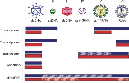
Regulation of virus host range is of particular importance. For best therapeutic benefit, gene therapy vehicles should be targeted specifically such that they transduce or infect target cells while avoiding sequestration in other organs or toxicity from infection of unwanted cells. Many methods have been used to target the tissue tropism for gene therapy. However, many of these apply only to nonreplicating vectors and almost all tend to be viral class specific (Figure 1).
Figure 1.
Targeting techniques applicable by viral class (where I–VI indicate Baltimore classification). Blue bars represent efficient targeting in replication-defective vectors. Red bars represent efficient targeting in replication-competent viruses. dsDNA, double-stranded DNA; ssDNA, single-stranded DNA.
Current methods to target tropism include transcriptional targeting whereby host transcription factors are employed to select for specific tissues or cell types,1 transductional targeting,2 whereby viruses are modified to be selective for specific cells at the level of entry and translational targeting that exploits defective interferon (IFN) signaling in cancer cells.3 Though all the aforementioned modalities prove very efficacious under certain circumstances, they are decidedly lacking in a number of ways.
Transcriptional targeting applies only to viral vectors that rely upon the host DNA polymerase for replication purposes and is not applicable to a large majority of emerging vectors, as they are often RNA viruses driven by viral RNA–dependent polymerases. Transductional targeting is theoretically possible for all viruses, but requires a large amount of space to accommodate coding sequences for retargeted attachment proteins within the viral genome and is often extremely inefficient. Translational targeting has only been applied to vectors employed for cancer gene therapy, and only when there is a defective IFN response within the particular cancer.
All targeting paradigms to date have been tailored very specifically to particular, singular vector systems. Very few, if any, targeting methods can be applied to all viral vectors, replicating or otherwise. Targeting viruses to be microRNA (miRNA) responsive, however, may be the first blanket method of altering tissue tropism. miRNA targeting involves engineering the viral genome to contain miRNA target (miRT) elements that can then be recognized and regulated by endogenous cellular miRNAs or, possibly, viral miRNAs. Viruses of each Baltimore class should be susceptible to miRNA-mediated attack, though at different places within the viral life cycle. miRNA-mediated targeting should avoid any size restrictions, as miRT elements are not traditionally in excess of 24 nt. While miRTs could possibly decrease antigen presentation that could dampen an antiviral immune response, miRNA targeting should appease many safety concerns such as those arising when modifying viruses by transductional means (which theoretically can increase pathogenicity by expanding host range to cells that are not normally susceptible to viral infection) because tropism is being restricted. Here we describe the strategies that have and can be used to engineer viruses to be recognized and regulated by miRNAs.
miRNAs: Biogenesis and Regulatory Functions
miRNAs are ~22-nt regulatory RNAs that act post-transcriptionally to influence a diverse and expansive array of cellular functions. First identified in Caenorhabditis elegans for their role in specifying cell fate,4 miRNAs are now known to act, among other functions, in disease pathogenesis,5 cancer,6 and the inflammatory response.7 Through base pairing with complementary regions [most often in the 3′ untranslated region (3′UTR) of cellular messenger RNA (mRNA)], miRNAs can act to suppress mRNA translation or, upon high-sequence homology, cause the catalytic degradation of mRNA.
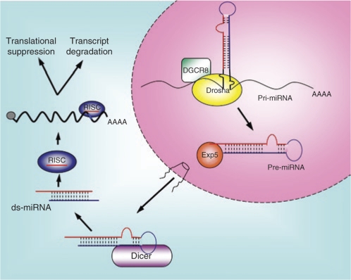
Cellular miRNAs are derived from RNA polymerase II (pol II) transcribed RNA in the nucleus, most often from intronic or UTRs of mRNA.8 miRNAs originate from larger precursor molecules characterized by a requisite secondary structure referred to as the primary miRNA that includes an imperfect ~80-nt stem loop (Figure 2).9 This secondary structure is recognized and cleaved by the nuclear RNase III Drosha when coupled with its essential nuclear cofactor DGCR8, and this cleavage gives rise to a precursor miRNA, essentially a ~60-nt hairpin loop.10 The precursor miRNA is then exported out of nucleus by the exportin-5 pathway11 and is recognized and cleaved by a second cellular RNase III, Dicer.12 Dicer cleavage liberates a duplex 20–24-nt RNA intermediate (double-stranded miRNA), and helicase activity of the Dicer complex then allows the incorporation of one of the RNA strands (typically the one with greater 5′ thermal instability) into the RNA-induced silencing complex (RISC).13 This RNA (the mature miRNA), then acts to guide the recognition of target mRNAs,14 while the “passenger strand” is degraded.
Figure 2.
Biogenesis and processing of human microRNAs (miRNAs). DGCR8, DeGeorge syndrome critical region 8 (protein); ds-miRNA, double-stranded microRNA; Exp5, exportin-5; pre-miRNA, precursor microRNA; pri-miRNA, primary microRNA; RISC, RNA-induced silencing complex.
Sequence complementarity in the 7-nt “seed region” [base pair (bp) 2–8 of the miRNA] is essential for recognition of miRNA and its target.12 In humans, it is predicted that there are >400 miRNAs,15 many of which can be expressed in excess of 1,000 copies/cell.16 A large number of these miRNAs are differentially expressed in different tissues and cell lineages such that they regulate tissue-specific gene expression.17
Lately, much focus has come upon the interplay between viruses and miRNAs. Well known for their ability to act in the antiviral response of plants and invertebrates, miRNAs were first assumed to play a similar role in higher metazoans. Debate is still ongoing, however, as to the role of mammalian miRNAs in the antiviral response: some claim definite antiviral activities for host miRNAs18,19 while other carefully conducted studies dispute this claim.20,21 It has been proposed that viruses are under selective pressure to avoid sequence homology to cellular miRNAs in tissues in which they replicate.22 However, at least one virus has been shown to utilize a tissue-specific miRNA to enhance its replication in target tissue.23
Because of the highly differential tissue expression of many miRNAs, it was proposed that cellular miRNAs could be exploited to mediate tissue-specific targeting of gene therapy vectors.24 By engineering tandem copies of target elements perfectly complementary to tissue-specific miRNAs within viruses and vectors, multiple groups have shown that host miRNAs can regulate exogenously introduced transgene expression24,25 and even viral gene products.26,27,28 These demonstrations not only show the potential for miRNA-mediated direction of tissue tropism, but lend credence to the potential ability of miRNAs to act on mammalian viruses in vivo, though viruses may then be evolutionarily directed to avoid sequence complementarity.
Target sequences for cellular-encoded miRNAs have recently been used in the design of targeted gene therapy vectors and safer oncolytic viruses with strong initial results. Viral miRNAs, however, have not yet been utilized in the design of engineered viruses. With road maps emerging from basic science, it is likely that virally encoded miRNAs could soon be used in gene therapy applications.
Here, we examine ways in which miRNAs have and can be employed to target viral tissue tropism and examine the feasibility of utilizing virally encoded miRNAs to decrease viral immunogenicity, as well as increase persistence of gene expression for improved therapeutic benefit of replacement gene therapy.
Viruses and RNA Interference
In many ways, miRNA-mediated silencing might be called the endogenous cellular RNA interference (RNAi). Once outside the nucleus, miRNA biogenesis becomes virtually indistinguishable from RNAi.29 The largest difference between endogenous miRNAs and introduced small interfering RNAs (siRNAs) is in the amount of homology that directs RISC to act on a target. miRNAs often interact with much less homology to suppress the translation of cellular mRNA, while siRNAs are designed to be perfectly complementary to the target such that transcript cleavage is more likely to occur.
In fact, RNAi has been optimized using information derived from the cellular processing of miRNAs. While first-generation gene silencing used transfected si/shRNAs,30 suppression of gene expression was too transient for many systems. Gene transfer vectors encoding short hairpin RNAs (shRNAs) driven by pol III promoters formed second-generation gene-silencing systems.31 However, shortly thereafter it was found that the use of pol II promoters as used in miRNA biogenesis were often equally, if not more, efficient in functional gene silencing.32 Now in addition to transcribing shRNAs in the same method as miRNAs, expression cassettes containing the flanking regions of well-characterized miRNAs are often used to further enhance the efficacy of exogenously introduced shRNAs.33
With the great success of RNAi in functional analyses, it followed that RNAi could work equally well as an antiviral therapy. In vitro, it appears that all viruses may be susceptible to RNAi.34 With almost every class of virus now having been tested (and antiviral effects seen in all cases), it has become clear that RNAi does inhibit viral replication. Because of this, it was thought that siRNAs might be delivered transiently for acute viral infections (particularly for respiratory viruses) and retro/lentiviral vectors might provide a method to counteract effects of the persistent infections of hepatitis B virus, hepatitis C virus, and human immunodeficiency virus for clinical benefit. And indeed, there are clinical trials underway that employ RNAi for these very means.35
Despite in vitro success with RNAi as an antiviral therapy, many barriers exist to true therapeutic benefit. Foremost among these is viral escape from siRNAs. It is now clear that viruses can escape siRNA silencing, not just through mutation to avoid sequence complementarity to the introduced antiviral siRNAs,36 but also by altering the secondary structure around the areas in which there is homology between virus and siRNA.37 In addition to escape, toxicity and delivery are barriers to the success of RNAi as an antiviral therapeutic. These same challenges encumber the use of miRNAs for targeting means in gene therapy.
Range of Target Elements
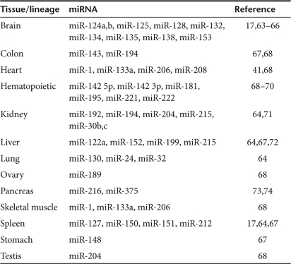
Target elements to multiple miRNAs engineered within a single vector can increase gene silencing in a single cell lineage38 or act to silence in multiple tissues.39 The exact target of choice becomes very specific to the cell type and tissue one wishes to focus upon. Most tissues such as brain, lung, spleen, liver, and others have multiple miRNAs that are much more abundant than in any other cell type (Table 1). Skeletal muscle, for example, has an increased abundance of miR-1, miR-133a, and miR-206 over all other cell types.40 Heart muscle is differentiated by increased expression of the three skeletal muscle miRNAs, as well as an additional heart-specific miRNA, miR-208.41 Every tissue has a unique miRNA expression profile that can be used as a database for the design of targeted gene therapy vectors.
Table 1.
Tissue-specific miRNA expression
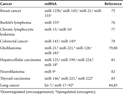
In addition to having tissue-specific signatures, miRNAs are also known to, in certain instances, have cancer-specific signatures (Table 2). Oncogenic miRNAs are found to be highly enriched in tumors, while tumor suppressor miRNAs are ubiquitously expressed in normal tissue, but specifically downregulated in cancer.42 miRNAs can even be associated with the exact stage of a cancer, some perhaps acting to suppress metastasis.43 Tumor suppressor miRNAs are known to be downregulated (or completely deleted) in breast,44 lung,45 and colorectal cancers46 and expression of perhaps the most well defined of the tumor suppressor miRNAs, miR-15 and miR-16, are altogether lost in chronic lymphocytic leukemia.47 Incorporation of tumor suppressor target elements within a vector could restrict expression in normal, untransformed tissue while allowing expression in tumors lacking these miRNAs. Targeting by this means could theoretically be transferred to a vector with toxicity to any cell type, providing a potential ubiquitous new way of conferring specificity.
Table 2.
Oncogenic and oncosuppresive miRNAs
While miRNAs almost always act to mediate post-transcriptional silencing, in at least one case, viral replication has actually been shown to be contingent upon a cellular miRNA.23 Hepatitis C Virus replicates exclusively in the liver. While in vivo this was attributed to liver-specific receptor expression, virus propagation in vitro that circumvented virus–receptor interactions (by way of replicon RNA transfection) was still exceedingly difficult. It has now been shown that the liver-specific miR-122 positively affects the accumulation of hepatitis C virus RNA.
While a mechanism for this positive regulation by miR-122 has not yet been defined, the concept has large implications for the design of miRNA targeted vectors. Oncogenic miRNAs could mediate the targeting of cancer-specific vectors and the wide database for tissue-specific miRNAs could now not only be used for vector restriction from those tissues, but actual targeted expression.
Designing the Regulatory Insert
Rough outlines are beginning to emerge on the best way to employ miRNAs for targeting (or restriction purposes). It has been shown that while four tandem copies of a single target work better than two copies or one copy, it does not hold that increasing numbers of targets will always translate to increased gene silencing.39 In actuality, increasing the number of miRTs within a single transcript has been shown to cause a decrease in miRNA function.48 In these cases, multiple RISCs, all guided by the same miRNA guide strands may bind to a single transcript creating what some have termed to be a “sponge” effect, whereby miRTs sequester all-sequence complementary miRNAs, thus severely impairing function. miRNA-mediated suppression of vector-delivered gene expression substantially decreases in this situation and may be more pronounced in the case of imperfect complementarity between target and miRNA. In addition, the normal function of these cellular miRNAs can be completely inhibited. In what may be an extreme example, impaired endogenous miRNA function by oversaturation of the exportin-5 pathway was shown to cause a fatal liver toxicity in mice.49
Much work remains to be done on the exact number of copies and the spacing elements between tandem copies of miRT elements that will prove most efficacious for vector targeting. In addition, to avoid problems for rescue and manufacturing, production cell lines should either lack the miRNA to which the corresponding target has been engineered or be treated with miRNA inhibitors.
Tissue-specific miRNAs are expressed at different copy numbers in different cell lineages, and it has been proposed that a threshold copy of miRNAs must be reached to achieve appreciable gene silencing.39 miRNA abundance alone does not necessarily make a good target, however. Even when delivered in equimolar amounts, different miRNAs will regulate the expression of a perfectly homologous target engineered into the same position in a transcript to different extents.38 While it is not clear why, the actual miRNA sequence itself does appear to affect the extent to which a target RNA is regulated. In some cases, it appears that perfect sequence matches may not be ideal.
The reigning dogma in miRNA regulation had always been the higher degree of homology between miRNA and target, the better. A perfect match in the seed sequence is obligatory, while higher degree of complementarity tends to increase the likelihood of catalytic mRNA cleavage between bp 10 and 11 by Argonaute 2 in the Dicer complex.12 However, in the absence of a formal proof, it remains a possibility that some miRTs might prove better as imperfect matches, engineered to include miRNA/miRT mismatch or even bulge sequences. In addition to questions as to whether the ideal sequence to best regulate a vector is the position in which a target(s) should be introduced.
In nature, most miRTs have been found within the 3′UTR of cellular mRNAs. It is well known that siRNAs can be quite effective when targeted against any region of an mRNA, whether coding or not. As RNAi and miRNA regulation are quite similar, it seems likely that target elements introduced within any part of a vector genome have the potential to work, as long as the region in which it was present was actually transcribed and found outside the nucleus. Studies whereby targets were introduced within the 5′UTR of reporter mRNAs showed that there was no great difference in silencing efficiency.50 It may be that the ideal spot for miRTs insertion relies not so much on the defined location (whether 5′ or 3′ untranslated or coding sequence), but upon the secondary structure of that location. Many studies have shown that target sequences present within regions with a great deal of secondary structure are ill recognized by miRNAs.51,52
miRNA-Mediated Targeting of Nonreplicating Vectors
Efforts to engineer viral RNA to be stably expressed or translated tissue-specifically have been underway for many years. In two examples, UTRs of two genes (fibroblast growth factor-2 and cyclooxegenase-2)53,54 were engineered into replication-competent adenoviruses to confer cancer-selective viral replication. While often the mechanism that made these RNA stability elements confer tissue specificity has been unclear, with developments in the understanding of miRNA function, it became apparent that cellular miRNAs present in some tissues presented a means to confer selective RNA stability.
With the demonstration that viruses are susceptible to RNAi-mediated attack, and with RNAi assuming more of the characteristics of true endogenously encoded miRNAs, it followed that viruses might be engineered to become miRNA responsive by engineering them to contain target elements for miRNAs.
In the first example of such engineering, it was hypothesized that poor specificity of a systemically delivered lentiviral vector caused the transduction of (hematopoietic) antigen-presenting cells such that an immune response was then generated against the delivered transgene. By engineering four perfect copies of a hematopoietic-specific miRT element within the 3′UTR of a transgene delivered by a lentiviral vector, the Naldini group was able to show that a hematopoietic-specific miRNA could recognize target elements within the transgene such that expression was squelched in hematopoietic cells, and thus no immune response to the transgene was generated.24 This study represented the first conclusive evidence that viral vectors could be engineered such that they were restricted from expressing in cells bearing cognate miRNAs both in vitro and in vivo. They then significantly extended these findings to show that tissue-specific miRNAs from a broad array of cell types could suppress the expression of transgenes bearing sequence complementary miRTs and that there was a relationship between miRNA abundance within a cell and the extent to which that miRNA could function to suppress gene expression.39
Similarly, it was thought that miRNAs highly enriched in the liver could perturb severe hepatotoxicity associated with a replication-deficient adenoviral vector. Adenovirus vectors encoding the thymidine kinase gene from herpes simplex virus can act as potent anticancer agents when used in combination with the prodrug ganciclovir.25 By incorporating tandem copies of the miRT for the liver-specific miR-122a, it has been shown that liver expression can be reduced up to 1,500-fold without affecting tumor transduction, resulting in roughly equivalent suicide gene–mediated cancer therapy with markedly reduced liver toxicity.
miRNAs represent a new paradigm for restricting tissue tropism of viral vectors. By increasing specificity, potency can potentially be increased without the worry of toxicity from transduction of off-target cells.
miRNA-Responsiveness in Replicating Viruses
Nonreplicating viral vectors transduce cells to express a transgene of interest aiming to mimic physiologically normal expression. In these cases, robust expression is wanted as long as it is not at the expense of normal cellular function. Replicating viruses used for oncolytic cancer therapy or for attenuated vaccines, however, act much differently. In these cases, robust viral expression is wanted to act lytically on cancer cells (i.e., oncolytic virotherapy) or generate a healthy immune response (vaccines).
Replicating viruses in many cases may represent a larger hurdle in terms of gene expression, able to completely overtake cellular machinery for DNA or RNA synthesis. In addition, the design for miRNA targeting may now have to be much more complex, as many viral genes are being expressed as opposed to a single transgene in the case of nonreplicating expression vectors. While miRTs themselves may be readily transferred without sequence change among viruses of different families and thus be one of the first pantropic targeting modalities, the actual placement may need to be very specific to the virus/family.
Picornaviruses represent possibly one of the least complex viral families. With a single-stranded positive-sense (+) RNA genome, targets placed anywhere within the viral genome also become part of the single-viral mRNA. Engineering the oncolytic picornavirus, coxsackievirus A21, to contain the muscle-specific miRTs corresponding to miR-133a and miR-206 within the 3′UTR effectively abolished replication in vitro in the presence of cognate miRNAs.38 In addition, it protected mice in vivo from developing fatal myositis associated with the wild-type virus. Because muscle-specific miRNAs were not present in melanoma and myeloma xenografts of these mice, the miRNA-targeted coxsackievirus A21 fully retained its oncolytic potency and was shown to be a fully curative therapy. In this study, target retention was shown in a majority of treated mice to be fully intact out to 45 days post-treatment (at which time no tumor remained). However, analysis of creatine kinase, a quantitative serum marker of muscle damage showed a small, but statistically significant increase in muscle damage that accumulated over time as a small number of mice mutated the miRT insert.
miRT mutation and deletion is of particular concern, and indeed has been shown in the regulation of the closely related picornavirus, poliovirus (PV). In an effort to control poliomyelitis caused by the virus, a miRNA-targeted PV that has been previously shown to be highly sensitive to the miRT insertion26 (though mutations through extended passage could overcome miRNA-mediated regulation) was investigated.55 It was hypothesized that incorporation of the neuronal miR-124T or the tumor suppressor let-7aT would curb viral replication in the brain, while still allowing for the generation of a protective immune response within mice transgenic for the PV receptor. Even with the incorporation of only two miRTs within the virus, PV receptor transgenic mice were shown to be completely protected from poliomyelitis when given up to 108 plaque-forming unit intramuscularly (intracerebral and intranasal administration were not reported), demonstrating proof of principle for the generation of safe de novo vaccines by way of miRT incorporation.
In a similar study, vesicular stomatitis virus was engineered to contain three copies of let-7a within the 3′UTR of the viral M gene, which is responsible for the inhibition of the cellular IFN response.27 let-7a is a member of the let-7 family of tumor suppressor miRNAs and should act to inhibit viral replication in a broad variety of normal cells. Through incorporation of its miRT, it was thought that tumor selectivity could be generated and, indeed, the viral output was decreased by three logs in vitro in cells expressing the let-7a miRNA. It is possible that targeting by this method could restrict acquired neuropathology, which is found commonly in animal models of vesicular stomatitis virus oncolysis. To examine this relationship, a total of six mice were inoculated intranasally with viruses containing miRTs with perfect matches to let-7a or with a mutated miRT insert and followed for 15 days, with only one mouse in either group succumbing to what is presumed to be neurotoxicity. In this regard, it is difficult to conclude definitively that neurovirulence was affected to any appreciable extent. The ability of this miRNA to provide a more pronounced response may have been limited by the placement of the miRT within the virus, among other factors. In this example, only one of five viral genes was targeted, by a target that did not fully extinguish the expression of a reporter gene in vitro.
Oncolytic adenoviruses have now been engineered to be miRNA responsive as well as their replication-defective counterparts. In a recent study, incorporation of the liver-specific miR-122T within the viral gene E1A decreased E1A mRNA expression in vitro, which may markedly reduce liver toxicity in vivo, though this has not yet been examined.28 The development of an oncolytic adenovirus with reduced hepatotoxicity may be of particular importance, as it is perhaps the furthest clinically advanced virus.
Unanswered Questions
Negative-strand RNA viruses (i.e., vesicular stomatitis virus) might be intrinsically harder to control via miRNA targeting than other viruses such as coxsackievirus A21 or PV. Because picornaviruses contain a single (+) RNA genome, both viral mRNA and genome are potentially targeted by an miRT in the same orientation. Vesicular stomatitis virus, however, has genome and mRNA in different orientations and transcribes viral genes as distinct mRNAs.56 In addition, the genome is protected by a ribonucleoprotein complex that surrounds the viral RNA such that RISC-mediated attack may not occur.
Viral escape may present another obstacle for the success of miRNA-mediated targeting. In this respect, many of the problems with the use of siRNA as an antiviral therapeutic may also apply to the use of miRNA targeting for replication-competent viruses.57 Clearly, a miRNA-targeted virus whose replication is restricted in a specific tissue should be capable of gaining a replication advantage through deletion or mutation of the miRT. Thus, the mutation rate of the viral or cellular polymerase becomes the limiting factor. In general terms, the mutation rate of viruses is such that ssRNA virus>retrovirus>single-stranded DNA virus>double-stranded DNA virus, ranging from ~10−3 to 10−8 mutations/nucleotide/genomic replication.58
It is hypothesized that viruses may escape the intrinsic miRNA defense mechanism found in plants by having evolved to avoid homology to miRNAs that are present within those cells in which replication occurs.22 It is clear that viruses can escape siRNA-mediated silencing, and it has now been demonstrated that even in the absence of selective pressure viruses can escape miRNA targeting as well.38 While increasing miRT number and sites could potentially delay the emergence of escape mutants, this might also act to form miRNA sponges that could increase toxicity and limit performance. With both attenuated viral vaccines and oncolytics, however, an immune response against the virus will often clear viremia such that escape mutants may never become apparent.
Replicating viruses present additional, alternative, and in some cases more complex obstacles for the success of miRNA-mediated targeting. However, miRNAs can provide methods for the creation of safer, improved vaccines and increase the number of oncolytic viruses that can be safely employed for cancer gene therapy.
Virally Encoded miRNAs
The utilization of cellular miRNAs for targeting the tissue tropism of gene therapy vehicles has lately garnered much attention. A large database of cellular miRNAs exists,17 and the tissue distribution, abundance, and function of these miRNAs have been extensively investigated. Perhaps equally important, however, are virally encoded miRNAs.
Viral miRNAs identified thus far are very similar to cellular miRNAs in terms of biogenesis and function. Like cellular miRNAs, they are thought to recognize targets through imperfect homology and most often inhibit gene expression by translational suppression. However, they are more likely than cellular miRNAs to be derived from open-reading frames and intronic regions. Because all identified viral miRNAs are transcribed in the nucleus, they have all, thus far, come from double-stranded DNA viruses. Adenoviruses and polyomaviruses encode viral miRNAs, and the herpesvirus family encodes upward of 100 viral miRNAs.59 It is improbable that RNA viruses would encode miRNAs. Many RNA viruses replicate exclusively in the cytoplasm, necessitating a different biogenesis pathway were a miRNA to be encoded. In addition, an encoded miRNA could act in an inhibitory fashion against the complementary viral RNA strand.
Viral miRNAs can mediate the regulation of both cellular and viral gene expression, to different ends. To mediate latency, viral miRNAs have been shown to downregulate the expression of specific viral proteins. SV40, for example, downregulates the expression of the large T antigen to avoid the adaptive immune response.60 By reducing expression of large T antigen, presentation to cytotoxic T lymphocytes is reduced, as is the release of cytokines such as gamma IFN. Similarly, herpes simplex virus-1 has recently been shown to downregulate the expression of the ICP0 and is thought to act to establish viral latency through this means.61 In addition, it is thought that viral miRNAs can act in tumorigenesis and may influence the immune response to viral infection.62
What the viral miRNAs are theoretically engineered to encode is as important or more important than what they do encode. Just like cellular miRNAs, viral miRNAs have the potential for acting on cellular gene expression, as well as viral gene expression. Viruses have evolved proteins that act to inhibit or evade the host immune response or to prevent the infected cell from activating its apoptotic machinery. In many cases, these innate immune modulatory and apoptosis inhibitory proteins represent targets for viral engineering to generate recombinants that are unable to suppress innate immunity or apoptosis and which, therefore, replicate exclusively or selectively in tumor cells.
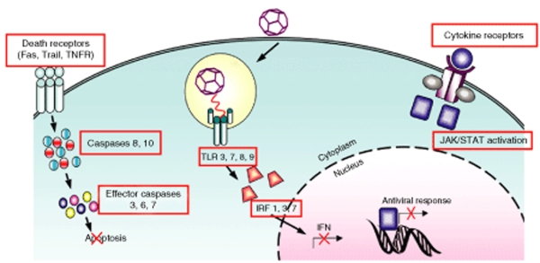
It seems likely that viral miRNAs could be designed against many targets (both cellular and viral) to enhance the performance of oncolytic viruses. Potential targets that would limit natural killer–mediated recognition limit viral antigen presentation, control the IFN response, suppress inflammatory cytokine release, and inhibit cellular apoptosis, could prove to be of particular importance (Figure 3).
Figure 3.
Potential targets of viral microRNAs. Red boxes represent potential targets. IFN, interferon; TLR, Toll-like receptor; TNFR, tumor necrosis factor receptor; TRAIL, tumor necrosis factor–related apoptosis-inducing ligand.
Conclusions
miRNA targeting is an effective approach for controlling the stability of vector-encoded mRNAs and of certain RNA viral genomes and is, therefore, rapidly emerging as a versatile new targeting strategy. The approach requires <100 bases of new vector sequence and, in contrast to previously developed targeting strategies, is potentially transferrable to all known viral and nonviral expression vectors as well as replication-competent viruses. Therefore, it is expected to be of major utility for the generation of targeted expression vectors, tumor-specific oncolytic viruses, and attenuated viral vaccines derived from serious pathogens. Because cellular miRNAs are not all equally abundant, and because flanking sequences influence the efficiency of target destruction, the precise molecular approach to miRNA targeting must be subtly different for each new vector or virus. Trial and error is, therefore, a necessary step at the current time in order to obtain optimal control of expression. Numerous miRTs exist, encompassing oncogenic (tumor-selective), oncosuppressive (selectively expressed in nontumor tissues), and tissue-specific miRNAs. These targets are already well characterized and most are highly conserved, even across species barriers. Therefore, it seems realistic to expect this targeting approach can be validated in disparate species, a distinct advantage over transductional targeting where targeting moieties against human cells are unlikely to function in nonhomologous species. One concern with the approach is that viruses can revert to their original tropism by acquiring mutations in their miRT sites, but such revertants are considered unlikely to be of clinical significance because they emerge late after the initial virus challenge by which time they must contend with a fully developed antiviral immune response.
In the future, exploitation of both cellular and viral miRNAs will likely be of great benefit for targeting gene therapy viruses and vectors, generation of new and safer vaccines, and, in addition, could provide a modality for increasing the persistence of nonintegrating gene therapy vehicles and the evasion of innate immunity.
REFERENCES
- Bazan-Peregrino M, Seymour LW., and , Harris AL. Gene therapy targeting to tumor endothelium. Cancer Gene Ther. 2007;14:117–127. doi: 10.1038/sj.cgt.7701001. [DOI] [PubMed] [Google Scholar]
- Waehler R, Russell SJ., and , Curiel DT. Engineering targeted viral vectors for gene therapy. Nat Rev Genet. 2007;8:573–587. doi: 10.1038/nrg2141. [DOI] [PMC free article] [PubMed] [Google Scholar]
- Barber GN. VSV-tumor selective replication and protein translation. Oncogene. 2005;24:7710–7719. doi: 10.1038/sj.onc.1209042. [DOI] [PubMed] [Google Scholar]
- Lee RC, Feinbaum RL., and , Ambros V. The C. elegans heterochronic gene lin-4 encodes small RNAs with antisense complementarity to lin-14. Cell. 1993;75:843–854. doi: 10.1016/0092-8674(93)90529-y. [DOI] [PubMed] [Google Scholar]
- Mattes J, Collison A., and , Foster PS. Emerging role of microRNAs in disease pathogenesis and strategies for therapeutic modulation. Curr Opin Mol Ther. 2008;10:150–157. [PubMed] [Google Scholar]
- Calin GA., and , Croce CM. MicroRNA signatures in human cancers. Nat Rev Cancer. 2006;6:857–866. doi: 10.1038/nrc1997. [DOI] [PubMed] [Google Scholar]
- Lindsay MA. microRNAs and the immune response. Trends Immunol. 2008;29:343–351. doi: 10.1016/j.it.2008.04.004. [DOI] [PubMed] [Google Scholar]
- Lee Y, Kim M, Han J, Yeom KH, Lee S, Baek SH, et al. MicroRNA genes are transcribed by RNA polymerase II. EMBO J. 2004;23:4051–4060. doi: 10.1038/sj.emboj.7600385. [DOI] [PMC free article] [PubMed] [Google Scholar]
- Cullen BR. Transcription and processing of human microRNA precursors. Mol Cell. 2004;16:861–865. doi: 10.1016/j.molcel.2004.12.002. [DOI] [PubMed] [Google Scholar]
- Gregory RI, Yan KP, Amuthan G, Chendrimada T, Doratotaj B, Cooch N, et al. The Microprocessor complex mediates the genesis of microRNAs. Nature. 2004;432:235–240. doi: 10.1038/nature03120. [DOI] [PubMed] [Google Scholar]
- Yi R, Qin Y, Macara IG., and , Cullen BR. Exportin-5 mediates the nuclear export of pre-microRNAs and short hairpin RNAs. Genes Dev. 2003;17:3011–3016. doi: 10.1101/gad.1158803. [DOI] [PMC free article] [PubMed] [Google Scholar]
- Bartel DP. MicroRNAs: genomics, biogenesis, mechanism, and function. Cell. 2004;116:281–297. doi: 10.1016/s0092-8674(04)00045-5. [DOI] [PubMed] [Google Scholar]
- Sontheimer EJ. Assembly and function of RNA silencing complexes. Nat Rev Mol Cell Biol. 2005;6:127–138. doi: 10.1038/nrm1568. [DOI] [PubMed] [Google Scholar]
- Schwarz DS, Hutvagner G, Haley B., and , Zamore PD. Evidence that siRNAs function as guides, not primers, in the Drosophila and human RNAi pathways. Mol Cell. 2002;10:537–548. doi: 10.1016/s1097-2765(02)00651-2. [DOI] [PubMed] [Google Scholar]
- Landgraf P, Rusu M, Sheridan R, Sewer A, Iovino N, Aravin A, et al. A mammalian microRNA expression atlas based on small RNA library sequencing. Cell. 2007;129:1401–1414. doi: 10.1016/j.cell.2007.04.040. [DOI] [PMC free article] [PubMed] [Google Scholar]
- Liang Y, Ridzon D, Wong L., and , Chen C. Characterization of microRNA expression profiles in normal human tissues. BMC Genomics. 2007;8:166. doi: 10.1186/1471-2164-8-166. [DOI] [PMC free article] [PubMed] [Google Scholar]
- Lagos-Quintana M, Rauhut R, Yalcin A, Meyer J, Lendeckel W., and , Tuschl T. Identification of tissue-specific microRNAs from mouse. Curr Biol. 2002;12:735–739. doi: 10.1016/s0960-9822(02)00809-6. [DOI] [PubMed] [Google Scholar]
- Lecellier CH, Dunoyer P, Arar K, Lehmann-Che J, Eyquem S, Himber C, et al. A cellular microRNA mediates antiviral defense in human cells. Science. 2005;308:557–560. doi: 10.1126/science.1108784. [DOI] [PubMed] [Google Scholar]
- Bennasser Y, Le SY, Benkirane M., and , Jeang KT. Evidence that HIV-1 encodes an siRNA and a suppressor of RNA silencing. Immunity. 2005;22:607–619. doi: 10.1016/j.immuni.2005.03.010. [DOI] [PubMed] [Google Scholar]
- Lin J., and , Cullen BR. Analysis of the interaction of primate retroviruses with the human RNA interference machinery. J Virol. 2007;81:12218–12226. doi: 10.1128/JVI.01390-07. [DOI] [PMC free article] [PubMed] [Google Scholar]
- Cullen BR. Is RNA interference involved in intrinsic antiviral immunity in mammals. Nat Immunol. 2006;7:563–567. doi: 10.1038/ni1352. [DOI] [PubMed] [Google Scholar]
- Cullen BR. Viruses and microRNAs. Nat Genet. 2006;38 suppl:S25–S30. doi: 10.1038/ng1793. [DOI] [PubMed] [Google Scholar]
- Jopling CL, Yi M, Lancaster AM, Lemon SM., and , Sarnow P. Modulation of hepatitis C virus RNA abundance by a liver-specific MicroRNA. Science. 2005;309:1577–1581. doi: 10.1126/science.1113329. [DOI] [PubMed] [Google Scholar]
- Brown BD, Venneri MA, Zingale A, Sergi Sergi L., and , Naldini L. Endogenous microRNA regulation suppresses transgene expression in hematopoietic lineages and enables stable gene transfer. Nat Med. 2006;12:585–591. doi: 10.1038/nm1398. [DOI] [PubMed] [Google Scholar]
- Suzuki T, Sakurai F, Nakamura S, Kouyama E, Kawabata K, Kondoh M, et al. miR-122a-regulated expression of a suicide gene prevents hepatotoxicity without altering antitumor effects in suicide gene therapy. Mol Ther. 2008;16:1719–1726. doi: 10.1038/mt.2008.159. [DOI] [PubMed] [Google Scholar]
- Vignuzzi M, Wendt E., and , Andino R. Engineering attenuated virus vaccines by controlling replication fidelity. Nat Med. 2008;14:154–161. doi: 10.1038/nm1726. [DOI] [PubMed] [Google Scholar]
- Edge RE, Falls TJ, Brown CW, Lichty BD, Atkins H., and , Bell JC. A let-7 MicroRNA-sensitive vesicular stomatitis virus demonstrates tumor-specific replication. Mol Ther. 2008;16:1437–1443. doi: 10.1038/mt.2008.130. [DOI] [PubMed] [Google Scholar]
- Ylösmäki E, Hakkarainen T, Hemminki A, Visakorpi T, Andino R., and , Saksela K. Generation of a conditionally replicating adenovirus based on targeted destruction of E1A mRNA by a cell type-specific microRNA. J Virol. 2008;82:11009–11015. doi: 10.1128/JVI.01608-08. [DOI] [PMC free article] [PubMed] [Google Scholar]
- Zeng Y, Yi R., and , Cullen BR. MicroRNAs and small interfering RNAs can inhibit mRNA expression by similar mechanisms. Proc Natl Acad Sci USA. 2003;100:9779–9784. doi: 10.1073/pnas.1630797100. [DOI] [PMC free article] [PubMed] [Google Scholar]
- Elbashir SM, Harborth J, Lendeckel W, Yalcin A, Weber K., and , Tuschl T. Duplexes of 21-nucleotide RNAs mediate RNA interference in cultured mammalian cells. Nature. 2001;411:494–498. doi: 10.1038/35078107. [DOI] [PubMed] [Google Scholar]
- Grimm D, Pandey K., and , Kay MA. Adeno-associated virus vectors for short hairpin RNA expression. Methods Enzymol. 2005;392:381–405. doi: 10.1016/S0076-6879(04)92023-X. [DOI] [PubMed] [Google Scholar]
- Giering JC, Grimm D, Storm TA., and , Kay MA. Expression of shRNA from a tissue-specific pol II promoter is an effective and safe RNAi therapeutic. Mol Ther. 2008;16:1630–1636. doi: 10.1038/mt.2008.144. [DOI] [PubMed] [Google Scholar]
- Chang K, Elledge SJ., and , Hannon GJ. Lessons from Nature: microRNA-based shRNA libraries. Nat Methods. 2006;3:707–714. doi: 10.1038/nmeth923. [DOI] [PubMed] [Google Scholar]
- Gitlin L., and , Andino R. Nucleic acid-based immune system: the antiviral potential of mammalian RNA silencing. J Virol. 2003;77:7159–7165. doi: 10.1128/JVI.77.13.7159-7165.2003. [DOI] [PMC free article] [PubMed] [Google Scholar]
- Haasnoot J, Westerhout EM., and , Berkhout B. RNA interference against viruses: strike and counterstrike. Nat Biotechnol. 2007;25:1435–1443. doi: 10.1038/nbt1369. [DOI] [PMC free article] [PubMed] [Google Scholar]
- Gitlin L, Stone JK., and , Andino R. Poliovirus escape from RNA interference: short interfering RNA-target recognition and implications for therapeutic approaches. J Virol. 2005;79:1027–1035. doi: 10.1128/JVI.79.2.1027-1035.2005. [DOI] [PMC free article] [PubMed] [Google Scholar]
- Westerhout EM, Ooms M, Vink M, Das AT., and , Berkhout B. HIV-1 can escape from RNA interference by evolving an alternative structure in its RNA genome. Nucleic Acids Res. 2005;33:796–804. doi: 10.1093/nar/gki220. [DOI] [PMC free article] [PubMed] [Google Scholar]
- Kelly EJ, Hadac EM, Greiner S., and , Russell SJ. Engineering microRNA responsiveness to decrease virus pathogenicity. Nat Med. 2008;14:1278–1283. doi: 10.1038/nm.1776. [DOI] [PubMed] [Google Scholar]
- Brown BD, Gentner B, Cantore A, Colleoni S, Amendola M, Zingale A, et al. Endogenous microRNA can be broadly exploited to regulate transgene expression according to tissue, lineage and differentiation state. Nat Biotechnol. 2007;25:1457–1467. doi: 10.1038/nbt1372. [DOI] [PubMed] [Google Scholar]
- Callis TE, Chen JF., and , Wang DZ. MicroRNAs in skeletal and cardiac muscle development. DNA Cell Biol. 2007;26:219–225. doi: 10.1089/dna.2006.0556. [DOI] [PubMed] [Google Scholar]
- van Rooij E, Sutherland LB, Qi X, Richardson JA, Hill J., and , Olson EN. Control of stress-dependent cardiac growth and gene expression by a microRNA. Science. 2007;316:575–579. doi: 10.1126/science.1139089. [DOI] [PubMed] [Google Scholar]
- Chen CZ. MicroRNAs as oncogenes and tumor suppressors. N Engl J Med. 2005;353:1768–1771. doi: 10.1056/NEJMp058190. [DOI] [PubMed] [Google Scholar]
- Tavazoie SF, Alarcón C, Oskarsson T, Padua D, Wang Q, Bos PD, et al. Endogenous human microRNAs that suppress breast cancer metastasis. Nature. 2008;451:147–152. doi: 10.1038/nature06487. [DOI] [PMC free article] [PubMed] [Google Scholar]
- Lowery AJ, Miller N, McNeill RE., and , Kerin MJ. MicroRNAs as prognostic indicators and therapeutic targets: potential effect on breast cancer management. Clin Cancer Res. 2008;14:360–365. doi: 10.1158/1078-0432.CCR-07-0992. [DOI] [PubMed] [Google Scholar]
- Jay C, Nemunaitis J, Chen P, Fulgham P., and , Tong AW. miRNA profiling for diagnosis and prognosis of human cancer. DNA Cell Biol. 2007;26:293–300. doi: 10.1089/dna.2006.0554. [DOI] [PubMed] [Google Scholar]
- Akao Y, Nakagawa Y., and , Naoe T. MicroRNA-143 and -145 in colon cancer. DNA Cell Biol. 2007;26:311–320. doi: 10.1089/dna.2006.0550. [DOI] [PubMed] [Google Scholar]
- Nicoloso MS, Kipps TJ, Croce CM., and , Calin GA. MicroRNAs in the pathogeny of chronic lymphocytic leukaemia. Br J Haematol. 2007;139:709–716. doi: 10.1111/j.1365-2141.2007.06868.x. [DOI] [PubMed] [Google Scholar]
- Ebert MS, Neilson JR., and , Sharp PA. MicroRNA sponges: competitive inhibitors of small RNAs in mammalian cells. Nat Methods. 2007;4:721–726. doi: 10.1038/nmeth1079. [DOI] [PMC free article] [PubMed] [Google Scholar]
- Grimm D, Streetz KL, Jopling CL, Storm TA, Pandey K, Davis CR, et al. Fatality in mice due to oversaturation of cellular microRNA/short hairpin RNA pathways. Nature. 2006;441:537–541. doi: 10.1038/nature04791. [DOI] [PubMed] [Google Scholar]
- Lytle JR, Yario TA., and , Steitz JA. Target mRNAs are repressed as efficiently by microRNA-binding sites in the 5′ UTR as in the 3′ UTR. Proc Natl Acad Sci USA. 2007;104:9667–9672. doi: 10.1073/pnas.0703820104. [DOI] [PMC free article] [PubMed] [Google Scholar]
- Long D, Lee R, Williams P, Chan CY, Ambros V., and , Ding Y. Potent effect of target structure on microRNA function. Nat Struct Mol Biol. 2007;14:287–294. doi: 10.1038/nsmb1226. [DOI] [PubMed] [Google Scholar]
- Vella MC, Reinert K., and , Slack FJ. Architecture of a validated microRNA::target interaction. Chem Biol. 2004;11:1619–1623. doi: 10.1016/j.chembiol.2004.09.010. [DOI] [PubMed] [Google Scholar]
- Stoff-Khalili MA, Rivera AA, Nedeljkovic-Kurepa A, DeBenedetti A, Li XL, Odaka Y, et al. Cancer-specific targeting of a conditionally replicative adenovirus using mRNA translational control. Breast Cancer Res Treat. 2008;108:43–55. doi: 10.1007/s10549-007-9587-7. [DOI] [PMC free article] [PubMed] [Google Scholar]
- Ahmed A, Thompson J, Emiliusen L, Murphy S, Beauchamp RD, Suzuki K, et al. A conditionally replicating adenovirus targeted to tumor cells through activated RAS/P-MAPK-selective mRNA stabilization. Nat Biotechnol. 2003;21:771–777. doi: 10.1038/nbt835. [DOI] [PubMed] [Google Scholar]
- Barnes D, Kunitomi M, Vignuzzi M, Saksela K., and , Andino R. Harnessing endogenous miRNAs to control virus tissue tropism as a strategy for developing attenuated virus vaccines. Cell Host Microbe. 2008;4:239–248. doi: 10.1016/j.chom.2008.08.003. [DOI] [PMC free article] [PubMed] [Google Scholar]
- Barber GN. Vesicular stomatitis virus as an oncolytic vector. Viral Immunol. 2004;17:516–527. doi: 10.1089/vim.2004.17.516. [DOI] [PubMed] [Google Scholar]
- Grimm D., and , Kay MA. Combinatorial RNAi: a winning strategy for the race against evolving targets. Mol Ther. 2007;15:878–888. doi: 10.1038/sj.mt.6300116. [DOI] [PMC free article] [PubMed] [Google Scholar]
- Duffy S, Shackelton LA., and , Holmes EC. Rates of evolutionary change in viruses: patterns and determinants. Nat Rev Genet. 2008;9:267–276. doi: 10.1038/nrg2323. [DOI] [PubMed] [Google Scholar]
- Gottwein E., and , Cullen BR. Viral and cellular microRNAs as determinants of viral pathogenesis and immunity. Cell Host Microbe. 2008;3:375–387. doi: 10.1016/j.chom.2008.05.002. [DOI] [PMC free article] [PubMed] [Google Scholar]
- Sullivan CS, Grundhoff AT, Tevethia S, Pipas JM., and , Ganem D. SV40-encoded microRNAs regulate viral gene expression and reduce susceptibility to cytotoxic T cells. Nature. 2005;435:682–686. doi: 10.1038/nature03576. [DOI] [PubMed] [Google Scholar]
- Umbach JL, Kramer MF, Jurak I, Karnowski HW, Coen DM., and , Cullen BR. MicroRNAs expressed by herpes simplex virus 1 during latent infection regulate viral mRNAs. Nature. 2008;454:780–783. doi: 10.1038/nature07103. [DOI] [PMC free article] [PubMed] [Google Scholar]
- Gottwein E, Mukherjee N, Sachse C, Frenzel C, Majoros WH, Chi JT, et al. A viral microRNA functions as an orthologue of cellular miR-155. Nature. 2007;450:1096–1099. doi: 10.1038/nature05992. [DOI] [PMC free article] [PubMed] [Google Scholar]
- Liu CG, Calin GA, Meloon B, Gamliel N, Sevignani C, Ferracin M, et al. An oligonucleotide microchip for genome-wide microRNA profiling in human and mouse tissues. Proc Natl Acad Sci USA. 2004;101:9740–9744. doi: 10.1073/pnas.0403293101. [DOI] [PMC free article] [PubMed] [Google Scholar]
- Sempere LF, Freemantle S, Pitha-Rowe I, Moss E, Dmitrovsky E., and , Ambros V. Expression profiling of mammalian microRNAs uncovers a subset of brain-expressed microRNAs with possible roles in murine and human neuronal differentiation. Genome Biol. 2004;5:R13. doi: 10.1186/gb-2004-5-3-r13. [DOI] [PMC free article] [PubMed] [Google Scholar]
- Schratt GM, Tuebing F, Nigh EA, Kane CG, Sabatini ME, Kiebler M, et al. A brain-specific microRNA regulates dendritic spine development. Nature. 2006;439:283–289. doi: 10.1038/nature04367. [DOI] [PubMed] [Google Scholar]
- Obernosterer G, Leuschner PJ, Alenius M., and , Martinez J. Post-transcriptional regulation of microRNA expression. RNA. 2006;12:1161–1167. doi: 10.1261/rna.2322506. [DOI] [PMC free article] [PubMed] [Google Scholar]
- Shingara J, Keiger K, Shelton J, Laosinchai-Wolf W, Powers P, Conrad R, et al. An optimized isolation and labeling platform for accurate microRNA expression profiling. RNA. 2005;11:1461–1470. doi: 10.1261/rna.2610405. [DOI] [PMC free article] [PubMed] [Google Scholar]
- Baskerville S., and , Bartel DP. Microarray profiling of microRNAs reveals frequent coexpression with neighboring miRNAs and host genes. RNA. 2005;11:241–247. doi: 10.1261/rna.7240905. [DOI] [PMC free article] [PubMed] [Google Scholar]
- Chen CZ, Li L, Lodish HF., and , Bartel DP. MicroRNAs modulate hematopoietic lineage differentiation. Science. 2004;303:83–86. doi: 10.1126/science.1091903. [DOI] [PubMed] [Google Scholar]
- Felli N, Fontana L, Pelosi E, Botta R, Bonci D, Facchiano F, et al. MicroRNAs 221 and 222 inhibit normal erythropoiesis and erythroleukemic cell growth via kit receptor down-modulation. Proc Natl Acad Sci USA. 2005;102:18081–18086. doi: 10.1073/pnas.0506216102. [DOI] [PMC free article] [PubMed] [Google Scholar]
- Sun Y, Koo S, White N, Peralta E, Esau C, Dean NM, et al. Development of a micro-array to detect human and mouse microRNAs and characterization of expression in human organs. Nucleic Acids Res. 2004;32:e188. doi: 10.1093/nar/gnh186. [DOI] [PMC free article] [PubMed] [Google Scholar]
- Fu H, Tie Y, Xu C, Zhang Z, Zhu J, Shi Y, et al. Identification of human fetal liver miRNAs by a novel method. FEBS Lett. 2005;579:3849–3854. doi: 10.1016/j.febslet.2005.05.064. [DOI] [PubMed] [Google Scholar]
- Sood P, Krek A, Zavolan M, Macino G., and , Rajewsky N. Cell-type-specific signatures of microRNAs on target mRNA expression. Proc Natl Acad Sci USA. 2006;103:2746–2751. doi: 10.1073/pnas.0511045103. [DOI] [PMC free article] [PubMed] [Google Scholar]
- Poy MN, Eliasson L, Krutzfeldt J, Kuwajima S, Ma X, Macdonald PE, et al. A pancreatic islet-specific microRNA regulates insulin secretion. Nature. 2004;432:226–230. doi: 10.1038/nature03076. [DOI] [PubMed] [Google Scholar]
- Iorio MV, Ferracin M, Liu CG, Veronese A, Spizzo R, Sabbioni S, et al. MicroRNA gene expression deregulation in human breast cancer. Cancer Res. 2005;65:7065–7070. doi: 10.1158/0008-5472.CAN-05-1783. [DOI] [PubMed] [Google Scholar]
- Eis PS, Tam W, Sun L, Chadburn A, Li Z, Gomez MF, et al. Accumulation of miR-155 and BIC RNA in human B cell lymphomas. Proc Natl Acad Sci USA. 2005;102:3627–3632. doi: 10.1073/pnas.0500613102. [DOI] [PMC free article] [PubMed] [Google Scholar]
- Calin GA, Dumitru CD, Shimizu M, Bichi R, Zupo S, Noch E, et al. Frequent deletions and down-regulation of micro- RNA genes miR15 and miR16 at 13q14 in chronic lymphocytic leukemia. Proc Natl Acad Sci USA. 2002;99:15524–15529. doi: 10.1073/pnas.242606799. [DOI] [PMC free article] [PubMed] [Google Scholar]
- Michael MZ, O' Connor SM, van Holst Pellekaan NG, Young GP., and , James RJ. Reduced accumulation of specific microRNAs in colorectal neoplasia. Mol Cancer Res. 2003;1:882–891. [PubMed] [Google Scholar]
- Ciafrè SA, Galardi S, Mangiola A, Ferracin M, Liu CG, Sabatino G, et al. Extensive modulation of a set of microRNAs in primary glioblastoma. Biochem Biophys Res Commun. 2005;334:1351–1358. doi: 10.1016/j.bbrc.2005.07.030. [DOI] [PubMed] [Google Scholar]
- Chan JA, Krichevsky AM., and , Kosik KS. MicroRNA-21 is an antiapoptotic factor in human glioblastoma cells. Cancer Res. 2005;65:6029–6033. doi: 10.1158/0008-5472.CAN-05-0137. [DOI] [PubMed] [Google Scholar]
- Murakami Y, Yasuda T, Saigo K, Urashima T, Toyoda H, Okanoue T, et al. Comprehensive analysis of microRNA expression patterns in hepatocellular carcinoma and non-tumorous tissues. Oncogene. 2006;25:2537–2545. doi: 10.1038/sj.onc.1209283. [DOI] [PubMed] [Google Scholar]
- Laneve P, Di Marcotullio L, Gioia U, Fiori ME, Ferretti E, Gulino A, et al. The interplay between microRNAs and the neurotrophin receptor tropomyosin-related kinase C controls proliferation of human neuroblastoma cells. Proc Natl Acad Sci USA. 2007;104:7957–7962. doi: 10.1073/pnas.0700071104. [DOI] [PMC free article] [PubMed] [Google Scholar]
- He H, Jazdzewski K, Li W, Liyanarachchi S, Nagy R, Volinia S, et al. The role of microRNA genes in papillary thyroid carcinoma. Proc Natl Acad Sci USA. 2005;102:19075–19080. doi: 10.1073/pnas.0509603102. [DOI] [PMC free article] [PubMed] [Google Scholar]
- Takamizawa J, Konishi H, Yanagisawa K, Tomida S, Osada H, Endoh H, et al. Reduced expression of the let-7 microRNAs in human lung cancers in association with shortened postoperative survival. Cancer Res. 2004;64:3753–3756. doi: 10.1158/0008-5472.CAN-04-0637. [DOI] [PubMed] [Google Scholar]
- Hayashita Y, Osada H, Tatematsu Y, Yamada H, Yanagisawa K, Tomida S, et al. A polycistronic microRNA cluster, miR-17-92, is overexpressed in human lung cancers and enhances cell proliferation. Cancer Res. 2005;65:9628–9632. doi: 10.1158/0008-5472.CAN-05-2352. [DOI] [PubMed] [Google Scholar]


