Abstract
Fracture of the os peroneus with retraction of the peroneus longus tendon can lead to weakness, instability, and progressive foot deformity. Treatment recommendations vary and include simple immobilization, repair of the fractured ossicle, excision of part or all of the fractured ossicle with repair of the tendon and tenodesis with the peroneus brevis tendon. We present two patients treated with excision of the proximal fragment and repair of the tendon to the distal fragment with relief of pain and restoration of function. The distal fragment was captured with a looped suture which allowed avoidance of a plantar exposure while still achieving an adequate repair. We also describe a technique for retinaculoplasty of the inferior peroneal retinaculum which we believe important to prevent postoperative adhesions to the tendon.
Level of Evidence: Level V, expert opinion. See Guidelines for Authors for a complete description of levels of evidence.
Introduction
Fracture of the os peroneus is typically associated with traumatic or atraumatic rupture of the peroneus longus tendon. Nonsurgical treatment is poorly tolerated in active patients because disruption of the peroneus longus tendon results in substantial loss of eversion strength and loss of plantar flexion strength of the first metatarsal. Neglect may result in development of a cavovarus foot due to muscular imbalance [17].
An os peroneum occurs in 5% to 26% of the population based on radiographic and anatomic studies [2, 10, 18, 20]. It can be a useful radiographic marker in peroneus longus tendon disruption, as the ossicle is visible on plain radiographs. The most common site of peroneus longus tendon pathology is at the region of the cuboid notch where the tendon enters the osseous groove in the plantar cuboid [2, 15]. Brandes and Smith [2] described this as Zone C in their classification of peroneus longus tendon ruptures. Here the tendon is subjected to high tensile and compressive forces and variably demonstrates a bony sesamoid or cartilaginous thickening [7]. When fractured, the proximal fragment of the os peroneus typically migrates to the inferior extensor retinaculum where it becomes entrapped [11, 12]. Migration of the proximal fragment of 6 mm or more is consistent with complete rupture of the peroneus longus tendon [4].
Repair of the peroneus longus tendon in Zone C presents certain technical difficulties, particularly when a fracture of an os peroneus has occurred. The os peroneus is typically a sizeable fragment ranging from 6 mm to 12 mm [7]. Described techniques for repair of the peroneus longus where a os peroneus fracture has occurred include primary repair of the fracture, excision of the bony fragments with direct repair of the tendon ends, and tenodesis of the peroneus longus to the peroneus brevis tendon [10, 14, 20]. Obtaining adequate exposure of the distal tendon segment can be problematic. The tendon traverses the plantar aspect of the foot and is deep to the muscular and neurovascular structures traveling transverse to the axis of these structures. Adequate repair may require an extensile plantar exposure that can have substantial morbidity in itself. The os peroneus sits laterally and inferiorly to the cuboid. As the tendon courses distally, it extends into the plantar peroneal tunnel. Adequate visualization is obviated by the abductor digiti minimi muscle, which is a small and friable and must be mobilized for retraction. The tunnel is roofed by the cuboid and a thin layer of fibrous tissue. Plantar to the tendon lay the main structural ligaments of the lateral midfoot joints including the long cuboideometatarsal ligaments and extensions of the long plantar ligament which extend to the third and fourth metatarsal bases. It has been our experience that in cases where an os peroneum is fractured and the distal fragment excised that a plantar exposure dividing some or all of these ligaments is necessary to gain adequate exposure for a strong repair [15, 17]. Repair of these ligaments must be accomplished during closure and can be difficult, requiring a more protected postoperative protocol. Division of the plantar lateral ligaments in this way risks the development of lateral column instability. The lateral plantar artery and nerve cross the mid axis of the cuboid plantarly and must be identified and kept from harm’s way to avoid intrinsic muscle paralysis and distal sensory disturbance. Of equal concern is the lateral plantar cutaneous nerve of the foot which directly crosses the surgical field if a plantar approach is used. We have seen injury to this nerve can cause a painful neuroma and reflex sympathetic dystrophy has been reported with open plantar repair [11].
The os peroneus can be sizeable, often measuring a centimeter or more and excision of the fracture fragments may leave a considerable gap that is difficult to repair. Tenodesis of the peroneal tendons has been suggested, but sacrifices the ability to independently plantarflex the first metatarsal and may lead to the development of a dorsal bunion or transfer lesion to the lesser metatarsals [18, 20].
We describe two patients in whom a peroneus longus tendon rupture occurred in conjunction with a fracture of the os peroneus. We present a simplified repair technique where the distal osseous fragment is captured with a looped suture. The technique allows for preservation of the distal fragment and can be accomplished without a plantar exposure. In addition, we believe the correction of an underlying calcaneal varus deformity with a calcaneal osteotomy was important in the second patient. We also describe our technique for retinaculoplasty of the inferior extensor retinaculum which we believe important to prevent adhesions of the repaired tendon.
Patient 1
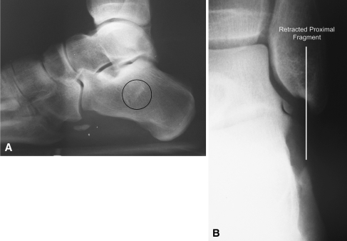
A 38-year-old man complained of pain over the lateral aspect of his foot following an inversion injury while playing baseball. The patient was initially seen at an emergency department where radiographs were reportedly negative. He was given an ankle brace and crutches, and presented to the office 2 weeks following the injury with complaints of pain and swelling with walking and standing. On physical exam, there was edema and ecchymosis over the lateral calcaneus with tenderness of the peroneal tendons at the peroneal tubercle. Eversion strength was 3/5 and resisted eversion caused considerable pain and discomfort. Radiographic examination revealed a fractured os peroneum with proximal migration of the fragment at the peroneal tubercle (Fig. 1A–B). MRI was performed and showed complete rupture of the peroneus longus tendon with the distal os peroneus fragment plantar to the cuboid.
Fig. 1A–B.
(A) Radiographs from Patient 1 demonstrate a fractured os peroneum with migration of proximal fragment to peroneal tubercle (circle). (B) An anterior/posterior radiograph of ankle (line) showing proximal migration of the fractured os peroneus which has retracted with the ruptured peroneal longus tendon.
At the time of surgery, the proximal tendon end had retracted further to the level of the fibula where the os peroneum was entrapped. The peroneus brevis tendon was intact, with no evidence of tearing or degeneration. A 6-mm fragment of the os peroneum was present distally. The distal fragment was drilled with a K wire and repaired as described below.
At 21 months postoperatively the patient had 5/5 strength in eversion and excellent strength in plantar flexion of the first metatarsal head. The patient was able to return to recreational athletics at 8 months postsurgery. A final AOFAS hindfoot score of 97 was calculated based on physical examination and patient responses at the final visit.
Patient 2
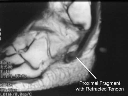
A 43-year-old woman presented with complaints of pain and swelling at the lateral aspect of her foot which started spontaneously 2 months prior to evaluation at our clinic. The patient denied injury or any inciting trauma. Prior to presentation she was evaluated by her primary care physician who treated her with immobilization in a boot walker and nonsteroidal antiinflammatory medications. The patient’s symptoms continued to worsen. When she was first seen in our clinic, she complained of pain with walking and standing for even short periods of time. She also had complaints of instability and giving way in the ankle when not using the boot walker. Physical examination revealed symmetric, bilateral calcaneal varus alignment of 5°. She had marked tenderness over the left peroneal tendons and swelling over the lateral aspect of the left foot with weakness in eversion. Eversion strength was rated as 3/5 on the initial evaluation. Radiographic and MRI examination revealed a fractured os peroneum with proximal migration to the level of the peroneal tubercle. MRI examination revealed a complete rupture of the peroneus longus tendon and a fractured os peroneus fragment plantar at the level of the inferior peroneal retinaculum (Fig. 2).
Fig. 2.
MRI from Patient 2 shows the proximally retracted fragment of os peroneus that has become entrapped in the inferior peroneal retinaculum at the level of the peroneal tubercle.
A lateral incision was made of the inferior peroneal retinaculum, and the retinaculum incised for exposure. The peroneus brevis tendon was not involved. A fractured os peroneum was identified retracted to the level of the peroneal tubercle. The proximal fracture fragment measured 2 mm and was excised with the degenerative tendon sharply excised as well. The distal fragment of the os peroneum was 5 mm in length. It was drilled with a K wire and the tendon repaired to the bony fragment using the lasso technique. A lateral closing wedge osteotomy of the calcaneus was also performed to correct the patient’s underlying calcaneal varus deformity to 5° of valgus.
At the last followup examination 17 months postoperatively the patient had 5/5 strength in eversion and 5/5 plantar flexion strength of the first metatarsal head. Calcaneal alignment was 5° of valgus measured while weight bearing. A final radiograph visit showed complete healing of the calcaneal osteotomy and no migration of the remaining os peroneum fragment. The patient reported no limitations with activities of daily living, although had pain localized to the screw head used to fix the calcaneal osteotomy if she stood or walked for more than 4 hours. She declined to undergo screw removal as she did not feel her symptoms warranted further surgery. An AOFAS hindfoot score of 88 was calculated at the final visit following interview and physical examination.
Operative Technique
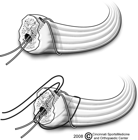
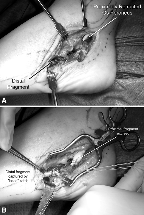
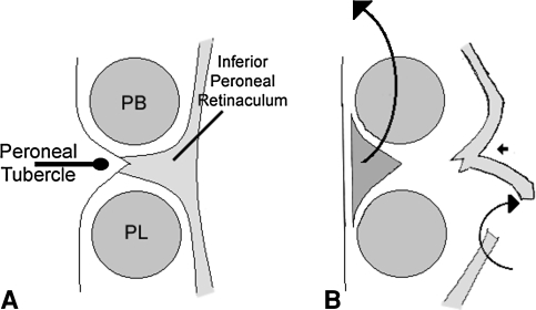
An incision was made over the lateral aspect of the foot in line with the peroneal tendons. Dissection was carried through the subcutaneous tissues, with care taken to identify and protect the sural nerve. The inferior peroneal retinaculum was divided from the fibula distally to expose the peroneal tendons. In one patient, the peroneus longus tendon had retracted to the level of the peroneal tubercle and in one it had retracted to the distal fibula where the proximal fragment of the ruptured os peroneum had become entrapped. The distal os peroneum fragment was freshened with a curette to healthy appearing cancellous bone. A drill hole was made in the distal fragment with a 0.62 Kirschner wire. Two sutures (2-O Fiberloop; Arthrex, Naples, FL) were passed through the drill holes as shown so that the os peroneum was “lassoed” by the closed end of the loop (Fig. 3). This afforded four strands of suture for repair with minimal trauma to the distal tendon fibers. The proximal osseous fragment was excised in both patients and the proximal tendon secured with a locking 2-O suture nonabsorbable (Fiberwire; Arthrex). An end-to-end tendon repair was performed with the ankle held in slight eversion (Fig. 4A–B). The repaired tendon was larger in diameter than a normal tendon and a retinaculoplasty of the inferior peroneal retinaculum was performed in both patients to reconstruct the inferior retinaculum without compression or stenosis over the area of repaired tendon [17]. The peroneal tubercle was excised and the raphe of the inferior extensor retinaculum that inserts on the peroneal tubercle was split to lengthen it and repaired side to side (Fig. 5A–B).
Fig. 3.
The distal fragment is “lassoed” by drilling a hole in the fragment with a Kirschner wire and passing a looped suture through the hole. A four-strand suture repair can be achieved by reversing the direction of the suture for the second pass. (Reprinted with permission and ©Cincinnati SportsMedicine Research and Education Foundation 2008.)
Fig. 4A–B.
Surgical repair in patient 2 is shown. (A) The fractured os peroneum has retracted to the level of the fibula. (B) The distal fragment has been secured with 4 strands of suture for repair.
Fig. 5A–B.
(A) The inferior peroneal retinaculum in cross section with the peroneal tubercle is shown. PB = peroneus brevis, PL = peroneus longus. (B) The retinaculoplasty is performed prior to closure to prevent adhesions and decompress the overlying peroneal retinaculum. The peroneal tubercle is excised. The inferior retinaculum can be lengthened by splitting the thick decussating fibers of the retinaculum where it attaches to the peroneal tubercle. (Reprinted with permission and ©Mosby-Elsevier from Sammarco VJ, Sammarco GJ. Injuries to the tibialis anterior, peroneal tendons, and long flexors of the toes. In: Porter DA, Schon LC, eds. Baxter’s The Foot and Ankle in Sports. Philadelphia, PA: Mosby-Elsevier; 2008:121–146.)
Postoperative rehabilitation included both patients immobilized in a short-leg cast for 3 weeks. A range-of-motion boot walker with the hinges locked at neutral dorsal flexion was used after 3 weeks and passive and active range of motion started under the guidance of a physical therapist. Resisted eversion was not allowed until 6 weeks postoperatively. The patients were allowed to bear weight at 6 weeks and at that time the hinges in the boot were unlocked to allow full dorsiflexion and plantar flexion. Formal physical therapy was continued for an additional 6 weeks with two sessions per week using an established protocol [16]. The goals were to improve ankle and subtalar motion so that motion was equal to the uninvolved side. Strengthening of the peroneal tendons was accomplished primarily with eversion exercises using elastic bands and weights. Proprioceptive training was initiated at 6 weeks and included used of a Biomechanical Ankle Platform System (Jelaga Inc., Jasper, MI). Therapeutic modalities to improve swelling and pain included ice and a compressive stocking, which was discontinued 3 months after the surgery.
Discussion
Fracture of the os peroneum represents a disruption of the peroneus longus tendon. If the fracture is nondisplaced, treatment with immobilization and nonweightbearing may prove adequate [18]. In patients where the ossicle fractures and retracts proximally, repair is recommended [6]. Treatment recommendations and techniques of repair are limited to small series and expert opinion. This manuscript is similarly limited by the small number of patients. While it is our opinion that this technique affords better fixation of the distal tendon segment, this has not been established with mechanical testing.
Degenerative tears and ruptures of the peroneal tendons are poorly tolerated and often require surgery. In patients where tendon ruptures are left untreated, secondary deformity may develop including varus of the hindfoot and first metatarsal elevatus can occur [6, 17, 18]. The described repair technique has two distinct advantages over more standard repair strategies. The technique described here simplifies capture of the distal os peroneus fragment and affords stable fixation to the terminal portion of the peroneus longus tendon. The “lasso stitch” is performed where a looped nonabsorbable suture captures and secures the distal os peroneum fragment. This technique allows for a four-strand suture repair while reducing tendon damage that can occur from multiple needle passes and tissue strangulation. The technique is particularly useful as it can be accomplished without extending the exposure onto the plantar aspect of the foot which may otherwise be necessary for adequate distal tendon fixation. Dissection into the plantar peroneal tunnel is not without its morbidity. The os peroneus can be a sizeable fragment, measuring up to 1.2 cm. In our opinion primary excision of the proximal and distal parts of a fractured os peroneus can cause overtightening of the repair and may increase risk of subsequent failure. Excision of the fractured os peroneum with subsequent repair of the tendon often leaves a substantial gap in the tendon that may preclude an end-to-end repair of the tendon without an intercalated graft. Sammarco reported the use of a free fascial graft in one patient where after excision of a large os peroneum, a 1-cm gap was present that required reconstruction with a free tendon graft with better pain relief and strength than excision of the fragments with direct repair performed in another patient [15]. Addressing foot deformity and adequate decompression of the retinaculum are also important considerations [2, 6].
Treatment of the fractured, displaced os peroneum has been described primarily in small series and in larger series of peroneus longus tendon pathology (Table 1). Nondisplaced fragments can be treated with immobilization, although good results have been reported where patients with displacement were treated nonoperatively [1, 3, 13, 18]. One concern of nonoperative treatment in patients with displaced fractures is eversion weakness, as reported by Bianchi et al. [1] Brandes and Smith noted os peroneus to exist in six of 22 patients with peroneus longus tendon pathology who were treated surgically [2]. Of these six, two had partial tendon tears allowing the tendon to elongate with subsequent proximal migration of part of the ossicle to the peroneal tubercle, and one had a complete tear. All three were repaired, although results from these three patients were not separated from the overall group.
Table 1.
Treatment of fractures of os peroneus
| Author(s) | Number of patients | Treatment | Followup (months) | Results | Complications |
|---|---|---|---|---|---|
| Brav and Chewning [3] (1949) | 1 | Nonoperative | 2 | Satisfactory | None |
| Grisolia [5] (1963) | 1 | OP excision and PL repair | 6 | Satisfactory | None |
| Mains and Sullivan [8] (1973) | 1 | Proximal OP excision and PL suture to distal OP fragment | 18 | Satisfactory | None |
| Tehranzadeh et al. [19] (1984) | 1 | Suture repair of os peroneus | 22 | Satisfactory | None |
| Peacock et al. [11] (1986) | 1 | Suture repair of proximal and distal fragments | 12 | Poor | RSD |
| Pessina [13] (1988) | 1 | Nonoperative | 16 | Satisfactory but eversion weakness | None |
| Bianchi et al. [1] (1991) | 3 | Nonoperative | Not reported | Satisfactory in 2 patients. Poor in 1 patient with eversion weakness | None |
| Peterson and Stinson [14] (1992) | 5 | OP excision and PL repair | 12–28 | Excellent in 4 patients Good in 1 patient | None |
| Sobel et al. [18] (1994) | 10 total, 6 with OP disruption | Nonoperative in 2; OP excision and PL repair in 2; OP excision and PL to PB tenodesis in 2 | Not reported | Nonop: 1 Excellent, 1 Good; OP excision/PL repair: 2 Good; OP excision/tenodesis: 1 Excellent, 1 Good | 2nd metatarsal stress fx in 1 pt treated by tenodesis |
| Sammarco [15] (1995) | 14 total, 3 with OP disruption | OP excision and PL repair in 2; OP excision and fascial graft in 1 | 2–20 | OP excision/PL repair: 1 Poor, 1 unrelated death 2 mos postop; OP excision/fascial graft: Excellent | None |
| Patterson and Cox [10] (1999 ) | 1 | OP excision and PL to PB tenodesis | 6 | Excellent | None |
| Okazaki et al. [9] (2003) | 1 | OP excision and PL repair | 16 | Satisfactory | None |
OP = os peroneum; PL = peroneus longus; PB = peroneus brevis.
Previous series lack specific attention to treatment off the inferior peroneal retinaculum. Anecdotally, some surgeons will simply leave this retinaculum unrepaired if repair will cause stenosis. While repair of the retinaculum is recommended, we believe repairs with decompression should be performed simultaneously. We have seen one patient where failure to repair the inferior peroneal retinaculum resulted in painful snapping of the peroneus longus tendon as it subluxated inferiorly over the calcaneus. We have found the technique described here an adequate method of achieving both a stable reconstruction of the retinaculum and an adequate decompression of Zone C peroneal tendon pathology.
Footnotes
Each author certifies that he or she has no commercial associations (eg, consultancies, stock ownership, equity interest, patent/licensing arrangements, etc) that might pose a conflict of interest in connection with the submitted article.
Each author certifies that his or her institution has approved the reporting of these cases, that all investigations were conducted in conformity with ethical principles of research, and that informed consent for participation in the study was obtained.
References
- 1.Bianchi S, Abdelwahab IF, Tegaldo G. Fracture and posterior dislocation of the os peroneum associated with rupture of the peroneus longus tendon. Can Assoc Radiol J. 1991;42:340–344. [PubMed] [Google Scholar]
- 2.Brandes CB, Smith RW. Characterization of patients with primary peroneus longus tendinopathy: a review of twenty-two cases. Foot Ankle Int. 2000;21:462–468. doi: 10.1177/107110070002100602. [DOI] [PubMed] [Google Scholar]
- 3.Brav EA, Chewning JB. Fracture of the os peroneum; a case report. Mil Surg. 1949;105:369–371. [PubMed] [Google Scholar]
- 4.Brigido MK, Fessell DP, Jacobson JA, Widman DS, Craig JG, Jamadar DA, Holsbeeck MT. Radiography and US of os peroneum fractures and associated peroneal tendon injuries: initial experience. Radiology. 2005;237:235–241. doi: 10.1148/radiol.2371041067. [DOI] [PubMed] [Google Scholar]
- 5.Grisolia A. Fracture of the os peroneum: review of the literature and report of one case. Clin Orthop Relat Res. 1963;28:213–215. [PubMed] [Google Scholar]
- 6.Heckman DS, Reddy S, Pedowitz D, Wapner KL, Parekh SG. Operative treatment for peroneal tendon disorders. J Bone Joint Surg Am. 2008;90:404–418. doi: 10.2106/JBJS.G.00965. [DOI] [PubMed] [Google Scholar]
- 7.Le Minor JM. Comparative anatomy and significance of the sesamoid bone of the peroneus longus muscle (os peroneum) J Anat. 1987;151:85–99. [PMC free article] [PubMed] [Google Scholar]
- 8.Mains DB, Sullivan RC. Fracture of the os peroneum. A case report. J Bone Joint Surg Am. 1973;55:1529–1530. [PubMed] [Google Scholar]
- 9.Okazaki K, Nakashima S, Nomura S. Stress fracture of an os peroneum. J Orthop Trauma. 2003;17:654–656. doi: 10.1097/00005131-200310000-00010. [DOI] [PubMed] [Google Scholar]
- 10.Patterson MJ, Cox WK. Peroneus longus tendon rupture as a cause of chronic lateral ankle pain. Clin Orthop Relat Res. 1999;365:163–166. doi: 10.1097/00003086-199908000-00021. [DOI] [PubMed] [Google Scholar]
- 11.Peacock KC, Resnick EJ, Thoder JJ. Fracture of the os peroneum with rupture of the peroneus longus tendon. A case report and review of the literature. Clin Orthop Relat Res. 1986;202:223–226. [PubMed] [Google Scholar]
- 12.Peacock KC, Resnick EJ, Thoder JJ. Rupture of the peroneus longus tendon. Report of three cases. J Bone Joint Surg Am. 1990;72:306–307. [PubMed] [Google Scholar]
- 13.Pessina R. Os peroneum fracture. A case report. Clin Orthop Relat Res. 1988;227:261–264. [PubMed] [Google Scholar]
- 14.Peterson DA, Stinson W. Excision of the fractured os peroneum: a report on five patients and review of the literature. Foot Ankle. 1992;13:277–281. doi: 10.1177/107110079201300509. [DOI] [PubMed] [Google Scholar]
- 15.Sammarco GJ. Peroneus longus tendon tears: acute and chronic. Foot Ankle Int. 1995;16:245–253. doi: 10.1177/107110079501600501. [DOI] [PubMed] [Google Scholar]
- 16.Sammarco V. Principles and Techniques in Rehabilitation of the Ahtlete’s Foot: Part II - Rehabilitation of Tendon Injuries. Techniques in Foot and Ankle Surgery. 2003;2:144–150.(Abstract).
- 17.Sammarco VJ, Sammarco GJ. Injuries to the tibialis anterior, peroneal tendons, and long flexors of the toes. In: Porter DA, Schon L, eds. Baxter’s The Foot and Ankle in Sport, 2nd Ed. Philadelphia, PA: Mosby-Elsevier; 2008:121–146.
- 18.Sobel M, Pavlov H, Geppert MJ, Thompson FM, DiCarlo EF, Davis WH. Painful os peroneum syndrome: a spectrum of conditions responsible for plantar lateral foot pain. Foot Ankle Int. 1994;15:112–124. doi: 10.1177/107110079401500306. [DOI] [PubMed] [Google Scholar]
- 19.Tehranzadeh J, Stoll DA, Gabriele OM. Case report 271. Posterior migration of the os peroneum of the left foot, indicating a tear of the peroneal tendon. Skeletal Radiol. 1984;12:44–47. doi: 10.1007/BF00373176. [DOI] [PubMed] [Google Scholar]
- 20.Thompson FM, Patterson AH. Rupture of the peroneus longus tendon. Report of three cases. J Bone Joint Surg Am. 1989;71:293–295. [PubMed] [Google Scholar]


