Abstract
A rare case of multiple malignant tumors (poorly differentiated squamous cell carcinoma and high grade osteosarcoma) arising in an ovarian dermoid cyst of a 55 year old female is reported. To the best of our knowledge, this is the first well documented example of such an unusual combination of tumors arising in a dermoid cyst. The osteosarcoma and squamous cell carcinoma appear to arise in the background of benign teratomatous environment of a dermoid cyst rather than from “pure” mixed mesodermal tumors of the ovary. The tumors did not appear to have well demarcated boundaries with a junction or close intermingling of both cell types, features less favorable for collision tumor or carcinosarcoma. Despite extensive surgery with negative surgical margins and combination chemotherapy, the patient had recurrence of the tumor within four months and she died secondary to septicemia to chemotherapy and bilateral pulmonary emboli shortly after.
Keywords: Malignant dermoid, teratoma, osteosarcoma, squamous cell carcinoma
Introduction
Malignant change in a benign cystic teratoma has been well documented, but the simultaneous occurrence of malignancy from two different somatic cell types is almost exceptional [1-4]. With only a handful being reported thus far, [5-8] this case report is intended to illustrate pathology of this rare case and shed light on the differential diagnosis of this unusual entity with current literature review.
The following case was evaluated and reported in compliance with the University of South Florida's Institutional Review Board Policy #311.
Case Presentation
A 55-year-old, gravida 1, paral, postmeno-pausal, Caucasian woman, presented with right lower quadrant pain. Abdominal CT showed an 11 × 11 × 5 cm mass arising from a pelvic mass with peripheral calcification. The patient underwent exploratory laparotomy.
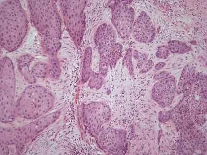
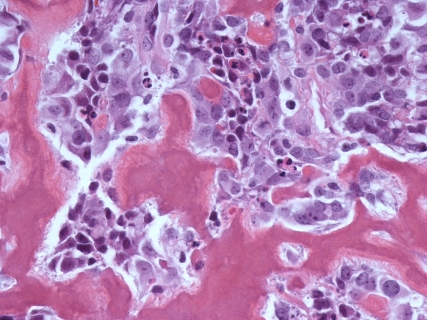
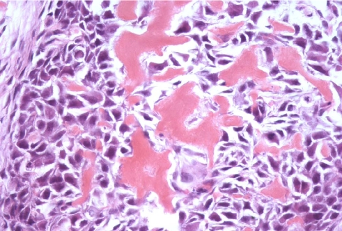
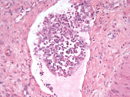
The gross specimen of “right ovarian mass” consisted of an intact cystic structure, 632 grams and 11.8 × 11.6 × 5.9 cm. The cyst was unilocular, filled with opaque greenish creamy fluid. The wall thickness varied from 0.5 to 2.5 cm and was calcified at the thickest areas. The inner lining ranges from shaggy to moderately trabeculated. Little, if any, ovarian parenchyma was identified grossly. Histological examination revealed a biphasic malignant high-grade tumor with epithelial and sarcomatous features. The epithelial component was composed predominantly of squamous cell carcinoma with invasion and desomoplastic reaction of the stroma (Figure 1). In addition, there was extensive necrosis and poorly differentiated areas with spindle cell morphology in the background of benign teratomatous elements. More interestingly, there were nodular areas with sheets of malignant undifferentiated epithelioid cells in association with immature osteoid (Figure 2). Figure 3 shows squamous cell carcinoma and osteosarcoma adjacent to each other. A few remnants of ovarian stroma were encountered. Features of vascular space invasion were also observed (Figure 4). The teratomatous component was cystic and mature with benign epithelium. Dr. Young at Massachusetts General Hospital reviewed the case in consultation with the original pathologist for this case and reported that it is a high grade squamous cell carcinoma with transformation to osteosarcoma arising from a dermoid cyst.
Figure 1.
Hematoxylin and eosin (H&E) of invasive squamous cell carcinoma with surrounding desmoplastic reaction (×100)
Figure 2.
Hematoxylin and eosin (H&E) of malignant osteoblasts with immature osteoid deposition (×400).
Figure 3.
Hematoxylin and eosin (H&E) of an area of tumor that has squamous cell carcinoma and osteosarcoma adjacent to each other (×400).
Figure 4.
Hematoxylin and eosin (H&E) of malignant tumor cells in the lumen of a blood vessel (100×).
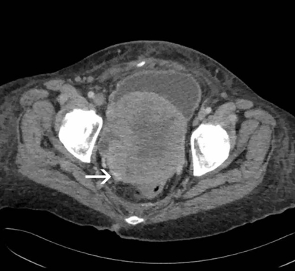
Post operatively patient was started on combination chemotherapy but the abdominal CT four months later had recurrence of the tumor in the pelvis (Figure 5) with liver metastasis. The patient expired five months from the date of diagnosis.
Figure 5.
Contrast-enhanced CT demonstrates a large recurrent solid pelvic mass with internal necrosis and subtle mineralization along its posterolateral margin (arrow).
Discussion
Mature cystic teratoma or dermoid cyst is the most common ovarian germ cell neoplasm and most common childhood ovarian tumor comprising approximately 25% of all ovarian tumors [9]. They have a very wide age range but usually occur during the reproductive years. Approximately 90% are unilateral and with a variable size of 5-10cm. Microscopically, they are usually unilocular cysts containing tissue from all three germ cells with teeth, bone, and neural tissue being found [9,10]. Secondary malignancies may develop in any of the tissues derived from the three germ layers-the ectoderm, meso-derm and endoderm. A nodular prominence is usually present in the wall of the cyst at the junction between the teratoma and normal ovarian tissue and this has been called a Rokitansky protuberance [10]. The greatest cellular variety is found in this area and hence should be thoroughly sampled to rule out malignant components.
Malignant transformation within a mature dermoid cyst is a rare event, less than 2% of all such lesions. Approximately 1% of mature cystic teratomas have malignant somatic-type tissue elements [11]. Risk factors for malignancy in a mature cystic teratoma include age over 45 years, tumor diameter greater than 10cm, and rapid growth [11]. The most common malignancy has been reported as squamous cell carcinoma followed by carcinoid tumor and transitional cell carcinoma. In their publication, Al-Rayyan et al concluded that regardless of the type of malignancy, the tumor size, the extent of the disease and the patient's age were the major factors governing the survival in these patients. They recommended maintaining a higher suspicion of malignancy in managing dermoid cysts occurring in patients over the age of 45 years, especially if rapidly growing or very large tumors greater than 10cm [11]. In addition, Kelly and Scully with their detailed report of cases of malignancy arising in dermoid cyst felt that thickening of the cyst wall and adherence to surrounding structures should also raise high index of suspicion for malignancy in a dermoid cyst [3]. The best outcome for their patients was when the tumor was completely excised and staging and treatment planning at the initial surgery or as soon as possible after the diagnosis. Squamous cell carcinoma if confined to the ovary had better outcome than if there were peritoneal extension [11]. When malignant transformation has occurred within a teratoma, treatment is usually tailored toward the transformed histology [11]. Recent studies indicated that a combination of adjuvant chemotherapy and radiotherapy might improve survival in these patients. Irrespective of the tumor type, or the size of tumor, they concluded that the prognosis is better if tumor was limited to one ovary and with an intact capsule [9,11,12]. Burgess et a I also reported the importance of exploring both ovaries as the incidence of teratoma has a bilateral proclivity [12].
Stromal tumors arising from the mesodermal germ cells of a mature dermoid cyst are rarer than their epithelial counterparts but have been reported [5,12]. The incidence of osteosarcoma alone arising from a mature dermoid cyst to date consists of four cases (Table 1). Ngwalle et al described the typical case occurring in a postmenopausal woman with an osteosarcoma arising in benign dermoid cyst [5]. The same age range has been observed for other soft tissue osteosarcomas which also exclusively affected adults older than 50 years [13]. This is not the same for osteosarcomas of the bone, a tumor of growing individuals with a peak incidence in the second decade of life [5]. The prognosis in all of the cases of osteosarcoma in dermoid cyst was poor, which is similar to osteosarcoma of soft tissue. In Ngwalle et al's case, it seemed to have a better prognosis and the postulation could have been due to the limited involvement of the ovary and low stage at the time of surgery [5].
Table 1.
Reported osteosarcomas in dermoid cyst
| Case | Investigators | Age (yrs) | Side | Size (cm) | Outcome |
|---|---|---|---|---|---|
| 1 | Stowe et al (1952) | 67 | Right | 15 | Died in 5 months |
| 2 | Ngwalle et al (1990) | 52 | Right | 10×9×8 | NED (3 years; then lost to follow up) |
| 3 | Ajitkumar et al (1999) | 80 | Left | Not reported | Died in 2 months |
| 4 | Aygun et al (2003) | 14 | Left | 22 × 17 | Local relapse in 7 months |
NED: No evidence of disease
A combination of different malignancies arising in the same dermoid cyst or the incidence of more than one malignancy arising from one ovary is exceptionally rare. But different combinations have been reported in the literature (Table 2) Hanada et al described the multiple malignancy arising from a dermoid cyst of the ovary namely squamous cell carcinoma and myxoid fibrous histiocytoma in 1981 [2]. In 1996, a case report of carcinosarcoma arising in the dermoid cyst of the ovary by Arora and Haldane comprising of adenocarcinoma and leiomyosarcoma was reported [1]. To date, to the best of our knowledge, a unique combination of squamous cell carcinoma and osteosarcoma arising in mature dermoid cyst of the ovary has never been reported before. The treatment of multiple malignancies arising from dermoid cyst consists of aggressive cancer surgery followed with combination chemotherapy and radiotherapy. The prognosis is dismal and the patients usually succumb to their disease within months [1].
Table 2.
Clinical and histological features and outcome of patients reported with multiple malignancies in dermoid cyst
| Case | Investigators | Age (yrs) | Side | Size (cm) | Pathology | Outcome |
|---|---|---|---|---|---|---|
| 1 | Burgess et al (1954) | 47 | Left | 19×10.5 | Leiomyosarcoma & Osteosarcoma | Not reported |
| 2 | Kelley et al (1961) | 51 | Right | 15 ×10 × 9 | SCC & trabecular adenoca. | NED(65 months following surgery) |
| 3 | Hanada et al (1981) | 75 | Right | 21×15×8 | SCC & myxoid variant of MFH | NED (21 months following surgery) |
| 4 | Tyagi et al (1993) | 60 | Right | 15×14× 8 | SCC & leiomyosarcoma | NED (1 year after surgery) |
| 5 | Arora et al (1996) | 78 | Left | 14 | Adenocarcinoma & leiomyosarcoma | Not reported |
| 6 | Present case | 55 | Right | 11.8×11.6×5.9 | Squamous cell carcinoma & osteosarcoma | Died in 5 months |
SCC- Squamous Cell Carcinoma; MFH -malignant fibrous histiocytoma; NED- no evidence of disease.
The distinction between “pure” malignant mixed mesodermal (carcinosarcoma) tumors of the ovary versus collision tumors arising in a benign dermoid cyst versus a squamous cell carcinoma with sarcomatous transformation is of academic interest. Carcinosarcoma is diagnosed when both the carcinoma and sarcoma are present without any benign teratoma tissue [14]. The coexistence of two malignancies in the background of benign teratomatous tissue especially in a postmenopausal woman eliminates this diagnosis. In our case, benign epithelium, squamous cell and osteosarcoma were clearly coexistent in the dermoid cyst. No immature elements were identified despite through examination and multiple sections of the specimen, ruling out to some extent the possibility of these tumors arising in an immature teratoma. Also immature teratomas usually occur in younger age group and typically the malignant transformation occurs chiefly in postmenopausal women.
Collision tumors occur when two different malignancies arise metachronously in the same organ at two separate areas and usually have a common point of meeting or contact but for the most part are separate from each other. In the present case, there was no such area of common contact point despite extensive sampling. There were no areas of two separate well defined point of origin with a common meeting point.
Pathogenesis of malignant transformation of one somatic cell type to another is not clearly. In our patient, the mature dermoid cyst most likely has undergone the transformation of the totipotential germ cells toward the formation of squamous cell carcinoma that might have later dedifferentiated into osteosarcoma.
Acknowledgments
The authors thank Ms. Angela Reagan for her assistance in this manuscript submission and dedication to graduate medical education at Moffitt Cancer Center.
References
- 1.Arora DS, Haldane S. Carcinoma arising in a dermoid cyst of the ovary. J Clin Pathol. 1996;49:519–521. doi: 10.1136/jcp.49.6.519. [DOI] [PMC free article] [PubMed] [Google Scholar]
- 2.Hanada M, Tsujimura T, Shimizu H. Multiple malignancies (Squamous cell carcinoma and sarcoma) arising in a dermoid cyst of the ovary. Acta Pathol. Jpn. 1981;31(4):681–688. doi: 10.1111/j.1440-1827.1981.tb02763.x. [DOI] [PubMed] [Google Scholar]
- 3.Tyagi SP, Maheshwari V, Tyagi N, Tewari K. Double malignancy in a benign cystic teratoma of the ovary( A case report) Indian Journal of Cancer. 1993;(30):140–142. [PubMed] [Google Scholar]
- 4.Kelly RR, Scully RE. Cancer developing in dermoid cyst of the ovary- A report of 8 cases, including a carcinoid and a leiomyosarcoma. Cancer. 1961;(14):989–1000. doi: 10.1002/1097-0142(196109/10)14:5<989::aid-cncr2820140512>3.0.co;2-u. [DOI] [PubMed] [Google Scholar]
- 5.Ngwalle K, Hirakawa T, Tsuneyoshi M, Enjoji M. Osteosarcoma arising in a benign dermoid cyst of the ovary. Gyne Onco. 1990;37:143–147. doi: 10.1016/0090-8258(90)90324-e. [DOI] [PubMed] [Google Scholar]
- 6.Aygun B, Kimpo M, Lee T, Valderrama E, Leoni-das J, Karayalcin G. An adolescent with ovarian osteosarcoma arising in a cystic teratoma. Jour Pediatric hematology/Oncology. 2003;25(5):410–413. doi: 10.1097/00043426-200305000-00012. [DOI] [PubMed] [Google Scholar]
- 7.Stowe LM. Watt JY. Osteogenic sarcoma of the ovary. Am J Obstet Gynecol. 1952;64:423–6. doi: 10.1016/0002-9378(52)90322-0. [DOI] [PubMed] [Google Scholar]
- 8.Ajithkumar TV, Abraham EK, Krishnan Nair M. Osteosarcoma arising in a mature cystic teratoma of the ovary. J Exp Clin Cancer Res. 1999;18:89–91. [PubMed] [Google Scholar]
- 9.Mills SE. Sex Cord-Stromal, Steroid cells, and Germ cell Tumors of the Ovary. In: Stacey E Mills., editor. Sternberg's Diagnostic Surgical Pathology. Fifth edition. Lippincott Williams & Wil-kins; 2010. pp. 2330–2337. [Google Scholar]
- 10.Kurman RJ. Blaustein's Pathology of the female genital tract. Fifth edition. New York: Springer. Verlag; 2002. Germ Cell Tumors of the Ovary; pp. 994–1006. [Google Scholar]
- 11.Al-Rayyan ES, Duqoum W, Sawalha MS, Nasci- mento MC, Pather S, Dalrymple CJ, Carter JR. Secondary malignancies in ovarian dermoid. cyst Saudi Med J. 2009;30(4):524–528. [PubMed] [Google Scholar]
- 12.Burgess G, Shutter H. Malignancy originating in ovarian dermoids. Report of three cases. Obstet and Gynecol. 1954;(4):567–571. [PubMed] [Google Scholar]
- 13.Weiss SW, Goldblum JR. Enzinger & Weiss's Soft Tissue Tumors. Fifth Edition. Osseous Soft Tissue Tumors; pp. 1051–1059. [Google Scholar]
- 14.Cabibi D, Martorana A, Cappello F, Barresi E, Gangi C, Rodolico V. Carcinosarcoma of monoclonal origin arising in a dermoid cyst of ovary: a care report. BMC Cancer. 2006:6–47. doi: 10.1186/1471-2407-6-47. [DOI] [PMC free article] [PubMed] [Google Scholar]


