Abstract
Osteoporosis is a low bone mass disease most often caused by an increase in bone resorption not compensated by a similar hike in bone formation1. Since gut–derived serotonin (GDS) inhibits bone formation2, we asked whether hampering its biosynthesis could treat osteoporosis through an anabolic mechanism. To that end we synthesized and used LP533401, a small molecule inhibitor of Tph1, the initial enzyme in GDS biosynthesis. Oral administration once daily for up to 6 weeks of this small molecule prevents the development of and fully rescues, in a dose–dependent manner, osteoporosis in ovariectomized rodents because of an isolated increase in bone formation. These results provide a proof of principle that inhibiting GDS biosynthesis could become a novel anabolic treatment for osteoporosis.
Gut-derived serotonin (GDS) is a powerful inhibitor of osteoblast proliferation and bone formation that does not affect bone resorption2. Thus inhibiting GDS biosynthesis could be a means to treat low bone mass conditions through a bone anabolic mode of action. LP533401 is a small molecule inhibitor of Tph1, the initial enzyme in the GDS biosynthesis, currently being tested at a dose of 100 mg per kg body weight for treatment of irritable bowel syndrome and no overt deleterious effects have been reported3. In vivo pharmacokinetic studies in rodents showed that LP533401 level in the brain is negligible following oral administration, indicating that it is virtually unable to cross the blood-brain barrier3,4. Thus, LP533401 appears to be a good tool to test the therapeutic potential of inhibiting GDS biosynthesis for low bone mass diseases.
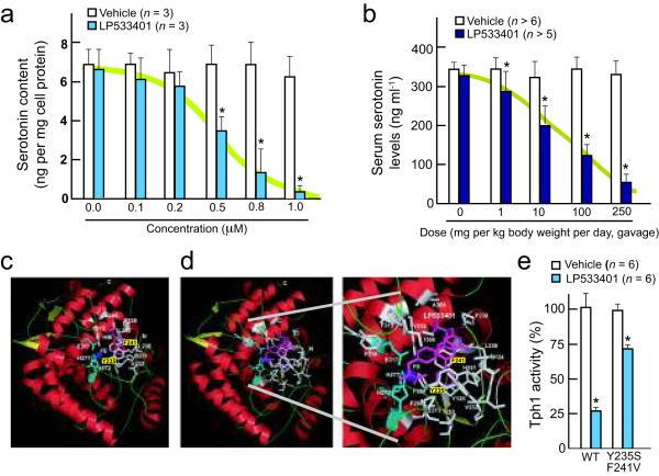
We used the available chemical description to synthesize LP533401 and verified its structural identity by multiple analyses4 (Supplementary Fig. 1). To evaluate LP533401 efficacy in inhibiting serotonin biosynthesis we treated Tph1–expressing cells (RBL2H3 cells) for 3 days with increasing amounts of LP533401 (0.1-1 μM). LP533401 completely inhibited serotonin production in these cells at a dose of 1 μM (Fig. 1a). Moreover, there was a dose–dependent decrease in serum serotonin levels in WT mice fed with LP533401 (Fig. 1b). This decrease in serum serotonin levels reached 30% of control values when using 250 mg per kg body weight per day of the compound although no change in brain serotonin content was observed (Supplementary Fig. S2). This latter point is important since brain– and gut–derived serotonin exert opposite influences on bone formation5.
Fig. 1. Analysis of LP533401 inhibition of Tph1 activity.
(a–b) In vitro (a) and in vivo (b) dose response of inhibition of serotonin synthesis by LP533401.
(c) Crystal structure of human TPH1 bound to 7,8–dihydro–L–biopterin co–factor (HBI, in magenta) and Fe(III) (in blue) (PDB ID: 1mlw). Amino acid side-chains interacting with HBI are in white and those binding the metal ion are in cyan.
(d) Left panel, crystal structure of human TPH1 docked to the generated 3D model of LP533401. LP533401 is in magenta and the metal ion is in blue. The side-chains of amino acid residues interacting with LP533401 and metal ion are in white and cyan respectively. The residues interacting with LP533401 include Val232, Tyr235, Leu236, Pro238, Phe241, His251, Ala309 and Tyr312. Right panel, Zoom image for the binding of LP533401 to TPH1. The structure figures have been made using PyMOL (http://www.pymol.org/).
(e) In vitro activity of wild–type or mutated (Y235S, F241V) TPH1 in the presence of LP533401 (0.01 μM).
All values are expressed as means ± SEM. * p < 0.05 vs vehicle.
To elucidate how LP533401 influences Tph1 activity we performed in silico and ex vivo analyses. The co–crystal structure of Tph1 enzymatic domain with its co–factor 7,8–dihydro–L–biopterin (HBI) (PDB ID: 1mlw)6 was used as a reference in studying interactions of LP533401 with Tph1 catalytic regions (Fig. 1c). A 3 dimensional (3D) model for LP533401 was generated from its chemical structure and docked onto the 3D structure of Tph1 to identify interactions between these two molecules7. Various generated docked conformers of LP533401 were analyzed and the one showing the lowest estimated free energy for binding (−9.46 kcal mol−1) selected for further analysis (Fig. 1d). This docked model of LP533401 and Tph1 interaction revealed that LP533401 interacts with two amino acids, Tyr235 and Phe241, near Tph1 catalytic site (Fig. 1d). To verify the importance of these residues in mediating LP533401 and Tph1 interaction we generated recombinant wild–type Tph1 or a mutated form in which Tyr235 and Phe241 were replaced by a serine and a valine, respectively. While LP533401 decreased the activity of wild–type Tph1 by more than 70%, the mutations markedly blunted this effect (Fig. 1e). These results indicate that, as predicted by the docked model, LP533401 inhibits Tph1 activity, in part, by interacting with residues Tyr235 and Phe241.
Next we tested the therapeutic relevance of LP533401 for low bone mass diseases, through the use of ovariectomized rodents that present8, as post-menopausal women do1, an increase in bone resorption of higher magnitude than the increase in bone formation also caused by gonadal failure.
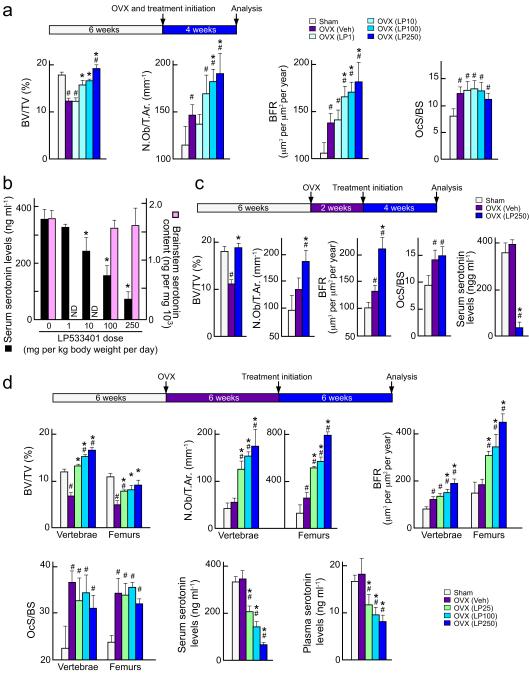
We first asked whether LP533401 could prevent ovariectomy–induced bone loss. Six week–old sham–operated or ovariectomized female C57Bl6/J mice were fed once daily with either vehicle or LP533401 at doses ranging from 1 to 250 mg per kg body weight per day from day 1 to 28 post–ovariectomy (Fig. 2a and Supplementary Fig. 3a). Osteoclast surface and serum deoxypyridinoline (Dpd) levels, a marker of bone resorption, were higher in ovariectomized mice, regardless of their treatment, than in sham–operated animals and, as a result, vehicle–treated ovariectomized mice developed a low bone mass (osteopenia) (Fig. 2a and Supplementary Fig. 3b). In contrast, mice treated with 250, 100 or even 10 mg per kg body weight per day of LP533401 had a higher bone mass than that of vehicle–treated ovariectomized mice (Fig. 2a). Consistent with the influence of GDS on osteoblast proliferation and bone formation, this increase in bone mass in the LP533401–treated ovariectomized mice was secondary to a major increase in bone formation parameters such as osteoblast numbers, bone formation rate, and osteocalcin serum levels (Fig. 2a and Supplementary Fig. 3c–d). We verified that although serum serotonin levels were decreased in a dose–dependent manner, brain serotonin content remained unaffected in LP533401–treated ovariectomized mice (Fig. 2b). These results established that LP533401 could prevent the development of ovariectomy–induced osteoporosis in the mouse. That a favorable effect on bone formation parameters is observed with a relatively small reduction in circulating serotonin levels (~30%) echoes what is observed in genetic model of decreased circulating serotonin levels. In that case the increase in bone formation parameters observed in heterozygous mutant mice is not statistically different from the one seen in homozygous mutant mice2. This suggests that there is a threshold for the reduction in serum serotonin levels beyond which the skeletal response is marginally increased.
Fig. 2. LP533401 can prevent and rescue osteoporosis in ovariectomized mice.
(a–b) Histological analysis of L3–L4 vertebrae (a) and serum and brain serotonin levels (b) of sham–operated (sham) and ovariectomized (OVX) mice treated orally with vehicle (Veh) or the indicated dose (1, 10, 100 or 250 mg per kg body weight per day) of LP533401 (LP) for 4 weeks post–ovariectomy.
(c) Histological analysis of L3–L4 vertebrae of sham and OVX mice treated with vehicle or LP533401 (LP) from week 2 to 6 post–ovariectomy. (n =8–10 animals each group).
(d) Histological analysis of L3–L4 vertebrae or distal femurs of sham and OVX mice treated with vehicle or the indicated dose (25, 100 or 250 mg per kg body weight per day) of LP533401 (LP) from week 6 to 12 post–ovariectomy. (n=8–10 animals each group).
BV/TV, Bone volume over total volume; Nb.Ob/T.Ar, osteoblast number over trabecular area; BFR, bone formation rate; OcS/BS, osteoclast surface over bone surface; ND, not determined.
All values are expressed as means ± SEM. # p < 0.05 vs sham and * p < 0.05 vs OVX (Veh).
We then asked whether LP533401 could also rescue an existing ovariectomy–induced osteopenia. In an initial experiment, sham–operated or ovariectomized 6 week–old mice were left without treatment for only 2 weeks, and then treated with vehicle or a daily dose of LP533401 (250 mg per kg body weight per day) for 4 weeks (Fig. 2c). Vehicle-treated ovariectomized mice developed the expected osteopenia secondary to an increase in bone resorption parameters (Fig. 2c and Supplementary Fig. 3e). These parameters were also increased in LP533401–treated ovariectomized mice, however, in these latter animals the increase in bone formation parameters was of such amplitude that it normalized their bone mass (Fig. 2c). Serum serotonin levels were decreased by 80% but brain serotonin content was unaffected in LP533401–treated animals (Fig. 2c and Supplementary Fig. 3f).
Given the success of this initial experiment, we then asked whether LP533401 could rescue an osteopenia even if administered substantially later after ovariectomy and at lower doses. For that purpose 6 week–old mice, ovariectomized, were left untreated for 6 weeks before being treated with vehicle or LP533401 at either 25, 100 or 250 mg per kg body weight per day for another 6 weeks (Fig. 2d). LP533401, by increasing bone formation parameters, reversed the deleterious effects of ovariectomy on bone mass and increased it to levels similar (25 mg per kg body weight per day) or higher (100 or 250 mg per kg body weight per day) than that seen in sham–operated mice (Fig. 2d). This increase in bone mass affected vertebrae and long bones (Fig. 2d and Supplementary Fig. 3g) and was also observed in naive mice (Supplementary Fig. 4a). There was no change in bone length or width in any of the groups treated with LP533401 (Supplementary Fig. 4b–c). To rule out the possibility that LP533401, at the doses used in this study, exerts adverse effects on the gastroinstestinal tract or hemostasis we measured total gastrointestinal transit, colonic mobility and gastric emptying, three cardinal parameters of gastrointestinal homeostasis9. None of these parameters were affected in LP533401–treated mice, even when using 250 mg per kg body weight per day (Supplementary Figs. 4d–f). Similarly, platelet numbers and coagulation times were similar in vehicle– and LP533401–treated mice (Supplementary Figs. 4g–h). These results establish that LP533401 can rescue, through a bone anabolic mechanism, ovariectomy–induced osteoporosis in mice even when given at a low dose (25 mg per kg body weight per day) and late after ovariectomy and that it does so without deleterious consequences on hemostasis or intestinal motility.
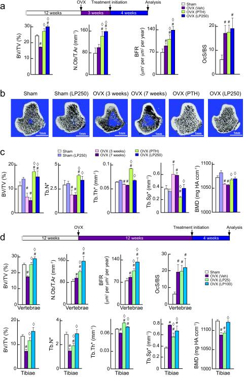
The rat is the rodent model of choice for post–menopausal osteoporosis and intermittent injections of parathyroid hormone (PTH), the standard to which any novel bone anabolic agent must be compared 10. Hence, we ovariectomized rats at 12 weeks of age and left them untreated for 3 or 12 more weeks so that they develop a severe osteopenia (Fig. 3 and Supplementary Fig. 5). Sham–operated or ovariectomized rats were then treated for 4 weeks with either vehicle, a relatively high dose of PTH (80 μg per kg per day, subcutaneous)11, or increasing amounts of LP533401 (25, 100 or 250 mg per kg per day, orally).
Fig. 3. LP533401 rescues osteoporosis in ovariectomized rats.
(a–c) Histomorphometric analysis of L2 vertebra (a) and micro–computed tomography analysis of the proximal tibiae (b,c) collected from rat sham-operated (sham) vehicle treated control, ovariectomized (OVX) and treated with vehicle, intermittent injections of PTH or 250 mg per kg body weight per day of LP533401 (LP) from week 3 to 7 post–ovariectomy (n =8–10 animals each group).
(d) Histomorphometric analysis of L2 vertebra and micro–computed tomography analysis of the proximal tibiae collected from rat sham-operated (sham) vehicle treated control, ovariectomized (OVX) and treated with vehicle, intermittent injections of PTH or LP533401 (25, 100 mg per kg body weight per day) from week 12 to 16 post–ovariectomy (n =8–10 animals each group).
BV/TV, Bone volume over trabecular volume; Tb.N*, trabecular number; Tb.Th*, trabecular thickness; Tb.Sp*, trabecular spacing; Nb.Ob/T.Ar, osteoblast number over trabecular area; BFR, bone formation rate; OcS/BS, osteoclast surface over bone surface.
All values are expressed as means ± SEM. * means the parameter is measured without a plate or rod model assumption, # p < 0.05 vs sham and ◇ p < 0.05 vs OVX (Veh).
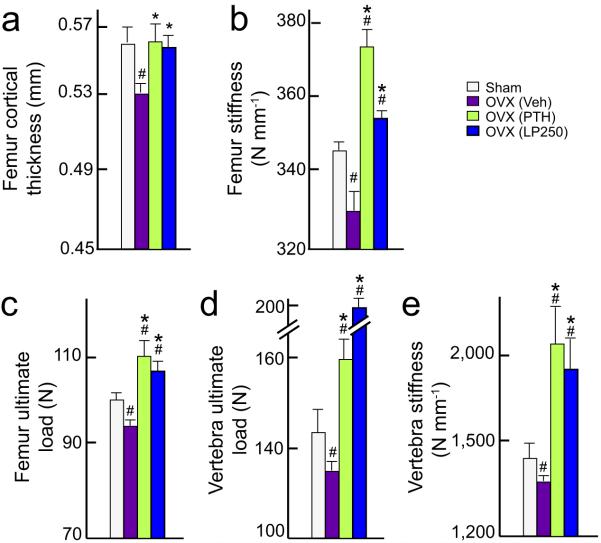
A histomorphometric analysis of vertebrae showed that LP533401 fully rescued the ovariectomy–induced osteopenia in rats, no matter whether it was given 3 or 12 weeks post–ovariectomy, and that it was efficacious even at the lowest dose used (25 mg per kg body weight per day) (Figs. 3a and 3d). This rescue was due to an increase in osteoblast number and bone formation rate while the osteoclasts surface per bone surface (OcS/BS) was similar in untreated, PTH– and LP533401–treated ovariectomized rats. To determine whether LP533401 also affects bone mass in long bones that are load bearing, we used micro–computed tomography. Ovariectomy profoundly reduced bone volume, trabecular number (Tb.N*), trabecular thickness (Tb.Th*) and increased trabecular spacing (Tb.Sp*) in long bones (Fig. 3b and c). LP533401, at all doses tested, increased Tb.N*, and Tb.Th*, and reduced Tb.Sp* in these bones thereby increasing, in a dose–dependent manner, bone mass in ovariectomized rats when compared to vehicle–treated controls (Figs. 3b–d). Illustrating the sensitivity of the serotonergic regulation of bone formation 25 mg per kg body weight per day of LP533401 rescued the ovariectomy–induced osteopenia, in rats as in mice, though it caused only a 35 to 40% reduction in serum serotonin levels (Figs. 2d, 3d and Supplementary Figs. 5h–I). While LP533401 appeared to be at least as efficient as the high dose PTH regimen used in this experiment in vertebrae the opposite is the case when looking at long bones (Compare Figs. 3a–c). Ovariectomy also lead to a decrease in cortical thickness, a key parameter of bone integrity12; this decrease at the femur mid–shaft was equally rescued by PTH and LP533401 (250 mg per kg body weight per day) (Fig. 4a). We also analyzed two micro–architectural parameters; endocortical and periosteal circumference, both parameters were, as expected, increased after OVX13; LP533401 and PTH treatments normalized them to a similar extent (Supplementary Fig. 6).
Fig. 4. Effect of LP533401 on bone biomechanical strength.
(a–e) Cortical thickness, stiffness and ultimate load analysis in femur and L3 vertebra collected from rat sham–operated (sham) vehicle treated control, ovariectomized (OVX) and treated with vehicle or intermittent injections of PTH or 250 mg per kg body weight per day of LP533401 (LP) from week 3 to 7 post–ovariectomy. (n =8–10 animals each group)
All values are expressed as means ± SEM. # p < 0.05 vs sham and * p < 0.05 vs OVX (Veh).
To measure bone quality, femur and vertebra samples from untreated, PTH– and LP533401–treated ovariectomized rats were subjected to a three-point bending test and a compression analysis, to determine maximal load and stiffness, two surrogates of bone quality14,15 (Figs. 4b–e). Both parameters were decreased after ovariectomy and restored to values seen in sham–operated rats by PTH and LP533401 (250 mg per kg body weight per day) treatments (Figs. 4b–e). Thus, daily oral administration of LP533401 could revert the bone loss and architectural deterioration caused by long–term gonadal failure in rats.
Given the progressive aging of the general population, post–menopausal osteoporosis is a growing public health concern. A major issue in the treatment of this disease is to identify safe anabolic agents that can increase bone formation on a long term basis and to such an extent that they compensate for the increase in bone resorption caused by menopause1. To date, only one drug fulfills this criteria, intermittent injections of PTH16,17. Therefore, there is an urgent need to identify additional bone anabolic agents1,10. We show here that an inhibitor of GDS synthesis acts as a bone anabolic agent that, when given to rodents for 4 to 6 weeks, can fully rescue gonadectomy–induced bone loss. That a high dose of PTH is more efficient than LP533401 in long bones but not in vertebrae raises the testable possibility that the two agents exert anabolic functions through different mechanisms. Although the data presented here are promising we remain fully aware that, since rodents do not remodel bones as much as humans do, our results will have to be confirmed in other species. Nevertheless, that this small molecule is taken orally, promotes only bone formation and, for the purpose of treating osteoporosis, is needed at a relatively small dose and only once daily, suggest that inhibitors of GDS synthesis have the potential to become a novel class of bone anabolic drugs to be added to the therapeutic arsenal against osteoporosis.
Supplementary Material
Acknowledgements
We thank M-T. Rached, Y–Y. Huang, N. Suda and G. Ren for help in experiments; Dr. D. Landry (Organic Chemistry, Columbia University) for providing LP533401 for initial stages of these experiments. Special thanks to Drs. S. Kousteni and J. Martin for helpful suggestions. This work was supported by grants from the US National Institutes of Health (V.K.Y, G.K., P.D.) and a Gideon and Sevgi Rodan fellowship from IBMS (V.K.Y.).
Appendix
Methods
Animals
We performed all procedures on animals approved by Columbia University Institutional Animal Care and Use Committee or by the Indian Institute of Science Animal ethics committee. Mice were purchased from the Jackson Laboratories, rats from the National Institute of Nutrition, India.
Bioinformatic analysis of LP533401 binding to Tph1
We generated a 3D model of LP533401 from its chemical structure using Corina direct (http://www.molecular-networks.com/software/corina). We docked LP533401 to human TPH1 using Autodock software7 and analyzed these docked models and the TPH1–HBI crystal structure (1mlw) using SWISS-PDB viewer and PyMOL (Supplementary Fig. 7). We used Perl scripts identified polar and apolar interactions involving amino acid residue side-chains with a threshold cut–off distance of 5.0 Å and 3.5 Å between atoms.
In vitro inhibition assays
We cloned human TPH1 in pMAL vector (New England Biolabs) and mutagenized residues Phe241 and Tyr235 using Site directed mutagenesis kit (Stratagene). Phe241 was mutated to Val241, Tyr235 to Ser235. TPH1 activity and in vitro inhibition assays using rat RBL2H3 cells as described3,15
Preventive and curative regimen in mice and rats
We subjected six week–old virgin female mice to either bilateral ovariectomy or sham operation. We treated mice from day 1 post–ovariectomy for 4 weeks with LP533401 (1, 10, 100 or 250 mg per kg body weight per day) or vehicle. Next we treated mice for 4 weeks starting 2 weeks post–ovariectomy with LP533401 (250 mg per kg body weight per day) or vehicle. lastly, we treated mice for 6 weeks starting 6 weeks post–ovariectomy with LP533401 (25, 100 or 250 mg per kg body weight per day) or vehicle. LP533401 was dissolved in polyethylene glycol/ 5% dextrose (40:60) (Sigma) and given daily by gavage at indicated doses.
We subjected virgin female Sprague–Dawley rats to either bilateral ovariectomy or sham operation, divided them into groups of sham-operated and ovariectomized animals and left them untreated for 3 or 12 weeks. We sacrificed one group of sham and one group of ovariectomized rats (n=10 per group) as baseline controls to determine bone loss before the start of LP533401 or PTH (ProSpec-Tany TechnoGene Ltd.) administration. The remaining animals (n=5–10 each group) received vehicle, LP533401 (25, 100 or 250 mg per kg body weight per day) or human PTH (80 μg per kg body weight per day) (daily subcutaneous injection) for 4 weeks. After sacrifice, we collected right tibia and lumbar vertebra 2 (L2), cleaned them of excess soft tissue, fixed overnight in 10% formalin (Fisher Scientific) and processed for μCT or histomorphometric analysis. We stored the right femur and L3 at −20 °C prior to biomechanical testing.
Bone and gastrointestinal functions analyses
We performed bone histomorphometry as described using the Osteomeasure Analysis System (Osteometrics, Atlanta, GA)12,18,19. We assessed bone volume over tissue volume (BV/TV) by Von Kossa/Von Gieson staining; Bone formation rate (BFR) by calcein double-labeling method18; osteoblasts and osteoclasts parameters by toluidine blue and tartrate–resistant acid phosphatase (TRAP) staining, respectively2. Using μCT analysis we assessed trabecular bone architecture of the proximal tibia20 (VivaCT 40, SCANCO Medical AG, Bassersdorf, Switzerland) and evaluated 3D morphological parameters14,15 by distance transformation (DT) of bone volume fraction (BV/TV), Tb.Th* (trabecular thickness), Tb.N* (trabecular number), Tb.Sp* (trabecular separation) and connectivity density (Conn.D). We analyzed gastrointestinal parameters as reported21.
Biochemistry and molecular biology
We collected blood samples from mice and rats through cardiac puncture in heparinized (Plasma) or non–heparinized (Serum) tubes, kept them on ice 7 min and centrifuged at 13,000 rpm for 10 min at 4 °C. We quantified blood and brain serotonin levels by ELISA (Serotonin kit, Fitzgerald) and HPLC2,22 respectively. We excluded samples showing any signs of hemolysis from the serotonin assay. We measured serum deoxypyridinoline cross–links using the Metra tDPD kit (Quidel corp.), osteocalcin using Ocn IRMA kits (Immutopics). We analyzed gene expression by real–time quantitative PCR (qPCR)2.
Hematological parameters
We measured blood clotting times 48 h after treatment with vehicle or different doses of LP533401 (25 or 250 mg per kg body weight per day) using a microhematocrit glass capillary tube. All evaluations started at 1100 h using a balanced latin square design. For platelet counts, we obtained blood using heparinized microcapillaries and immediately diluted 1:100 in saline. We counted platelets under a phase contrast microscope at x 400 magnification using a Neubauer hemocytometer.
Biomechanical Testing
We determined mechanical properties of right femurs and L3 vertebrae using three–point bending and axial unconfined compression23,24. The loading protocol included a 5 N pre–load for 3 min, followed by continuous load at 0.005 mm s−1 until failure. We recorded displacement and mechanical load and processed the data to determine the ultimate load (N) and stiffness (N per mm) of each femur and vertebra, respectively.
Statistical analyses
We assessed statistical significance by Student’s t test or one–way ANOVA followed by Newman–Keuls test for comparison between more than 2 groups. P < 0.05 was considered significant. All values are expressed as means ± SEM.
Footnotes
Conflict of interest
We and our collaborators have no conflict of interests to declare and Columbia University does not plan to license this drug.
References
- 1.Rodan GA, Martin TJ. Therapeutic approaches to bone diseases. Science. 2000;289:1508–1514. doi: 10.1126/science.289.5484.1508. [DOI] [PubMed] [Google Scholar]
- 2.Yadav VK, et al. Lrp5 controls bone formation by inhibiting serotonin synthesis in the duodenum. Cell. 2008;135:825–837. doi: 10.1016/j.cell.2008.09.059. [DOI] [PMC free article] [PubMed] [Google Scholar]
- 3.Liu Q, et al. Discovery and characterization of novel tryptophan hydroxylase inhibitors that selectively inhibit serotonin synthesis in the gastrointestinal tract. J Pharmacol Exp Ther. 2008;325:47–55. doi: 10.1124/jpet.107.132670. [DOI] [PubMed] [Google Scholar]
- 4.Shi ZC, et al. Modulation of peripheral serotonin levels by novel tryptophan hydroxylase inhibitors for the potential treatment of functional gastrointestinal disorders. J Med Chem. 2008;51:3684–3687. doi: 10.1021/jm800338j. [DOI] [PubMed] [Google Scholar]
- 5.Yadav VK, et al. A serotonin-dependent mechanism explains the leptin regulation of bone mass, appetite, and energy expenditure. Cell. 2009;138:976–989. doi: 10.1016/j.cell.2009.06.051. [DOI] [PMC free article] [PubMed] [Google Scholar]
- 6.Wang L, Erlandsen H, Haavik J, Knappskog PM, Stevens RC. Three-dimensional structure of human tryptophan hydroxylase and its implications for the biosynthesis of the neurotransmitters serotonin and melatonin. Biochemistry. 2002;41:12569–12574. doi: 10.1021/bi026561f. [DOI] [PubMed] [Google Scholar]
- 7.Morris GM, et al. AutoDock4 and AutoDockTools4: Automated docking with selective receptor flexibility. J Comput Chem. 2009 doi: 10.1002/jcc.21256. [DOI] [PMC free article] [PubMed] [Google Scholar]
- 8.Manolagas SC, Kousteni S, Jilka RL. Sex steroids and bone. Recent Prog Horm Res. 2002;57:385–409. doi: 10.1210/rp.57.1.385. [DOI] [PubMed] [Google Scholar]
- 9.Gershon MD, Tack J. The serotonin signaling system: from basic understanding to drug development for functional GI disorders. Gastroenterology. 2007;132:397–414. doi: 10.1053/j.gastro.2006.11.002. [DOI] [PubMed] [Google Scholar]
- 10.Bilezikian JP, et al. Targeting bone remodeling for the treatment of osteoporosis: summary of the proceedings of an ASBMR workshop. J Bone Miner Res. 2009;24:373–385. doi: 10.1359/jbmr.090105. [DOI] [PubMed] [Google Scholar]
- 11.Frolik CA, et al. Anabolic and catabolic bone effects of human parathyroid hormone (1-34) are predicted by duration of hormone exposure. Bone. 2003;33:372–379. doi: 10.1016/s8756-3282(03)00202-3. [DOI] [PubMed] [Google Scholar]
- 12.Parfitt AM, et al. Bone histomorphometry: standardization of nomenclature, symbols, and units. Report of the ASBMR Histomorphometry Committee. J Bone Miner Res. 1987;6:595–610. doi: 10.1002/jbmr.5650020617. [DOI] [PubMed] [Google Scholar]
- 13.Andersson N, et al. Pharmacological treatment of osteopenia induced by gastrectomy or ovariectomy in young female rats. Acta Orthop Scand. 2004;75:201–209. doi: 10.1080/00016470412331294465. [DOI] [PubMed] [Google Scholar]
- 14.Feldkamp LA, Goldstein SA, Parfitt AM, Jesion G, Kleerekoper M. The direct examination of three-dimensional bone architecture in vitro by computed tomography. J Bone Miner Res. 1989;4:3–11. doi: 10.1002/jbmr.5650040103. [DOI] [PubMed] [Google Scholar]
- 15.Gundersen HJ, Boyce RW, Nyengaard JR, Odgaard A. The Conneulor: unbiased estimation of connectivity using physical disectors under projection. Bone. 1993;14:217–222. doi: 10.1016/8756-3282(93)90144-y. [DOI] [PubMed] [Google Scholar]
- 16.Hodsman AB, et al. Parathyroid hormone and teriparatide for the treatment of osteoporosis: a review of the evidence and suggested guidelines for its use. Endocr Rev. 2005;26:688–703. doi: 10.1210/er.2004-0006. [DOI] [PubMed] [Google Scholar]
- 17.Vegni FE, Corradini C, Privitera G. Effects of parathyroid hormone and alendronate alone or in combination in osteoporosis. N Engl J Med. 2004;350:189–192. doi: 10.1056/NEJM200401083500219. author reply 189-192. [DOI] [PubMed] [Google Scholar]
- 18.Ducy P, et al. Leptin inhibits bone formation through a hypothalamic relay: a central control of bone mass. Cell. 2000;100:197–207. doi: 10.1016/s0092-8674(00)81558-5. [DOI] [PubMed] [Google Scholar]
- 19.Takeda S, et al. Leptin regulates bone formation via the sympathetic nervous system. Cell. 2002;111:305–317. doi: 10.1016/s0092-8674(02)01049-8. [DOI] [PubMed] [Google Scholar]
- 20.Hildebrand T, Laib A, Muller R, Dequeker J, Ruegsegger P. Direct three-dimensional morphometric analysis of human cancellous bone: microstructural data from spine, femur, iliac crest, and calcaneus. J Bone Miner Res. 1999;14:1167–1174. doi: 10.1359/jbmr.1999.14.7.1167. [DOI] [PubMed] [Google Scholar]
- 21.Chen JJ, et al. Maintenance of serotonin in the intestinal mucosa and ganglia of mice that lack the high affinity serotonin transporter (SERT): abnormal intestinal motility and the expression of cation transporters. J. Neurosci. 2001;21:6348–6361. doi: 10.1523/JNEUROSCI.21-16-06348.2001. [DOI] [PMC free article] [PubMed] [Google Scholar]
- 22.Mann JJ, et al. Relationship between central and peripheral serotonin indexes in depressed and suicidal psychiatric inpatients. Arch Gen Psychiatry. 1992;49:442–446. doi: 10.1001/archpsyc.1992.01820060022003. [DOI] [PubMed] [Google Scholar]
- 23.Lane NE, et al. Glucocorticoid-treated mice have localized changes in trabecular bone material properties and osteocyte lacunar size that are not observed in placebo-treated or estrogen-deficient mice. J Bone Miner Res. 2006;21:466–476. doi: 10.1359/JBMR.051103. [DOI] [PMC free article] [PubMed] [Google Scholar]
- 24.Turner CH, Burr DB. Basic biomechanical measurements of bone: a tutorial. Bone. 1993;14:595–608. doi: 10.1016/8756-3282(93)90081-k. [DOI] [PubMed] [Google Scholar]
Associated Data
This section collects any data citations, data availability statements, or supplementary materials included in this article.


