

Abstract
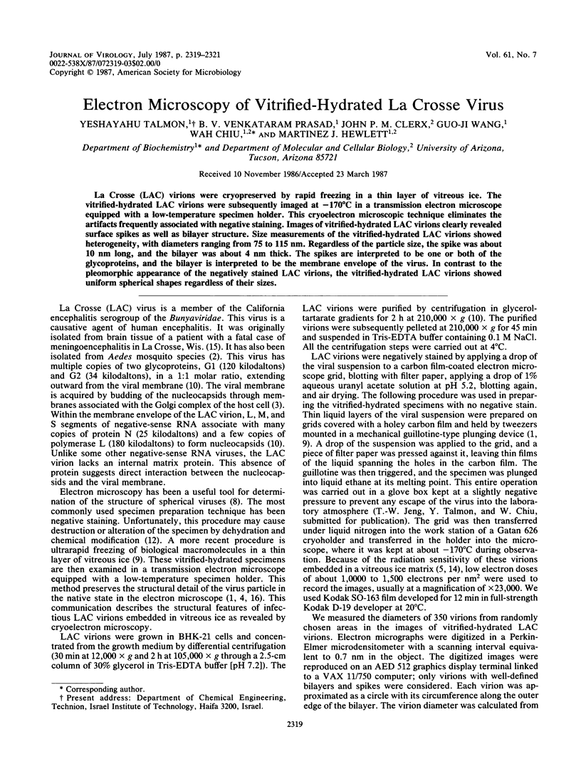
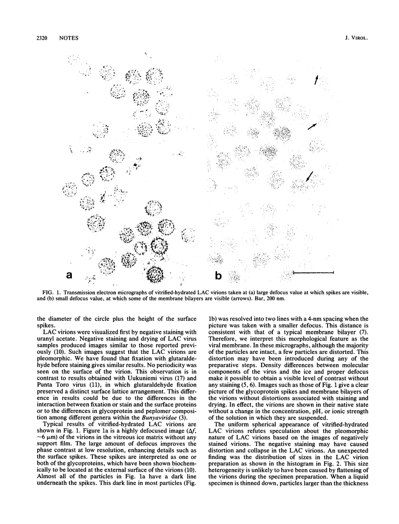
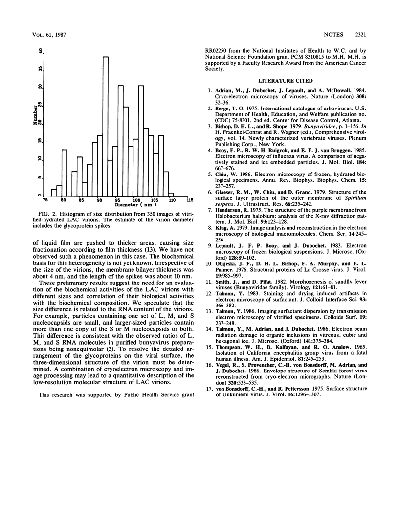
La Crosse (LAC) virions were cryopreserved by rapid freezing in a thin layer of vitreous ice. The vitrified-hydrated LAC virions were subsequently imaged at -170 degrees C in a transmission electron microscope equipped with a low-temperature specimen holder. This cryoelectron microscopic technique eliminates the artifacts frequently associated with negative staining. Images of vitrified-hydrated LAC virions clearly revealed surface spikes as well as bilayer structure. Size measurements of the vitrified-hydrated LAC virions showed heterogeneity, with diameters ranging from 75 to 115 nm. Regardless of the particle size, the spike was about 10 nm long, and the bilayer was about 4 nm thick. The spikes are interpreted to be one or both of the glycoproteins, and the bilayer is interpreted to be the membrane envelope of the virus. In contrast to the pleomorphic appearance of the negatively stained LAC virions, the vitrified-hydrated LAC virions showed uniform spherical shapes regardless of their sizes.
Full text
PDF
Images in this article
Selected References
These references are in PubMed. This may not be the complete list of references from this article.
- Adrian M., Dubochet J., Lepault J., McDowall A. W. Cryo-electron microscopy of viruses. Nature. 1984 Mar 1;308(5954):32–36. doi: 10.1038/308032a0. [DOI] [PubMed] [Google Scholar]
- Booy F. P., Ruigrok R. W., van Bruggen E. F. Electron microscopy of influenza virus. A comparison of negatively stained and ice-embedded particles. J Mol Biol. 1985 Aug 20;184(4):667–676. doi: 10.1016/0022-2836(85)90312-2. [DOI] [PubMed] [Google Scholar]
- Chiu W. Electron microscopy of frozen, hydrated biological specimens. Annu Rev Biophys Biophys Chem. 1986;15:237–257. doi: 10.1146/annurev.bb.15.060186.001321. [DOI] [PubMed] [Google Scholar]
- Glaeser R. M., Chiu W., Grano D. Structure of the surface layer protein of the outer membrane of Spirillum serpens. J Ultrastruct Res. 1979 Mar;66(3):235–242. doi: 10.1016/s0022-5320(79)90121-7. [DOI] [PubMed] [Google Scholar]
- Henderson R. The structure of the purple membrane from Halobacterium hallobium: analysis of the X-ray diffraction pattern. J Mol Biol. 1975 Apr 5;93(2):123–138. doi: 10.1016/0022-2836(75)90123-0. [DOI] [PubMed] [Google Scholar]
- Lepault J., Booy F. P., Dubochet J. Electron microscopy of frozen biological suspensions. J Microsc. 1983 Jan;129(Pt 1):89–102. doi: 10.1111/j.1365-2818.1983.tb04163.x. [DOI] [PubMed] [Google Scholar]
- Obijeski J. F., Bishop D. H., Murphy F. A., Palmer E. L. Structural proteins of La Crosse virus. J Virol. 1976 Sep;19(3):985–997. doi: 10.1128/jvi.19.3.985-997.1976. [DOI] [PMC free article] [PubMed] [Google Scholar]
- Smith J. F., Pifat D. Y. Morphogenesis of sandfly viruses (Bunyaviridae family). Virology. 1982 Aug;121(1):61–81. doi: 10.1016/0042-6822(82)90118-0. [DOI] [PubMed] [Google Scholar]
- THOMPSON W. H., KALFAYAN B., ANSLOW R. O. ISOLATION OF CALIFORNIA ENCEPHALITIS GROUP VIRUS FROM A FATAL HUMAN ILLNESS. Am J Epidemiol. 1965 Mar;81:245–253. doi: 10.1093/oxfordjournals.aje.a120512. [DOI] [PubMed] [Google Scholar]
- Vogel R. H., Provencher S. W., von Bonsdorff C. H., Adrian M., Dubochet J. Envelope structure of Semliki Forest virus reconstructed from cryo-electron micrographs. Nature. 1986 Apr 10;320(6062):533–535. doi: 10.1038/320533a0. [DOI] [PubMed] [Google Scholar]
- von Bonsdorff C. H., Pettersson R. Surface structure of Uukuniemi virus. J Virol. 1975 Nov;16(5):1296–1307. doi: 10.1128/jvi.16.5.1296-1307.1975. [DOI] [PMC free article] [PubMed] [Google Scholar]