Abstract
Propagation-defective vesicular stomatitis virus (VSV) vectors that encode a truncated G protein (VSV-Gstem) or lack the G gene entirely (VSV-ΔG) are attractive vaccine vectors because they are immunogenic, cannot replicate and spread after vaccination, and do not express many of the epitopes that elicit neutralizing anti-VSV immunity. To consider advancing nonpropagating VSV vectors towards clinical assessment, scalable technology that is compliant with human vaccine manufacturing must be developed to produce clinical trial material. Accordingly, two propagation methods were developed for VSV-Gstem and VSV-ΔG vectors encoding HIV gag that have the potential to support large-scale production. One method is based on transient expression of G protein after electroporating plasmid DNA into Vero cells and the second is based on a stable Vero cell line that contains a G gene controlled by a heat shock-inducible transcription unit. Both methods reproducibly supported production of 1×10⁷ to 1×10⁸ infectious units (I.U.s) of vaccine vector per ml. Results from these studies also showed that optimization of the G gene is necessary for abundant G protein expression from electroporated plasmid DNA or from DNA integrated in the genome of a stable cell line, and that the titers of VSV-Gstem vectors generally exceeded VSV-ΔG.
Keywords: Negative-strand RNA virus, VSV, Vero cells, Electroporation, Heat shock, Non-propagating
1. Introduction
Vesicular stomatitis virus (VSV) is a member of the Rhabdoviridae family, and accordingly, is an enveloped virus that contains a non-segmented, negative-strand RNA genome. The 11-kb genome contains 5 genes arranged sequentially 3′-N-P-M-G-L-5′ (Fig. 1a) that encode 5 known structural proteins (Jayakar et al., 2004; Rose and Whitt, 2001; Whelan et al., 2004). The nucleocapsid (N) protein encapsidates the genome whether the RNA is packaged in mature virions or is being actively transcribed and replicated in the infected cell cytoplasm by the viral-encoded RNA-dependent RNA polymerase. The polymerase is a multi subunit enzyme composed of the P (phosphoprotein) and L (large) protein. The matrix protein (M) lines the inner surface of the virus particle and promotes virion assembly and budding. Finally, multifunctional envelope glycoprotein (G) mediates cell attachment, membrane fusion, and likely promotes assembly of progeny virus particles (Jayakar et al., 2004; Swinteck and Lyles, 2008).
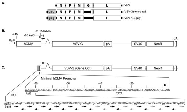
Figure 1. rVSV and plasmid DNA vectors.
Part A illustrates the structure of the rVSV vector genetic background and two propagation-defective vectors encoding gag from the first position in the rVSV genome. The rVSV-Gstem-gag1 encodes a truncated G protein composed of 41 amino acid residues of the extracellular domain (the Stem), the transmembrane region and the cytoplasmic tail. The rVSV-ΔG-gag1 vector lacks the G gene. Part B contains a map illustrating the structure of the expression plasmids that were used to transiently express various G genes following electroporation. The G genes were placed under control of the hCMV promoter and transcriptional enhancer region and the SV40 late poly-A signal (pA). The plasmid also contains the neomycin resistance gene (NeoR) driven by the SV40 promoter and transcriptional enhancer (SV40). The schematic in Part C illustrates the structure of the heat shock-inducible plasmid vector, which controls expression of the Gene Opt VSV Gin gene and was used to establish the Vero-HS4 cell line. The heat shock-inducible transcriptional control region is composed of a minimal hCMV promoter that extends to 86 nucleotides upstream (−86) of the transcription initiation signal (+1), and a repetitive array of nGAAn located between the BglII and AsiSI restriction enzyme sites, which forms multiple copies of the heat shock factor 1 (HSF-1) binding site (nGAAnnTTCnnGAAn). Black arrows in the lower portion of Part C represent perfect copies of nGAAn or its complement, whereas gray arrows identify imperfect nGAAn sequences.
Advancement in genetic engineering of rhabdoviruses (Finke and Conzelmann, 2005; Lawson et al., 1995; Schnell et al., 1994; Whelan et al., 1995) has made it possible to explore recombinant VSV (rVSV) as a vaccine vector. Many characteristics of VSV make it an attractive candidate for delivering human vaccines including: 1) VSV infection is not typically associated with human illness; 2) there is little pre-existing immunity in most human populations that might interfere with its use; 3) VSV infects and propagates efficiently in many cell types including cell lines suitable for manufacturing vaccines; 4) the non-segmented genome is stable and does not reassort; 5) VSV can accept one or more foreign gene inserts and direct high levels of expression upon infection; and 6) VSV infection is an efficient inducer of both cellular and humoral immunity (Bukreyev et al., 2006; Clarke et al., 2006; Finke and Conzelmann, 2005). A number of preclinical studies conducted with rVSV vectors have produced promising results; prototype vaccine vectors elicited potent immune responses against the encoded foreign antigen, and importantly, were found to be safe when administered to small laboratory animals and non-human primates (Grigera et al., 2000; Kahn et al., 2001; Roberts et al., 1999; Roberts et al., 1998; Rose et al., 2001; Rose et al., 2000; Schlereth et al., 2000). Notably, Rose et al. found that co-administration of two vaccine vectors, one encoding HIV-1 env and the other encoding SIV gag, elicited immune responses in vaccinated macaques that protected against challenge with a pathogenic SHIV (Rose et al., 2001).
The transition of a vaccine candidate from preclinical evaluation to clinical development focuses much greater emphasis on potential vaccination risk, and consequently, interest in vectors that are highly attenuated or propagation-defective. Propagation-defective rVSV vectors, encoding a variety of foreign antigens, have been produced in which the VSV G gene has been either deleted completely (rVSV-ΔG) or truncated to encode a G protein lacking most of the extracellular domain (rVSV-Gstem) (Clarke et al., 2006; Cooper et al., 2009; Kahn et al., 2001; Klas et al., 2006; Klas et al., 2002; Majid et al., 2006; Publicover et al., 2005). Studies conducted with small animal models have indicated that these vectors are immunogenic and may be particularly suited to eliciting antibody responses (Cooper et al., 2009; Kapadia et al., 2008; Publicover et al., 2005). In addition, the VSV-Gstem vector has been subjected to analysis in a murine neurovirulence model and found to cause little or no pathology when inoculated directly into the brain of neonatal mice (Cooper et al., 2009).
Although propagation-defective VSV vectors have produced promising results in preclinical studies, scalable propagation methods that can be validated and are compliant with regulatory agency guidelines governing human vaccine manufacture are needed before clinical evaluation of these vaccine candidates can be considered seriously. Development of a manufacturing process is complicated by the constraint that it must be based on continuous cell lines that are acceptable for vaccine production. Furthermore, providing genetic complementation for VSV G is problematic because the viral protein promotes membrane fusion and is cytotoxic (Rose and Whitt, 2001). Producing stable cell lines that express G protein from an inducible promoter is one potential solution, but leaky expression frequently results in toxicity and cell line instability, and the quantity of G synthesized after induction often is inadequate to promote efficient virus particle packaging particularly on a scale needed for vaccine manufacturing. One inducible cell line has been described (Schnell et al., 1997), but it is derived from BHK cells, which are not used for production of human vaccines because they produce contaminating intracisternal R-type particles (Blanchard et al., 2003; Chan, 1994; Compans et al., 1966; Shipman et al., 1969; Wang et al., 1999). Transient expression of G protein in transfected BHK or 293T cells (Majid et al., 2006; Takada et al., 1997) or electroporated Vero cells (Witko et al., 2006b) also has been used to propagate rVSV-ΔG and -Gstem vectors. These methods have been used to produce virus particles needed to support preclinical studies, but the yields of packaged virus or the complexity of producing a scalable process makes these procedures impractical for vaccine manufacturing. BHK cells, which produce intracisternal particles, or 293T cells, which encode SV40 T antigen (DuBridge et al., 1987), have been used in the transfection-based methods and are not preferred substrates for manufacture of live vaccines intended for use in humans. Therefore, to advance a propagation-defective VSV vector candidate into the clinic, a vaccine production process must be developed that meets a number of criteria including: 1) it must be scalable for development of a manufacturing process; 2) it has to be based on a continuous cell line substrate that is permissive for VSV infection and can be qualified for vaccine production; 3) materials used in the process are compliant with regulations governing vaccine production; 4) the abundance of G protein expression is adequate to promote efficient virus particle packaging, and 5) yields of infectious particles are sufficient to formulate vaccines that contain more than 1×10⁷ infectious units per ml, which has been demonstrated to be an immunogenic dose of VSV vector in a number of non-human primate studies (Daddario-DiCaprio et al., 2006a; Daddario-DiCaprio et al., 2006b; Egan et al., 2005; Egan et al., 2004; Feldmann et al., 2007; Jones et al., 2005; Rose et al., 2001).
Two procedures for G complementation are described below that support improved rVSV-ΔG and rVSV-Gstem packaging. Both are potentially scalable for manufacturing and both employ Vero cells, which are a well-characterized substrate for vaccine production and have been used to produce a licensed rotavirus vaccine (GlaxoSmithKline, 2008; Merck, 2006; Sheets, 2000). One approach is based on transient production of abundant G protein from electroporated plasmid DNA. The second method is based on development of stable cell lines that express G protein under control of a transcriptional control sequence regulated by the cellular heat shock response. Both methods have been used to propagate VSV Gstem and G vectors (Fig. 1) producing over 1×10⁷ infectious units (I.U.) per ml.
2. Materials and Methods
2.1 Cell culture
Vero cells were propagated in Dulbecco s modified minimal essential medium (DMEM) supplemented with 10% heat-inactivated fetal bovine serum (FBS) and 0.01 mg/ml gentamicin. The heat shock-inducible Vero-HS4 cell line, which expresses the Indiana serotype VSV G protein (Gin), was isolated after electroporating plasmid pHS-Gin (described below and Fig. 1B) into Vero cells. Briefly, Vero cells were propagated to near confluence in 150-cm2 flasks before being harvested and processed for conducting electroporation (Witko et al., 2006b). The cell suspension from a 150-cm2 flask was electroporated with approximately 25 μg of linearized (Bgl II; Fig. 1B) pHS-Gin DNA after which the cells were processed and distributed into three 10-cm2 culture dishes containing DMEM supplemented with 10% heat-inactivated FBS and 0.01 mg/ml gentamicin. Approximately 24 hours later, the medium was replaced with DMEM containing the same supplements plus 1 mg per ml neomycin (Geneticin from Invitrogen). The cells were maintained in medium containing neomycin until the monolayers were nearly confluent, at which time the cells were subcultured at a ratio of 1:100 to 1:5000 and propagated for about 2 weeks under selective conditions until isolated cell colonies were picked and expanded. The clonal cell lines were screened initially for VSV G expression by determining whether heat shock induction made it possible to propagate rVSV-Gstem vectors (Fig. 1A), which was determined by observing cytopathic effect (CPE) caused by viral replication. Positive cell lines then were screened more rigorously by performing Western blot analysis (Witko et al., 2006a) to confirm that G protein was expressed following heat shock, and by identifying cell lines that supported the most abundant production of rVSV-Gstem or -ΔG virus after infection.
2.2 Molecular cloning
Plasmid DNAs were prepared using standard molecular cloning procedures (Ausubel et al., 1987) and their structures were verified by nucleotide sequencing (Witko et al., 2006b). A modified pCI-Neo plasmid (Promega) that lacked a T7 RNA polymerase promoter (Witko et al., 2006b) was used to construct vectors in which the VSV Gin or the New Jersey serotype G (Gnj) were placed under the control of the hCMV promoter and enhancer. Three types of G expression plasmid were constructed that differed in the type of modifications that were applied to the cloned G sequences. The first type of expression plasmid was constructed simply by adding a Kozak consensus element (Kozak, 1991) at the translation initiation site of the native Gin and Gnj sequences. The second type of plasmid was generated with G genes that were subjected to RNA Optimization (RNA Opt; also sometimes referred to as “codon optimization”) using the strategy described by Jalah et al. (Jalah et al., 2007). This method is based on the observation that protein quantity is influenced by several mRNA attributes including stability, nuclear export efficiency, and the efficiency of translation initiation (Nasioulas et al., 1994; Schneider et al., 1997; Schwartz et al., 1992a; Schwartz et al., 1992b). Accordingly, synonymous codon changes were introduced that increased guanine and cytosine (G+C) content, eliminated potential splicing signals or other RNA processing elements, minimized repetitive sequences or other sequence motifs that had the potential to form extensive secondary structure, and eliminated known mRNA instability sequences. Optimal translation initiation and polyadenylation signals were incorporated as well.
The last type of modified G gene was produced by a related method that is referred to as “Gene Optimization” (Gene Opt), which included the following steps: i) the Backtranslate program (SeqWeb software suite, Accelrys Software, Inc) was used to produce G coding sequence composed of codons used at high frequency in human cells; ii) homopolymeric regions in the backtranslated sequence of more than 5 nucleotides were interrupted by exchanging sequences with synonymous codons; iii) splice donor and acceptor signals predicted by the webtool (http://www.fruitfly.org/seq_tools/splice.html) described by Reese et al. (Reese et al., 1997) were removed from the coding sequence by incorporating synonymous codons; iv) potential mRNA instability signals (Shaw and Kamen, 1986; Zubiaga et al., 1995) were eliminated by replacing sequence with synonymous codons; and, v) optimal translation initiation and termination signals were added (Kochetov et al., 1998; Kozak, 1991). The optimized G gene sequences had been deposited in Genbank and their Accession numbers are as follows: GU177824 (Indiana G, RNA optimized); GU177825 (Indiana G, gene/codon optimized); GU177826 (New Jersey G, RNA optimized).
A heat shock-inducible plasmid vector (pHS-Gin; Fig. 1C) was constructed by substituting much of the hCMV promoter and enhancer sequence (Meier and Stinski, 1996), between the Bgl II and Asi SI sites in pCI-Neo (Fig 1b), with multiple copies of the heat shock factor 1 (HSF-1) binding element (Kroeger and Morimoto, 1994). The Gene Opt Gin gene was cloned 3 of the heat shock inducible promoter.
2.3 G protein expression and VSV propagation
Propagation-defective rVSV-ΔG and rVSV-Gstem vectors used in these studies have been described before (Witko et al., 2006b) and their genomic structures are illustrated in Fig. 1. Two methods were used to package propagation-defective rVSV vectors. The first was based on transient expression of G protein after electroporation. Vero cells were electroporated with 25–50 μg of G expression plasmid (Witko et al., 2006b), and approximately 20 hours later, the monolayer of electroporated cells cultured in a T150-cm2 flask was infected with approximately 0.01 plaque-forming units (PFU) per cell. Virus particles were harvested 24–48 hours post-infection and quantified by plaque assay conducted with BHK-G cells (Schnell et al., 1997).
The second packaging and propagation method was based on using the inducible cell line (Vero-HS4) that expresses Gin after heat shock. Vero-HS4 cells were split the day before infection to achieve a nearly confluent monolayer the following day. The cells were then fed with fresh medium lacking neomycin and transferred to an incubator set at 43°C for 3–6 hours after which the cells were incubated at 37°C overnight. The cells were then infected with approximately 0.01 PFU per cell of propagation-defective VSV and allowed to incubate 24–48 hours before virus particles were harvested and quantified by plaque assay. Cells that were heat shocked but not infected were harvested and tested for G protein expression by Western blot analysis.
3. RESULTS
3.1 Packaging VSV particles in cells after electroporation of plasmids encoding G protein
VSV particle packaging procedures based on transient G expression have been used successfully to produce relatively small-scale quantities of rVSV-ΔG and rVSV-Gstem vectors needed for preclinical studies (Majid et al., 2006; Publicover et al., 2005; Witko et al., 2006b). For development of a manufacturing process, transient expression approaches are attractive because they can be applied to multiple cell types, and importantly, it is possible to directly use a validated cell line without further qualification or testing (i.e. adventitious agent testing, karyotyping, tumorigenicity testing, etc.), which will be needed to qualify a new stable inducible cell line. Although the published transient expression methods have been used successfully, their application to vaccine manufacture is restricted because they are based on cells that are not qualified for vaccine production (i.e. BHK) or packaging yields generally have been lower than 1×10⁷ I.U.s per ml (data not shown). To begin developing a method that can support vaccine manufacturing, process development was initiated with Vero cells and plasmid DNA electroporation. Vero cells were selected because qualified lines can be obtained for vaccine production, and because it is one of the few continuous cell lines used today to manufacture licensed vaccines (GlaxoSmithKline, 2008; Merck, 2006; Sheets, 2000). Electroporation was selected as the method for introducing DNA for several reasons including; equipment has been developed that can support manufacturing-scale DNA electroporation (Fratantoni et al., 2003), electroporation is one of the more effective methods to introduce plasmid DNA into Vero cells (Kaur et al., 2008; Surman et al., 2007; Witko et al., 2006b), and DNA electroporation can be performed without using transfection reagents that might be very costly, or contain components that are not acceptable for a vaccine manufacturing process.
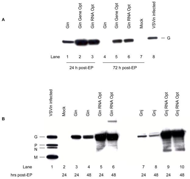
In addition to the efficiency of plasmid DNA introduction into Vero cells, the next most important variable limiting virus packaging yields was expected to be the abundance of G protein in transfected cells. Accordingly, expression plasmid optimization was investigated as a method to improve G expression. Gin or Gnj genes were designed using either the related RNA Opt or Gene Opt strategies described in the Methods, and were cloned into the pCI-Neo vector (Methods and Fig. 1B) under the control of the hCMV promoter and enhancer region (Boshart et al., 1985; Meier and Stinski, 1996). To investigate the effect of optimization, 50 μg of plasmid DNA was electroporated into approximately 1×10⁷ Vero cells and total cellular protein was harvested 24 or 72 hours post-electroporation (Fig. 2). Western blot analysis with VSV G-specific monoclonal antibody (Indiana serotype) polyclonal antiserum revealed that Gin protein abundance was increased significantly by either optimization method at 24 or 72 hours post-electroporation (post-EP) when compared to expression directed by the native Gin gene, and this effect was most pronounced at the later time point. In most experiments, it appeared that the Gin gene designed with the RNA Opt method was expressed somewhat more efficiently particularly at the 72 h time-point, although the magnitude of this effect was not rigorously evaluated since it was evident that either procedure would notably elevate Gin protein production for the purpose of providing complementation. The RNA Opt procedure was applied to the Gnj coding sequence as well and had a similar positive effect on expression (Fig. 2B).
Figure 2. Transient expression of VSV G following electroporation.
(A) Vero cells were harvested at 24 (lanes 1–3) or 72 (lanes 4–7) h post-electroporation (EP) with expression plasmids (Fig. 1B) containing various forms of the VSV G gene (native Gin gene, lanes 1 and 4; Gene Optimized Gin, lanes 2 and 5; RNA Optimized VSV Gin, lanes 3 and 6), after which protein extracts were prepared for analysis by Western blotting. G protein was detected with a G-specific monoclonal antibody (Roche). Extract prepared from Vero cells infected with a recombinant propagation-competent VSVin was used as a positive control (Lanes 8). (B) The description of this experiment is similar to Part A, except that the effect of RNA optimization on expression of both the Gin and Gnj genes was evaluated. Gnj was detected with rabbit anti-VSVnj polyclonal antiserum.
After finding that electroporated Vero cells produced abundant G protein, studies were performed to determine whether the increased concentrations of G enhanced viral particle packaging. Five independent experiments were conducted (Table 1) in which rVSV-ΔG and rVSV-Gstem vectors encoding HIV-1 gag were packaged in cells electroporated with different G expression plasmids. Several conclusions can be drawn from these studies. First, packaging of the rVSV-ΔG-gag1 or rVSV-Gstem-gag1 vectors was significantly improved ( p<0.05 or p<0.01, respectively) when an optimized plasmid was used for complementation producing 0.6 to 1.2 log10 I.U.s more per packaging run. This conclusion was supported by multiple trials with optimized Gin plasmids and by one performed with Gnj. The second conclusion was that the Gstem vector titers were generally higher than those produced by the ΔG vector, and packaging runs routinely exceeded 1×10⁷ I.U.s per ml and at times reached 1×10⁸ I.U.s per ml. Taken together, these results demonstrated that enhanced G expression did improve particle titers, and that the Gstem vector tended to package more efficiently than the ΔG counterpart.
Table 1.
Virus particle packaging
| Virus | G Gene | Virus Particle Titer (Log10 I.U./ml)a | |||||
|---|---|---|---|---|---|---|---|
| Expb 1 | Exp 2 | Exp 3 | Exp 4 | Exp 5 | Mean ± SDc | ||
| rVSV-ΔG-gag1 | VSV Gin | 6.6 | 5.9 | 6.4 | 6.8 | n.d.d | 6.6 ± 0.2 |
| Gin Gene Opt | 7.8 | 6.6 | 6.9 | 7.5 | n.d. | 7.2 ± 0.5* | |
| Gin RNA Opt | n.d. | n.d. | 6.9 | 7.5 | n.d. | 7.2* | |
| rVSV-Gstem-gag1 | VSV Gin | 7.1 | 6.7 | 7.0 | 7.6 | 7.3 | 7.1 ± 0.3 |
| Gin Gene Opt | 8.1 | 7.6 | 7.6 | 8.3 | n.d. | 7.9 ± 0.4** | |
| Gin RNA Opt | n.d. | n.d. | 7.7 | 8.1 | 8.1 | 8.0 ± 0.2** | |
| VSV Gnj | n.d. | n.d. | n.d. | n.d. | 7.3 | 7.3 | |
| Gnj RNA Opt | n.d. | n.d. | n.d. | n.d. | 8.1 | 8.1 | |
Titers of virus particles were determined using BHK-G cells
Exp; experiment
Data are expressed as mean titers ± standard deviation (SD); SD calculated if 3 or more trials were conducted. For statistical analysis by Student’s t-test, virus titers obtained with optimized genes were compared with corresponding native gene titers;
p<0.05;
p<0.01
n.d.; not done
3.2 Packaging VSV particles in stable inducible cell lines expressing G protein
Stable cell lines that express adequate concentrations of G protein to support virus particle packaging do provide practical advantages particularly for large-scale applications. Notably, they can be used to propagate rVSV-ΔG or -Gstem vectors without the handling needed to perform electroporation or transfection, which can be difficult to manage reproducibly when performed on a scale needed to support vaccine manufacturing. Although stable cell lines are a very attractive alternative, they can be difficult to produce and maintain particularly when the complementing gene product is toxic like VSV G. Attempts to produce Vero cells expressing G under control of tetracycline-responsive systems (Corbel and Rossi, 2002) failed, prompting investigation of additional approaches (data not shown).
A system based on induction of transcription by heat shock was an attractive alternative. Promoters controlling expression of cellular heat shock proteins (HSPs) have been used to control synthesis of a foreign protein (Rome et al., 2005), and it is known that Vero cells are tolerant of heat shock (Witko et al., 2006b). Moreover, induction by heat shock eliminates the need to use chemical compounds to control induction or repression of transcription that might need to be removed from the final vaccine preparation. Although it was appealing to make use of heat shock response, it is known that promoters controlling expression of the HSPs do exhibit significant basal activity (Rome et al., 2005) that might cause toxicity when controlling expression of VSV G protein. Therefore, a modified strategy was investigated to minimize basal promoter activity. A heat shock-inducible transcriptional control region was constructed by starting with a minimal promoter (Gossen and Bujard, 1992) derived from the hCMV immediate early region 1 transcriptional control region (Meier and Stinski, 1996) that was expected to exhibit low levels of basal activity (Fig 1C). To make it responsive to heat shock and promote an increased magnitude of response, multiple copies of the sequence 5′-NGAAN-3′ and its complement (5′-NTTCN-3′) were inserted 5′ of the minimal promoter generating multiple bindings sites (nGAAnnTTCnnGAAn) for heat shock factor 1 (HSF-1) (Wang and Morgan, 1994), which binds DNA as a trimer (Wu, 1995).
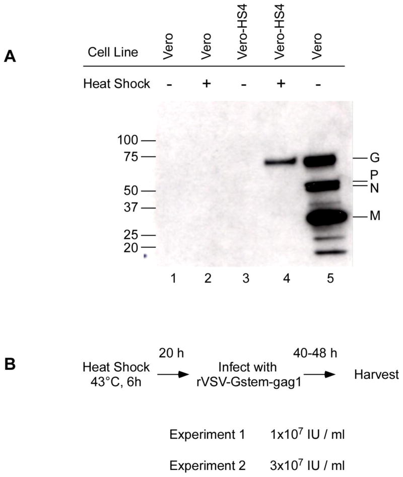
The VSV Gin Gene Opt protein-coding sequence was cloned 3′ of the modified promoter in a plasmid that also contained the NeoR selectable marker controlled by the SV40 promoter and enhancer (Fig. 1C). Cell lines were established by introducing linearized plasmid DNA into Vero cells by electroporation and applying G418 selection 24 hours later. Drug resistant cell colonies were isolated and expanded, and subsequently screened for their ability to support propagation of rVSV-Gstem vector after heat shock, which was determined simply by monitoring the cultures for cytopathic effect caused by replication. Positive cell lines were screened more rigorously by quantifying Gin expression induced by heat shock (data not shown) resulting in selection of the Vero-HS4 cell line for further evaluation. Inducible expression of G protein in Vero-HS4 cells is illustrated by the Western blot shown in Fig. 3A. Vero or Vero-HS4 cells were subjected to heat shock for 6 hours at 43°C then returned to 37°C for overnight incubation. Control cells were maintained at 37°C throughout the experiment. Proteins extracted from treated and control cells were separated by gel electrophoresis and transferred to a nitrocellulose membrane, which was incubated subsequently with an anti-VSV polyclonal antiserum. The results demonstrated that heat shock-induced Vero-HS4 cells synthesized detectable quantities of G protein whereas no protein expression was evident in Vero controls or Vero-HS4 cells that were not heat shocked. Scanning of the blot by a Densitometer showed that a ~ 50 fold induction of G expression was seen upon heat treatment (data not shown).
Figure 3. Expression of G protein and Packaging of rVSV-Gstem-gag1 using the heat shock inducible Vero-HS4 cell line.
(A) Vero (lanes 1and 2) or Vero-HS4 cells (lanes 3 and 4) were subjected to heat shock at 43°C for 6 hours before incubation overnight at 37°C. Approximately 24 hours after initiating heat shock, cell lysates were prepared and analyzed by Western blotting as described in Figure 2. Lysate prepared from Vero cells infected with rVSVin wt was included as a positive control (lane 5) and the viral polypeptides recognized by the antiserum are identified. The blot was probed with rabbit anti-VSVin antiserum. (B) The protocol for propagating rVSV-Gstem-gag1 with Vero-HS4 cells is illustrated at the top of Part B. Cells induced by heat shock and subsequently incubated at 37ºC to allow G protein synthesis were infected with rVSV-Gstem-gag1 at a multiplicity of infection of 0.01. The infected cells were incubated 40–48 hours at which time viral particles were harvested from the medium supernatant and infectious particle titers were determined using BHK-G cells. The I.U.s recovered in two independent experiments are shown.
The ability of Vero-HS4 cells to support propagation of the rVSVin-Gstem- gag1 vector was examined as well. Near confluent Vero-HS4 cell monolayers were heat shocked for 6 hours then incubated overnight at 37°C to allow time for expression of G protein. The monolayers were then infected with 0.01 I.U.s per cell of rVSVin-Gstem-gag1, which had been produced originally using the transient expression methods described above, and incubated approximately 20 – 40 hours at 37°C before virus was harvested and quantified. The yield from 2 independent experiments was 1×10⁷ and 3×10⁷ I.U.s per ml (Fig. 3B). Moreover, the Vero-HS4 cell line has been stable for more than 20 passages as determined by its ability to support Gstem propagation (data not shown). These results demonstrate that cell lines like Vero-HS4 can be used to develop a manufacturing process for vaccines based on rVSV-ΔG and rVSV-Gstem vectors.
4.0 DISCUSSION
A transient expression method, based on DNA electroporation, and a stable inducible cell line approach were developed for packaging propagation- defective VSV-ΔG and VSV-Gstem vectors. Both procedures were able to produce over 1×10⁷ I.U.s of rVSV-ΔG or rVSV-Gstem vector encoding HIV gag per ml of material harvested from infected Vero cell substrates. The highest titers were achieved with the rVSV-Gstem-gag1 vector, which reached 1×10⁸ I.U.s per ml in some transient expression trials. As mentioned earlier, achieving 1×10⁷ I.U.s per ml was a key benchmark since preclinical studies conducted with non- human primates indicated that manufacture of doses in this range will be required to advance a rVSV vector into the clinic (Daddario-DiCaprio et al., 2006a; Daddario-DiCaprio et al., 2006b; Egan et al., 2005; Egan et al., 2004; Feldmann et al., 2007; Jones et al., 2005; Rose et al., 2001).
Optimization of the G gene was an important factor in the success of both the transient expression and stable cell line approaches as demonstrated by a marked increase in G abundance in electroporated Vero cells (Fig. 2) that correlated with improvements in virus particle packaging (Table 1). In addition to using cis-acting signals that promote efficient translation (Kochetov et al., 1998; Kozak, 1991; Schwartz et al., 1992c), multiple elements of the optimization procedures probably contributed to their positive effect. For example, both the RNA Opt and Gene Opt strategies removed a number of sequences from the native G coding sequence that closely resembled consensus nuclear mRNA splicing signals. Removal of these sequences minimized unwanted nuclear splicing and probably increased the abundance of mRNA exiting the nucleus that contained a full-length G coding sequence. Optimization also significantly increased the G+C nucleotide content of the G protein mRNA. The native Gin sequence was 44% G+C whereas the RNA Opt (62%) and the Gene Opt (64%) versions were notably higher. Perhaps the higher G+C content of the optimized G transcripts better mimics highly translated human mRNAs (Kochetov et al., 1998; Zhang, 1998) or increases the abundance of steady-state message in the cytoplasm by mechanisms that might include improved nuclear export or greater mRNA stability (Graf et al., 2000; Kudla et al., 2006; Maldarelli et al., 1991; Nasioulas et al., 1994; Olsen et al., 1992; Schwartz et al., 1992a; Schwartz et al., 1992b; Zubiaga et al., 1995). Regardless of the exact mechanism, the most dramatic effect was seen 3 days after DNA electroporation (Fig. 2A) indicating that optimization promoted sustained accumulation of G protein.
Ternette and colleagues (Ternette et al., 2007a; Ternette et al., 2007b) also have observed significant improvement in protein expression from transfected plasmid DNAs encoding G proteins from VSV and respiratory syncytial virus after applying an optimization strategy, and a similar positive effect has been produced after applying the RNA and Gene Opt strategies to VSV M (our unpublished data). Although this is a small sampling, these examples indicate that efficient expression of paramyxovirus or rhabdovirus genes from cellular RNA polymerase II promoters can be improved if the protein coding sequences are subjected to some form of coding sequence optimization. This probably is related to the fact that these viruses replicate exclusively in the cytoplasm and have evolved coding sequences that are not designed for synthesis and export by nuclear processes in eukaryotic cells.
The virus packaging methods were developed to package rVSV-Gstem and rVSV-ΔG vectors, although additional applications are possible. It is likely that other propagation-defective negative-strand RNA virus vectors lacking their native attachment proteins can be packaged with VSV G on their surface. In fact VSV G protein has been shown to substitute as an attachment protein for recombinant replication-competent measles virus and respiratory syncytial virus (Oomens et al., 2003; Spielhofer et al., 1998) indicating that it will function similarly in the context of a propagation-defective vector. VSV G protein also is used widely to pseudo-type retrovirus particles thereby providing an attachment protein that can mediate infection of a broad spectrum of cell types (Cronin et al., 2005; Yee et al., 1994). For large-scale manufacturing, genetic complementation provided by a stable inducible cell line is preferred because it requires far less manipulation than transient expression approaches. Moreover, induction controlled by heat shock provides additional advantages because gene expression can be turned on by a simple temperature shift and no chemical compounds are added to the medium to control gene expression. This eliminates the need to replace medium in a large culture vessel to remove compounds that are used to repress transcription, and might also eliminate the need to purify the packaged virus to remove a chemical inducer from the final vaccine product. It also is likely that gains in virus particle production can be realized if the heat shock system is subjected to a systematic evaluation of variables that can effect induction of G expression and VSV maturation such as: 1) heat shock temperature and duration; 2) timing of infection following heat shock; 3) incubation temperature following heat shock; 4) multiplicity of infection; and 5) timing of virus particle harvest.
The hybrid heat shock element/minimal CMV promoter seemed to be an important component of the stable cell line approach. The basal level of expression in Vero-HS4 cells was low, which likely contributed to the stability of this line, but the magnitude of induction was high, promoting efficient production of infectious rVSV-Gstem or -ΔG particles. The combination of multimerized heat shock elements linked to a minimal CMV promoter was probably the major factor in determining the effective balance between basal activity and efficient induction, although it should be noted that the site of chromosomal integration also may contribute (Barnes and Dickson, 2006). It also should be mentioned that the basal expression was low but not undetectable, and a hardy cell line like Vero might tolerate low levels of G expression better than some other cell types. In instances where tighter control over the basal activity is needed, the minimal CMV promoter could be modified to further reduce basal expression or other less active promoters could be tested to identify those that have very low levels of basal expression, but remain responsive to heat shock when linked to heat-shock-responsive elements (Emiliusen et al., 2001).
Finally, it is worth noting that the rVSV-Gstem-gag1 vector generally packaged more efficiently than the corresponding ΔG vector. The rVSV-Gstem vectors were constructed because earlier studies indicated that the Gstem polypeptide could promote more efficient virion morphogenesis (Jeetendra et al., 2003; Jeetendra et al., 2002; Robison and Whitt, 2000). The mechanism by which the Stem polypeptide improves packaging efficiency is not known, but it might help recruit viral nucleocapsids to sites along the membrane where viruses assemble and bud or perhaps it increases the quantity of I.U.s in the harvest because incorporation of the Gstem polypeptide enhances the infectivity or stability of the virus particle (Jeetendra et al., 2003; Jeetendra et al., 2002; Robison and Whitt, 2000; Swinteck and Lyles, 2008; Zhou and Blissard, 2008). Regardless of the precise mechanism through which the Gstem polypeptide works, the improved yields of infectious particles can be significant particularly for applications like a vaccine manufacturing process where achieving maximum yields are critical.
Acknowledgments
This work was sponsored in part by an HIV-1 Vaccine Design and Development Team contract from the National Institutes of Health and the National Institute of Allergy and Infectious Diseases (HVDDT NO1-A1-25458), and the National Cancer Institute Intramural Research Program at the Center for Cancer Research. We thank Julia Li for performing statistical analysis, Alan Gordon for insightful comments and suggestions and Ellen Murphy and Yury Matsuka for critical review of the manuscript.
Footnotes
Publisher's Disclaimer: This is a PDF file of an unedited manuscript that has been accepted for publication. As a service to our customers we are providing this early version of the manuscript. The manuscript will undergo copyediting, typesetting, and review of the resulting proof before it is published in its final citable form. Please note that during the production process errors may be discovered which could affect the content, and all legal disclaimers that apply to the journal pertain.
References
- Ausubel FM, Brent R, Kingston RE, Moore DD, Siedman JG, Smith JA, Struhl K, editors. Current Protocols in Molecular Biology. Greene Publishing Associates and Wiley Interscience; New York: 1987. [Google Scholar]
- Barnes LM, Dickson AJ. Mammalian cell factories for efficient and stable protein expression. Curr Opin Biotechnol. 2006;17:381–6. doi: 10.1016/j.copbio.2006.06.005. [DOI] [PubMed] [Google Scholar]
- Blanchard E, Brand D, Roingeard P. Endogenous virus and hepatitis C virus-like particle budding in BHK-21 cells. J Virol. 2003;77:3888–9. doi: 10.1128/JVI.77.6.3888-3889.2003. author reply 3889. [DOI] [PMC free article] [PubMed] [Google Scholar]
- Boshart M, Weber F, Jahn G, Dorsch-Hasler K, Fleckenstein B, Schaffner W. A very strong enhancer is located upstream of an immediate early gene of human cytomegalovirus. Cell. 1985;41:521–30. doi: 10.1016/s0092-8674(85)80025-8. [DOI] [PubMed] [Google Scholar]
- Bukreyev A, Skiadopoulos MH, Murphy BR, Collins PL. Nonsegmented negative-strand viruses as vaccine vectors. J Virol. 2006;80:10293–306. doi: 10.1128/JVI.00919-06. [DOI] [PMC free article] [PubMed] [Google Scholar]
- Chan SY. Characterization of recombinant BHK-21 endogenous particles: R-type particles. Biologicals. 1994;22:121–5. doi: 10.1006/biol.1994.1018. [DOI] [PubMed] [Google Scholar]
- Clarke DK, Cooper D, Egan MA, Hendry RM, Parks CL, Udem SA. Recombinant vesicular stomatitis virus as an HIV-1 vaccine vector. Springer Semin Immunopathol. 2006;28:239–253. doi: 10.1007/s00281-006-0042-3. [DOI] [PMC free article] [PubMed] [Google Scholar]
- Compans RW, Holmes KV, Dales S, Choppin PW. An electron microscopic study of moderate and virulent virus-cell interactions of the parainfluenza virus SV5. Virology. 1966;30:411–26. doi: 10.1016/0042-6822(66)90119-x. [DOI] [PubMed] [Google Scholar]
- Cooper D, Witko SE, Calderon PC, Wright KJ, Johnson JE, Guo M, Kotash CS, Nowak RM, Natuk RJ, Hendry RM, Udem SA, Parks CL. Evaluation Neurovirulence, Immunogenicity, and In Vitro Packaging of Two Types of Propagation-Defective Vesicular Stomatitis Virus Vaccine Vectors. 2009 In preparation. [Google Scholar]
- Corbel SY, Rossi FM. Latest developments and in vivo use of the Tet system: ex vivo and in vivo delivery of tetracycline-regulated genes. Curr Opin Biotechnol. 2002;13:448–52. doi: 10.1016/s0958-1669(02)00361-0. [DOI] [PubMed] [Google Scholar]
- Cronin J, Zhang XY, Reiser J. Altering the tropism of lentiviral vectors through pseudotyping. Curr Gene Ther. 2005;5:387–98. doi: 10.2174/1566523054546224. [DOI] [PMC free article] [PubMed] [Google Scholar]
- Daddario-DiCaprio KM, Geisbert TW, Geisbert JB, Stroher U, Hensley LE, Grolla A, Fritz EA, Feldmann F, Feldmann H, Jones SM. Cross-protection against Marburg virus strains by using a live, attenuated recombinant vaccine. J Virol. 2006a;80:9659–66. doi: 10.1128/JVI.00959-06. [DOI] [PMC free article] [PubMed] [Google Scholar]
- Daddario-DiCaprio KM, Geisbert TW, Stroher U, Geisbert JB, Grolla A, Fritz EA, Fernando L, Kagan E, Jahrling PB, Hensley LE, Jones SM, Feldmann H. Postexposure protection against Marburg haemorrhagic fever with recombinant vesicular stomatitis virus vectors in non-human primates: an efficacy assessment. Lancet. 2006b;367:1399–404. doi: 10.1016/S0140-6736(06)68546-2. [DOI] [PubMed] [Google Scholar]
- DuBridge RB, Tang P, Hsia HC, Leong PM, Miller JH, Calos MP. Analysis of mutation in human cells by using an Epstein-Barr virus shuttle system. Mol Cell Biol. 1987;7:379–87. doi: 10.1128/mcb.7.1.379. [DOI] [PMC free article] [PubMed] [Google Scholar]
- Egan MA, Chong SY, Megati S, Montefiori DC, Rose NF, Boyer JD, Sidhu MK, Quiroz J, Rosati M, Schadeck EB, Pavlakis GN, Weiner DB, Rose JK, Israel ZR, Udem SA, Eldridge JH. Priming with plasmid DNAs expressing interleukin-12 and simian immunodeficiency virus gag enhances the immunogenicity and efficacy of an experimental AIDS vaccine based on recombinant vesicular stomatitis virus. AIDS Res Hum Retroviruses. 2005;21:629–43. doi: 10.1089/aid.2005.21.629. [DOI] [PubMed] [Google Scholar]
- Egan MA, Chong SY, Rose NF, Megati S, Lopez KJ, Schadeck EB, Johnson JE, Masood A, Piacente P, Druilhet RE, Barras PW, Hasselschwert DL, Reilly P, Mishkin EM, Montefiori DC, Lewis MG, Clarke DK, Hendry RM, Marx PA, Eldridge JH, Udem SA, Israel ZR, Rose JK. Immunogenicity of attenuated vesicular stomatitis virus vectors expressing HIV type 1 Env and SIV Gag proteins: comparison of intranasal and intramuscular vaccination routes. AIDS Res Hum Retroviruses. 2004;20:989–1004. doi: 10.1089/aid.2004.20.989. [DOI] [PubMed] [Google Scholar]
- Emiliusen L, Gough M, Bateman A, Ahmed A, Voellmy R, Chester J, Diaz RM, Harrington K, Vile R. A transcriptional feedback loop for tissue-specific expression of highly cytotoxic genes which incorporates an immunostimulatory component. Gene Ther. 2001;8:987–98. doi: 10.1038/sj.gt.3301470. [DOI] [PubMed] [Google Scholar]
- Feldmann H, Jones SM, Daddario-DiCaprio KM, Geisbert JB, Stroher U, Grolla A, Bray M, Fritz EA, Fernando L, Feldmann F, Hensley LE, Geisbert TW. Effective post-exposure treatment of Ebola infection. PLoS Pathog. 2007;3:e2. doi: 10.1371/journal.ppat.0030002. [DOI] [PMC free article] [PubMed] [Google Scholar]
- Finke S, Conzelmann KK. Recombinant rhabdoviruses: vectors for vaccine development and gene therapy. Curr Top Microbiol Immunol. 2005;292:165–200. doi: 10.1007/3-540-27485-5_8. [DOI] [PubMed] [Google Scholar]
- Fratantoni JC, Dzekunov S, Singh V, Liu LN. A non-viral gene delivery system designed for clinical use. Cytotherapy. 2003;5:208–10. doi: 10.1080/14653240310001479. [DOI] [PubMed] [Google Scholar]
- GlaxoSmithKline. FDA; 2008. Rotarix (Rotavirus Vaccine, Live, Oral) Oral Suspension. http://www.fda.gov/Cber/products/rotarix.htm. [Google Scholar]
- Gossen M, Bujard H. Tight control of gene expression in mammalian cells by tetracycline-responsive promoters. Proc Natl Acad Sci U S A. 1992;89:5547–51. doi: 10.1073/pnas.89.12.5547. [DOI] [PMC free article] [PubMed] [Google Scholar]
- Graf M, Bojak A, Deml L, Bieler K, Wolf H, Wagner R. Concerted action of multiple cis-acting sequences is required for Rev dependence of late human immunodeficiency virus type 1 gene expression. J Virol. 2000;74:10822–6. doi: 10.1128/jvi.74.22.10822-10826.2000. [DOI] [PMC free article] [PubMed] [Google Scholar]
- Grigera PR, Marzocca MP, Capozzo AV, Buonocore L, Donis RO, Rose JK. Presence of bovine viral diarrhea virus (BVDV) E2 glycoprotein in VSV recombinant particles and induction of neutralizing BVDV antibodies in mice. Virus Res. 2000;69:3–15. doi: 10.1016/s0168-1702(00)00164-7. [DOI] [PubMed] [Google Scholar]
- Jalah R, Rosati M, Kulkarni V, Patel V, Bergamaschi C, Valentin A, Zhang GM, Sidhu MK, Eldridge JH, Weiner DB, Pavlakis GN, Felber BK. Efficient systemic expression of bioactive IL-15 in mice upon delivery of optimized DNA expression plasmids. DNA Cell Biol. 2007;26:827–40. doi: 10.1089/dna.2007.0645. [DOI] [PubMed] [Google Scholar]
- Jayakar HR, Jeetendra E, Whitt MA. Rhabdovirus assembly and budding. Virus Res. 2004;106:117–32. doi: 10.1016/j.virusres.2004.08.009. [DOI] [PubMed] [Google Scholar]
- Jeetendra E, Ghosh K, Odell D, Li J, Ghosh HP, Whitt MA. The membrane-proximal region of vesicular stomatitis virus glycoprotein G ectodomain is critical for fusion and virus infectivity. J Virol. 2003;77:12807–18. doi: 10.1128/JVI.77.23.12807-12818.2003. [DOI] [PMC free article] [PubMed] [Google Scholar]
- Jeetendra E, Robison CS, Albritton LM, Whitt MA. The membrane-proximal domain of vesicular stomatitis virus G protein functions as a membrane fusion potentiator and can induce hemifusion. J Virol. 2002;76:12300–11. doi: 10.1128/JVI.76.23.12300-12311.2002. [DOI] [PMC free article] [PubMed] [Google Scholar]
- Jones SM, Feldmann H, Stroher U, Geisbert JB, Fernando L, Grolla A, Klenk HD, Sullivan NJ, Volchkov VE, Fritz EA, Daddario KM, Hensley LE, Jahrling PB, Geisbert TW. Live attenuated recombinant vaccine protects nonhuman primates against Ebola and Marburg viruses. Nat Med. 2005;11:786–90. doi: 10.1038/nm1258. [DOI] [PubMed] [Google Scholar]
- Kahn JS, Roberts A, Weibel C, Buonocore L, Rose JK. Replication-competent or attenuated, nonpropagating vesicular stomatitis viruses expressing respiratory syncytial virus (RSV) antigens protect mice against RSV challenge. J Virol. 2001;75:11079–87. doi: 10.1128/JVI.75.22.11079-11087.2001. [DOI] [PMC free article] [PubMed] [Google Scholar]
- Kapadia SU, Simon ID, Rose JK. SARS vaccine based on a replication-defective recombinant vesicular stomatitis virus is more potent than one based on a replication-competent vector. Virology. 2008;376:165–72. doi: 10.1016/j.virol.2008.03.002. [DOI] [PMC free article] [PubMed] [Google Scholar]
- Kaur J, Tang RS, Spaete RR, Schickli JH. Optimization of plasmid-only rescue of highly attenuated and temperature-sensitive respiratory syncytial virus (RSV) vaccine candidates for human trials. J Virol Methods. 2008 doi: 10.1016/j.jviromet.2008.07.012. [DOI] [PubMed] [Google Scholar]
- Klas SD, Lavine CL, Whitt MA, Miller MA. IL-12-assisted immunization against Listeria monocytogenes using replication-restricted VSV-based vectors. Vaccine. 2006;24:1451–61. doi: 10.1016/j.vaccine.2005.05.046. [DOI] [PubMed] [Google Scholar]
- Klas SD, Robison CS, Whitt MA, Miller MA. Adjuvanticity of an IL-12 fusion protein expressed by recombinant deltaG-vesicular stomatitis virus. Cell Immunol. 2002;218:59–73. doi: 10.1016/s0008-8749(02)00575-0. [DOI] [PubMed] [Google Scholar]
- Kochetov AV, Ischenko IV, Vorobiev DG, Kel AE, Babenko VN, Kisselev LL, Kolchanov NA. Eukaryotic mRNAs encoding abundant and scarce proteins are statistically dissimilar in many structural features. FEBS Lett. 1998;440:351–5. doi: 10.1016/s0014-5793(98)01482-3. [DOI] [PubMed] [Google Scholar]
- Kozak M. Structural features in eukaryotic mRNAs that modulate the initiation of translation. J Biol Chem. 1991;266:19867–70. [PubMed] [Google Scholar]
- Kroeger PE, Morimoto RI. Selection of new HSF1 and HSF2 DNA-binding sites reveals difference in trimer cooperativity. Mol Cell Biol. 1994;14:7592–603. doi: 10.1128/mcb.14.11.7592. [DOI] [PMC free article] [PubMed] [Google Scholar]
- Kudla G, Lipinski L, Caffin F, Helwak A, Zylicz M. High guanine and cytosine content increases mRNA levels in mammalian cells. PLoS Biol. 2006;4:e180. doi: 10.1371/journal.pbio.0040180. [DOI] [PMC free article] [PubMed] [Google Scholar]
- Lawson ND, Stillman EA, Whitt MA, Rose JK. Recombinant vesicular stomatitis viruses from DNA. Proc Natl Acad Sci U S A. 1995;92:4477–81. doi: 10.1073/pnas.92.10.4477. [DOI] [PMC free article] [PubMed] [Google Scholar]
- Majid AM, Ezelle H, Shah S, Barber GN. Evaluating replication-defective vesicular stomatitis virus as a vaccine vehicle. J Virol. 2006;80:6993–7008. doi: 10.1128/JVI.00365-06. [DOI] [PMC free article] [PubMed] [Google Scholar]
- Maldarelli F, Martin MA, Strebel K. Identification of posttranscriptionally active inhibitory sequences in human immunodeficiency virus type 1 RNA: novel level of gene regulation. J Virol. 1991;65:5732–43. doi: 10.1128/jvi.65.11.5732-5743.1991. [DOI] [PMC free article] [PubMed] [Google Scholar]
- Meier JL, Stinski MF. Regulation of human cytomegalovirus immediate-early gene expression. Intervirology. 1996;39:331–42. doi: 10.1159/000150504. [DOI] [PubMed] [Google Scholar]
- Merck . FDA; 2006. RotaTeq (Rotavirus Vaccine, Live, Oral, Petavalent) http://www.fda.gov/cber/products/rotateq.htm. [Google Scholar]
- Nasioulas G, Zolotukhin AS, Tabernero C, Solomin L, Cunningham CP, Pavlakis GN, Felber BK. Elements distinct from human immunodeficiency virus type 1 splice sites are responsible for the Rev dependence of env mRNA. J Virol. 1994;68:2986–93. doi: 10.1128/jvi.68.5.2986-2993.1994. [DOI] [PMC free article] [PubMed] [Google Scholar]
- Olsen HS, Cochrane AW, Rosen C. Interaction of cellular factors with intragenic cis-acting repressive sequences within the HIV genome. Virology. 1992;191:709–15. doi: 10.1016/0042-6822(92)90246-l. [DOI] [PubMed] [Google Scholar]
- Oomens AG, Megaw AG, Wertz GW. Infectivity of a human respiratory syncytial virus lacking the SH, G, and F proteins is efficiently mediated by the vesicular stomatitis virus G protein. J Virol. 2003;77:3785–98. doi: 10.1128/JVI.77.6.3785-3798.2003. [DOI] [PMC free article] [PubMed] [Google Scholar]
- Publicover J, Ramsburg E, Rose JK. A single-cycle vaccine vector based on vesicular stomatitis virus can induce immune responses comparable to those generated by a replication-competent vector. J Virol. 2005;79:13231–8. doi: 10.1128/JVI.79.21.13231-13238.2005. [DOI] [PMC free article] [PubMed] [Google Scholar]
- Reese MG, Eeckman FH, Kulp D, Haussler D. Improved splice site detection in Genie. J Comput Biol. 1997;4:311–23. doi: 10.1089/cmb.1997.4.311. [DOI] [PubMed] [Google Scholar]
- Roberts A, Buonocore L, Price R, Forman J, Rose JK. Attenuated vesicular stomatitis viruses as vaccine vectors. J Virol. 1999;73:3723–32. doi: 10.1128/jvi.73.5.3723-3732.1999. [DOI] [PMC free article] [PubMed] [Google Scholar]
- Roberts A, Kretzschmar E, Perkins AS, Forman J, Price R, Buonocore L, Kawaoka Y, Rose JK. Vaccination with a recombinant vesicular stomatitis virus expressing an influenza virus hemagglutinin provides complete protection from influenza virus challenge. J Virol. 1998;72:4704–11. doi: 10.1128/jvi.72.6.4704-4711.1998. [DOI] [PMC free article] [PubMed] [Google Scholar]
- Robison CS, Whitt MA. The membrane-proximal stem region of vesicular stomatitis virus G protein confers efficient virus assembly. J Virol. 2000;74:2239–46. doi: 10.1128/jvi.74.5.2239-2246.2000. [DOI] [PMC free article] [PubMed] [Google Scholar]
- Rome C, Couillaud F, Moonen CT. Spatial and temporal control of expression of therapeutic genes using heat shock protein promoters. Methods. 2005;35:188–98. doi: 10.1016/j.ymeth.2004.08.011. [DOI] [PubMed] [Google Scholar]
- Rose JK, Whitt MA. Rhabdoviradae: The viruses and their replication. In: Knipe DM, Howley PM, editors. Fields Virology. Vol. 1. Lippincott Williams and Wilkins; Philadelphia: 2001. pp. 1221–1244. [Google Scholar]
- Rose NF, Marx PA, Luckay A, Nixon DF, Moretto WJ, Donahoe SM, Montefiori D, Roberts A, Buonocore L, Rose JK. An effective AIDS vaccine based on live attenuated vesicular stomatitis virus recombinants. Cell. 2001;106:539–49. doi: 10.1016/s0092-8674(01)00482-2. [DOI] [PubMed] [Google Scholar]
- Rose NF, Roberts A, Buonocore L, Rose JK. Glycoprotein exchange vectors based on vesicular stomatitis virus allow effective boosting and generation of neutralizing antibodies to a primary isolate of human immunodeficiency virus type 1. J Virol. 2000;74:10903–10. doi: 10.1128/jvi.74.23.10903-10910.2000. [DOI] [PMC free article] [PubMed] [Google Scholar]
- Schlereth B, Rose JK, Buonocore L, ter Meulen V, Niewiesk S. Successful vaccine-induced seroconversion by single-dose immunization in the presence of measles virus-specific maternal antibodies. J Virol. 2000;74:4652–7. doi: 10.1128/jvi.74.10.4652-4657.2000. [DOI] [PMC free article] [PubMed] [Google Scholar]
- Schneider R, Campbell M, Nasioulas G, Felber BK, Pavlakis GN. Inactivation of the human immunodeficiency virus type 1 inhibitory elements allows Rev-independent expression of Gag and Gag/protease and particle formation. J Virol. 1997;71:4892–903. doi: 10.1128/jvi.71.7.4892-4903.1997. [DOI] [PMC free article] [PubMed] [Google Scholar]
- Schnell MJ, Johnson JE, Buonocore L, Rose JK. Construction of a novel virus that targets HIV-1-infected cells and controls HIV-1 infection. Cell. 1997;90:849–57. doi: 10.1016/s0092-8674(00)80350-5. [DOI] [PubMed] [Google Scholar]
- Schnell MJ, Mebatsion T, Conzelmann KK. Infectious rabies viruses from cloned cDNA. Embo J. 1994;13:4195–203. doi: 10.1002/j.1460-2075.1994.tb06739.x. [DOI] [PMC free article] [PubMed] [Google Scholar]
- Schwartz S, Campbell M, Nasioulas G, Harrison J, Felber BK, Pavlakis GN. Mutational inactivation of an inhibitory sequence in human immunodeficiency virus type 1 results in Rev-independent gag expression. J Virol. 1992a;66:7176–82. doi: 10.1128/jvi.66.12.7176-7182.1992. [DOI] [PMC free article] [PubMed] [Google Scholar]
- Schwartz S, Felber BK, Pavlakis GN. Distinct RNA sequences in the gag region of human immunodeficiency virus type 1 decrease RNA stability and inhibit expression in the absence of Rev protein. J Virol. 1992b;66:150–9. doi: 10.1128/jvi.66.1.150-159.1992. [DOI] [PMC free article] [PubMed] [Google Scholar]
- Schwartz S, Felber BK, Pavlakis GN. Mechanism of translation of monocistronic and multicistronic human immunodeficiency virus type 1 mRNAs. Mol Cell Biol. 1992c;12:207–19. doi: 10.1128/mcb.12.1.207. [DOI] [PMC free article] [PubMed] [Google Scholar]
- Shaw G, Kamen R. A conserved AU sequence from the 3′ untranslated region of GM-CSF mRNA mediates selective mRNA degradation. Cell. 1986;46:659–67. doi: 10.1016/0092-8674(86)90341-7. [DOI] [PubMed] [Google Scholar]
- Sheets R. History and characterization of the vero cell line. FDA; 2000. http://www.fda.gov/ohrms/dockets/ac/00/backgrd/3616b1a.pdf. [Google Scholar]
- Shipman C, Jr, Vander Weide GC, Ma BI. Prevalence of type R virus-like particles in clones of BHK-21 cells. Virology. 1969;38:707–10. doi: 10.1016/0042-6822(69)90192-5. [DOI] [PubMed] [Google Scholar]
- Spielhofer P, Bachi T, Fehr T, Christiansen G, Cattaneo R, Kaelin K, Billeter MA, Naim HY. Chimeric measles viruses with a foreign envelope. J Virol. 1998;72:2150–9. doi: 10.1128/jvi.72.3.2150-2159.1998. [DOI] [PMC free article] [PubMed] [Google Scholar]
- Surman SR, Collins PL, Murphy BR, Skiadopoulos MH. An improved method for the recovery of recombinant paramyxovirus vaccine candidates suitable for use in human clinical trials. J Virol Methods. 2007;141:30–3. doi: 10.1016/j.jviromet.2006.11.024. [DOI] [PubMed] [Google Scholar]
- Swinteck BD, Lyles DS. Plasma membrane microdomains containing vesicular stomatitis virus M protein are separate from microdomains containing G protein and nucleocapsids. J Virol. 2008;82:5536–47. doi: 10.1128/JVI.02407-07. [DOI] [PMC free article] [PubMed] [Google Scholar]
- Takada A, Robison C, Goto H, Sanchez A, Murti KG, Whitt MA, Kawaoka Y. A system for functional analysis of Ebola virus glycoprotein. Proc Natl Acad Sci U S A. 1997;94:14764–9. doi: 10.1073/pnas.94.26.14764. [DOI] [PMC free article] [PubMed] [Google Scholar]
- Ternette N, Stefanou D, Kuate S, Uberla K, Grunwald T. Expression of RNA virus proteins by RNA polymerase II dependent expression plasmids is hindered at multiple steps. Virol J. 2007a;4:51. doi: 10.1186/1743-422X-4-51. [DOI] [PMC free article] [PubMed] [Google Scholar]
- Ternette N, Tippler B, Uberla K, Grunwald T. Immunogenicity and efficacy of codon optimized DNA vaccines encoding the F-protein of respiratory syncytial virus. Vaccine. 2007b;25:7271–9. doi: 10.1016/j.vaccine.2007.07.025. [DOI] [PubMed] [Google Scholar]
- Wang G, Mulligan MJ, Baldwin DN, Linial ML. Endogenous virus of BHK-21 cells complicates electron microscopy studies of foamy virus maturation. J Virol. 1999;73:8917. doi: 10.1128/jvi.73.10.8917-8917.1999. [DOI] [PMC free article] [PubMed] [Google Scholar]
- Wang Y, Morgan WD. Cooperative interaction of human HSF1 heat shock transcription factor with promoter DNA. Nucleic Acids Res. 1994;22:3113–8. doi: 10.1093/nar/22.15.3113. [DOI] [PMC free article] [PubMed] [Google Scholar]
- Whelan SP, Ball LA, Barr JN, Wertz GT. Efficient recovery of infectious vesicular stomatitis virus entirely from cDNA clones. Proc Natl Acad Sci U S A. 1995;92:8388–92. doi: 10.1073/pnas.92.18.8388. [DOI] [PMC free article] [PubMed] [Google Scholar]
- Whelan SP, Barr JN, Wertz GW. Transcription and Replication of Nonsegmented Negative-Strand RNA Viruses. In: Kawaoka Y, editor. Biology of Negative-Strand RNA Viruses: The Power of Reverse Genetics. Springer; New York: 2004. pp. 61–119. [DOI] [PubMed] [Google Scholar]
- Witko SE, Kotash C, Sidhu MS, Udem SA, Parks CL. Inhibition of measles virus minireplicon-encoded reporter gene expression by V protein. Virology. 2006a;348:107–19. doi: 10.1016/j.virol.2005.12.019. [DOI] [PubMed] [Google Scholar]
- Witko SE, Kotash CS, Nowak RM, Johnson JE, Boutilier LA, Melville KJ, Heron SG, Clarke DK, Abramovitz AS, Hendry RM, Sidhu MS, Udem SA, Parks CL. An efficient helper-virus-free method for rescue of recombinant paramyxoviruses and rhadoviruses from a cell line suitable for vaccine development. J Virol Methods. 2006b;135:91–101. doi: 10.1016/j.jviromet.2006.02.006. [DOI] [PubMed] [Google Scholar]
- Wu C. Heat shock transcription factors: structure and regulation. Annu Rev Cell Dev Biol. 1995;11:441–69. doi: 10.1146/annurev.cb.11.110195.002301. [DOI] [PubMed] [Google Scholar]
- Yee JK, Friedmann T, Burns JC. Generation of high-titer pseudotyped retroviral vectors with very broad host range. Methods Cell Biol. 1994;43(Pt A):99–112. doi: 10.1016/s0091-679x(08)60600-7. [DOI] [PubMed] [Google Scholar]
- Zhang MQ. Statistical features of human exons and their flanking regions. Hum Mol Genet. 1998;7:919–32. doi: 10.1093/hmg/7.5.919. [DOI] [PubMed] [Google Scholar]
- Zhou J, Blissard GW. Display of heterologous proteins on gp64null baculovirus virions and enhanced budding mediated by a vesicular stomatitis virus G-stem construct. J Virol. 2008;82:1368–77. doi: 10.1128/JVI.02007-07. [DOI] [PMC free article] [PubMed] [Google Scholar]
- Zubiaga AM, Belasco JG, Greenberg ME. The nonamer UUAUUUAUU is the key AU-rich sequence motif that mediates mRNA degradation. Mol Cell Biol. 1995;15:2219–30. doi: 10.1128/mcb.15.4.2219. [DOI] [PMC free article] [PubMed] [Google Scholar]


