

Abstract
CD4+ helper T cells specific for human immunodeficiency virus type 1 (HIV-1) are associated with control of viremia. Nevertheless, vaccines have had limited effectiveness thus far, in part because sequence variability and other structural features of the HIV envelope glycoprotein deflect the immune response. Previous studies indicated that CD4+ T-cell epitope dominance is controlled by antigen three-dimensional structure through its influence on antigen processing and presentation. In this work, three disulfide bonds in the outer domain of gp120 were individually deleted in order to destabilize the local three-dimensional structure and enhance the presentation of nearby weakly immunogenic epitopes. However, upon immunization of groups of BALB/c mice, the CD4+ T-cell response was broadly reduced for all three variants, and distinct epitope profiles emerged. For one variant, antibody titers were sharply increased, and the antibody exhibited significant CD4-blocking activity.
The development of an effective vaccine against HIV has been hampered by an incomplete understanding of the correlates of protection against the virus. It is generally accepted that a robust antibody response and cytotoxic T-lymphocyte (CTL) response are required to control the disease and to prevent progression to AIDS (2, 17, 19, 20, 36, 38-42). Both of these arms of the immune system require help from CD4+ helper T cells (1, 27, 48). However, several important aspects of the CD4+ helper T-cell response remain poorly defined; these include the factors that determine epitope immunodominance in the CD4+ T-cell response, the relationship of specificity in the CD4+ T-cell response to specificity in the antibody and CD8+ responses, and the investment made by HIV (or any pathogen) to control the CD4+ T-cell response.
Previous studies of mice showed that antigen structure modulates antigen processing and presentation of CD4+ helper T-cell epitopes (3-6, 9, 10, 23, 24, 43). Immunodominant CD4+ helper T-cell epitopes raised in response to immunization with the HIV envelope glycoprotein gp120 were found adjacent to flexible loops between elements of secondary structure (10). This was rationalized by the fact that flexible loops more readily conform to protease active sites and therefore are preferentially cleaved by proteases during antigen processing (10, 14, 15). Helper T-cell epitopes of gp120 in humans infected with HIV were also found flanking flexible loops (30). Dominant epitopes were located in the outer domain, an average of 12 residues C-terminal to flexible loops. In the less immunogenic inner domain, epitopes were found an average of five residues N-terminal to conserved regions of the protein, once again placing the epitopes C-terminal to flexible loops (30). These results suggested that antigen structure plays a significant role in the shaping of the helper T-cell response against HIV gp120 in both mice and humans.
In reviewing previous studies mapping the helper T-cell response to gp120, we noted a marked absence of CD4+ T-cell responses to regions of the outer domain that coincided with the locations of highly conserved disulfide bonds (Fig. 1). Disulfide bonds have previously been shown to interfere with presentation of nearby helper T-cell epitopes (13, 26). Thus, we hypothesized that disulfide bonds stabilized these regions of the protein, protecting them from proteolysis. This resulted in the exclusion of these regions from presentation to helper T cells. We further hypothesized that the deletion of these disulfide bonds would result in the production of new helper T-cell epitopes by creating localized regions of flexibility that could now be processed and presented to T cells. The creation of new helper T-cell epitopes could also potentially lead to changes in the antibody response.
FIG. 1.

Gaps in helper T-cell epitope frequency in the outer domain of HIV gp120 coincide with the locations of disulfide bonds. The graph illustrates the frequencies of responses by residue for the combined profiles from immunized BALB/c and CBA mice (gray area) and for a group of seven HIV-infected human subjects (black line) (10, 30).
For the present work, we constructed three disulfide-bond variants of gp120 by replacing paired cysteines in the outer domain with alanines. Characterization of the variants revealed that the proteins were structurally distinct from one another and from wild-type gp120. Groups of 10 BALB/c mice immunized with these proteins produced patterns of helper T-cell responses that were very different from each other and from that of a group of 10 BALB/c mice immunized with wild-type gp120. In general, the T-cell response was reduced in mice immunized with the variant proteins. For one of the variants, anti-gp120 antibody titers were increased and exhibited CD4-blocking activity.
MATERIALS AND METHODS
Plasmids.
Wild-type gp120 from strain 89.6, including a C-terminal His6 tag, was cloned into the pFastBac-1 vector as previously described (10). Variants gp120dss298, gp120dss378, and gp120dss385 were constructed from this original plasmid by mutating codons for paired cysteines into codons for alanines, using a Stratagene site-directed mutagenesis kit. The sequences of the primers used to construct each variant are shown in Table S1 in the supplemental material.
Proteins.
Wild-type gp120 from HIV strain 89.6 and the gp120 variant proteins were expressed in High Five cells (Invitrogen) infected at a multiplicity of infection of 5 for 72 h. Supernatants from these infected cultures were harvested and stored at −80°C until use. Proteins were initially purified from the culture supernatant by use of a Galanthus nivalis lectin (GNL) column (Vector Laboratories). The protein was further purified from the eluate of the GNL column by use of a HisTrap affinity column (GE Healthcare). The purified protein was then stored in phosphate-buffered saline (PBS) with 10% glycerol at −80°C until needed. Upon thawing of the sample, the concentration of protein in the sample was assessed using a bicinchoninic acid assay (Thermo Scientific) per the manufacturer's instructions.
Peptides.
The 45 peptides (20-mers overlapping by 10 residues) spanning residues 30 to 489 of the simian-human immunodeficiency virus (SHIV) 89.6p Env gp120 sequence were obtained from the NIH AIDS Research & Reference Reagent (ARRR) Program. The SHIV 89.6p Env gp120 sequence differs from the HIV 89.6 Env gp120 sequence at 13 positions (affecting 17 peptides) (see Table S2 in the supplemental material). For T-cell proliferation assays, peptides were dissolved in dimethyl sulfoxide (DMSO), sterile distilled water, or PBS, pH 8.4, as directed by the data sheet from the ARRR Program. For enzyme-linked immunosorbent assay (ELISA), peptides were dissolved in DMSO and used at a concentration of 5 μg/ml in freshly prepared 0.1 M Na2HCO3.
Antibodies.
Monoclonal antibodies (MAbs) 17B, E51, 21E, 15E, C11, and 23G were produced from hybridomas as previously described (33, 44, 45, 49). Monoclonal antibodies F425 A1g8, F425 B4e8, and F105 were provided by Marshall Posner and Lisa Cavacini through the NIH ARRR Program. IgG1 b12 was provided by Dennis Burton and Paul Parren through the NIH ARRR Program, while 2G12 was provided by Hermann Katinger through the same reagent program. All antibodies were used at a concentration of 5 μg/ml.
Limited proteolysis.
Each reaction mix of 10 μl contained 10 μg of wild-type or variant gp120 and 0.12 μg of trypsin (Sigma) or cathepsin S (a kind gift of M. McGrath, Axys Pharmaceuticals, Inc.). Reaction mixtures with trypsin were incubated for 15 or 30 min on ice and stopped by the addition of 1 μl of 100 mM phenylmethylsulfonyl fluoride (Sigma). Reaction mixtures with cathepsin S were incubated for 30 min at room temperature and stopped by the addition of NuPAGE gel-loading buffer (Invitrogen) and boiling. Samples were analyzed by nonreducing sodium dodecyl sulfate-polyacrylamide gel electrophoresis, using a 4 to 12% NuPAGE SDS-morpholineethanesulfonic acid (SDS-MES) gel (Invitrogen).
CD.
Protein was diluted to 5 μM with 10 mM sodium phosphate buffer (pH 7.0) in a 0.1-cm cell. Wavelength scans were performed in a Jasco J-810 circular dichroism (CD) spectrophotometer operating at room temperature. Data analysis was performed by subtracting the CD spectrum of buffer alone from the CD spectrum of the protein solutions. The corrected spectrum was converted to molar ellipticity by dividing the corrected CD intensity by the product of the number of residues in the protein and the concentration of the protein, in decimolar.
Immunization.
Six- to 8-week-old female BALB/c mice from Charles River Laboratory were used for this study. One week after arrival at the animal facility, the mice were immunized with 20 μg of protein antigen plus 5 μg of mutant (R192G) heat-labile toxin as adjuvant (kindly provided by John Clements) in a total volume of 20 μl. The mice received two boosts of the same mixture at 2-week intervals. The mice were sacrificed 1 week after the last boost. Cardiac blood and spleen were collected from each mouse immediately after sacrifice.
T-cell proliferation assay.
T cells were isolated as previously described (10). Briefly, spleens were dissociated using a 40-μm cell strainer (Falcon BD). The cells were pelleted via centrifugation, and red blood cells were lysed using RBC lysing buffer (Sigma) per the manufacturer's instructions. The purified splenocytes were isolated by centrifugation and immediately plated in a 96-well plate at a density of 4 × 10⁵ cells in 190 μl. Peptide was added in a volume of 10 μl to achieve a concentration of 20 μg/ml. Proliferation was assayed by adding 1 μCi of [3H]thymidine on day 3 of culture and harvesting the cells after 18 h. Cell proliferation was measured using a scintillation counter. The mean value for two assays for each condition was divided by the mean value for unstimulated cultures to obtain the stimulation index (SI). In separate experiments, an SI of 2 corresponded to a proliferation level that was >2 standard deviations above the mean proliferation of unstimulated cultures. Thus, an SI of 2 was considered a positive response.
Statistical analysis.
T-cell epitope profiles were compared using Pearson correlation (GraphPad Prism) of paired stimulation indices. The significance of a difference in the numbers of mice responding to a particular peptide was tested with a two-by-two contingency table (GraphPad Prism). Mean monoclonal antibody reactivities of disulfide-bond variants were compared to those of wild-type gp120 by the t test. Mean serum titers and levels of CD4-binding inhibition were compared by one-way analysis of variance (ANOVA) with Dunnett's posttest for multiple comparisons (GraphPad Prism).
RESULTS AND DISCUSSION
Previous studies of HIV gp120 have shown that the patterns of helper T-cell epitope frequency in immunized mice (10) and in HIV-infected patients (30) correlate with features of antigen three-dimensional structure. In general, high-frequency epitopes are found adjacent to flexible loops and excluded from the structured core of the protein. Here we sought to test the hypothesis that infrequent responses in the structural core are due to disulfide bonds that stabilize the structure. We predicted that the deletion of these disulfide bonds would create regions of local instability, which in turn would lead to the generation of new immunodominant epitopes within the protein core.
Characterization of variant gp120 proteins.
Disulfide bonds in the outer domain of HIV gp120 (strain 89.6) were abolished by substituting paired alanines for cysteines. The resulting variants (gp120dss298, gp120dss378, and gp120dss385) were named according to the residue number of the N-terminal cysteine that was replaced in each variant. His6-tagged versions of the wild-type protein and the three variants were expressed in High Five insect cells and purified by lectin-affinity and nickel-affinity chromatography. The proteins migrated in nonreducing SDS-PAGE gels as smears approaching 98 kDa. This migration is consistent with simpler glycoforms being incorporated into insect cells than into mammalian cells. Nevertheless, the glycosylation sites utilized in insect and mammalian cells appear to be the same (50). Whereas gp120dss378 and gp120dss385 comigrated with the wild-type protein, gp120dss298 migrated slightly faster than the other proteins (Fig. 2, compare lane 4 with lanes 1, 7, and 10). This could be due to a difference in glycosylation or protein conformation. Complete deglycosylation under denaturing conditions yielded polypeptides of approximately 60 kDa for all four proteins, suggesting that the gene products were as expected from the predicted amino acid sequences (data not shown). Thus, removal of the disulfide bond at the base of the V3 loop in gp120dss298 appeared to have a more substantial effect on the folding and/or glycosylation of gp120 than did removal of either disulfide bond flanking V4.
FIG. 2.

Limited proteolytic digestion of wild-type and disulfide-bond-variant gp120s analyzed by nonreducing SDS-PAGE. (a) Variants gp120dss298 and gp120dss378 are more sensitive to trypsin than wild-type (WT) gp120, as indicated by the accelerated disappearance of intact polypeptide, whereas gp120dss385 retains wild-type resistance to trypsin. The distinctive release of a 14-kDa tryptic fragment from gp120dss378 (enclosed by a box) suggests that this fragment corresponds to a C-terminal tryptic fragment that was previously shown to be released by reduction of the C378-C441 disulfide bond (34). (b) Limited proteolysis with cathepsin S indicates that the variants are more sensitive to proteolysis than wild-type gp120, as indicated by the accelerated disappearance of intact polypeptide. The rank order of proteolytic sensitivity is gp120dss298 > gp120dss378 > gp120dss385.
Limited proteolysis has been used previously as a probe for structural features of a protein. Experimental studies have shown that preferred sites of proteolysis correspond to flexible, solvent-accessible regions on the protein (11, 12, 35). These studies have also shown that the well-structured regions of a protein are not susceptible to limited proteolysis. In this study, limited proteolysis with trypsin or cathepsin S was combined with nonreducing SDS-PAGE to probe the structure of the three variant proteins. Two of the disulfide-bond variants, gp120dss298 and gp120dss378, were degraded to a greater extent than was wild-type gp120 (as indicated by the disappearance of intact gp120 in lanes 5, 6, 8, and 9 versus lanes 2 and 3 in Fig. 2a and also in lanes 5 and 7 versus lane 3 in Fig. 2b). In contrast, the gp120dss385 variant retained a wild-type-like resistance to trypsin and near-wild-type resistance to cathepsin S (compare gp120 in lanes 11 and 12 versus lanes 2 and 3 in Fig. 2a and also in lane 9 versus lane 3 in Fig. 2b).
Trypsin digestion of the gp120dss378 variant yielded a fragment of approximately 14 kDa, which was absent from the digestion products of the other two variants (Fig. 2a, boxed region). The 14-kDa tryptic peptide was previously identified by Pollard et al. as a C-terminal fragment produced by cleavage of the envelope glycoprotein at residue 432 (34), which is located in the bridging sheet. In that study, the fragment was observed only when wild-type gp120 was reduced. Here we saw that deletion of the C378-C441 disulfide bond allowed this fragment to be released from nonreduced gp120dss378. Thus, the C378-C441 disulfide bond anchors the bridging sheet to the gp120 core.
Pollard et al. found that formation of the CD4-gp120 complex blocked proteolytic cleavage at residue 432 and, conversely, that prior cleavage at this site blocked formation of the CD4-gp120 complex. In light of the fact that gp120dss378 was unique in the ability to raise CD4-blocking antibodies (see below), these results suggest that the C378-C441 disulfide bond profoundly influences the conformation of the bridging sheet, potentially preventing formation or exposure of the epitope(s) that raises CD4-blocking antibodies.
CD spectroscopy can provide a comparative measure of the major types of regular protein secondary structure. The CD spectra for gp120dss378 and gp120dss385 were virtually indistinguishable from the CD spectrum of wild-type gp120 (Fig. 3). These proteins exhibited CD spectra that are typical of β-sheet-rich proteins, characterized by a single minimum near 210 nm. In contrast, gp120dss298 appeared to have a more α-helical structure than the wild-type protein, as indicated by more (negative) CD near 208 nm and the appearance of a shoulder at 222 nm. Taken together, these results suggest that gp120dss378 and gp120dss385 retain a wild-type-like distribution of secondary structure, while the secondary structure content of gp120dss298 is altered.
FIG. 3.

Circular dichroism spectra of wild-type and disulfide-bond-variant gp120s. The increased negative intensity for gp120dss298 indicates altered secondary structure.
In order to compare the tertiary structures of the wild-type and variant proteins, the binding of well-characterized monoclonal antibodies and soluble CD4 (sCD4) was compared (Fig. 4). In the most general terms, the patterns of binding suggest that the three disulfide-bond variants exhibit a range of difference from wild-type gp120, with gp120dss385 being most similar, gp120dss298 being the least similar, and gp120dss378 showing intermediate similarity.
FIG. 4.

Mean binding of antibodies and sCD4 to wild-type and disulfide-bond-variant gp120s, analyzed by ELISA. Reactions with monoclonal antibodies for well-known epitopes are grouped as indicated. Additional antibodies included 2G12 (glycan), C11 (discontinuous N- and C-terminal segments), and 23G (C-terminal segment). Mean reactions (n = 3) that were significantly different from the wild type (P < 0.05) are indicated with asterisks. Error bars indicate standard deviations. Binding of MAb was detected with goat anti-human IgG conjugated with horseradish peroxidase (HRP). Binding of sCD4 was detected with biotinylated guinea pig anti-CD4 and HRP-streptavidin.
For most of the MAbs tested, no significant difference in binding to gp120dss385 was observed compared with binding to the wild type. The exceptions were the CD4-binding-site (CD4BS) antibody F105 and the CD4-induced-site (CD4i) antibody 17B, for which there was significantly less binding than that observed for the wild type. Binding of sCD4 to gp120dss385 was approximately 80% of that to the wild type. These results indicate that gp120dss385 has retained a structure and stability that are very similar, but not identical, to those of wild-type gp120.
Variant gp120dss298 was the least like wild-type gp120. Although the protein retained the ability to bind all of the MAbs tested, it bound most of them very poorly. Only MAbs specific for the V3 loop bound to this variant at wild-type levels. Presumably, the conformation of the V3 loop is sufficiently independent of the rest of gp120 that deletion of the C298-C331 disulfide bond did not perturb the V3 epitopes. Binding of sCD4 was reduced to approximately 10% of the level observed with the wild type. These results indicate that the conformation of this variant is substantially altered in comparison to that of the wild type.
The gp120dss378 variant exhibited an intermediate conformational phenotype in comparison to the other two disulfide-bond variants. For example, binding of CD4BS MAb b12 and CD4i MAb E51 was reduced in comparison to that for wild-type gp120 or gp120dss385 but greater than that observed for gp120dss298. Binding of sCD4 was reduced to 60% of the wild-type level. Kwong et al. (22) noted that this region of the bridging sheet (residues 365 to 371 and 425 to 430) makes 57% of the intermolecular contacts between gp120 and CD4. As noted above, the C378-C441 disulfide bond stabilizes the bridging sheet. Thus, deletion of this disulfide bond disrupted the bridging sheet and, consequently, reduced binding of sCD4.
Helper T-cell epitope mapping.
Groups of 10 mice were immunized with wild-type or variant gp120. Helper T-cell epitopes were mapped for each mouse by splenocyte proliferation, which was stimulated by a series of 45 overlapping synthetic peptides spanning the length of gp120. Overall T-cell immunogenicities of the various gp120s were compared in two ways: mean total proliferation in response to the 45 peptides and mean number of stimulatory peptides (Fig. 5). Mean total peptide proliferation for mice immunized with gp120dss298 or gp120dss378 was significantly lower than that for mice immunized with wild-type gp120 (65% and 61%, respectively, of that for the wild type). Mean total proliferation for mice immunized with gp120dss385 was indistinguishable from that for mice immunized with wild-type gp120 (87% of the wild-type level). The mean numbers of stimulatory peptides for a single mouse in each group were lower for the mice immunized with gp120dss298 and gp120dss385 (4 peptides and 10 peptides, respectively, versus 26 peptides for those immunized with the wild type). The variability in the response within groups was too great to distinguish the mean number of stimulatory peptides for gp120dss378 (16 peptides) from that for the wild type. Thus, the disulfide-bond variants exhibited lower overall immunogenicity for T cells, and gp120dss298 was the least immunogenic for T cells.
FIG. 5.

Immunogenicity of wild-type and disulfide-bond-variant gp120s. (a) The mean total proliferation in response to 45 peptides was reduced for mice immunized with gp120dss298 or gp120dss378. (b) The mean number of stimulatory peptides (SI > 2) was reduced for mice immunized with gp120dss298 or gp120dss385. *, P < 0.05; **, P < 0.005.
In order to visualize the distribution of epitope frequencies in the amino acid sequence, the profiles of stimulatory peptides were converted to profiles of epitope scores by assigning to each residue of gp120 the number of mice responding to the peptide containing that residue (Fig. 6).
FIG. 6.

Profiles of helper T-cell responses following immunization with wild-type or disulfide-bond-variant gp120. Each graph presents a distinct profile in the foreground (black) for comparison to the wild-type profile in the background (gray). The topmost graph presents the profile for wild-type gp120 obtained by Dai et al. (10) in order to illustrate reproducibility. Note that the vertical scale for the profile of Dai et al. (at right) is compressed to facilitate comparison. Asterisks mark the locations of peptides that stimulated significantly different frequencies of responses from the two animal groups being compared. Circles with a slash indicate disulfide bonds detected in the variants.
In the topmost graph of Fig. 6, it can be seen that the T-cell epitope map obtained for wild-type gp120 was similar to the map obtained previously (r2 = 38%; P < 0.0001) (10). Thus, the disparate epitope profiles obtained with the disulfide-bond variants may be related to structural differences in the proteins. The map for gp120dss385 was similar to that for wild-type gp120 (r2 = 26%; P = 0.0004), but maps for the other two disulfide-bond variants were not similar to any other map. Thus, the variant that was structurally most similar to the wild type (gp120dss385) produced a T-cell epitope map that was most similar to that produced by wild-type gp120.
The frequency of a T-cell response to gp120dss298 was globally reduced in comparison to the response to wild-type gp120 (Fig. 6 and 7a). The frequencies of responses to peptides 2, 8, 14, 15, 17, 19, 20, 22, 23, 25, 27, 29, 30, 32, 38, and 44 were significantly lower in the gp120dss298-immunized mice than in the gp120-immunized mice. The reduced responses probably were not due to a failure of T-cell recognition because none of the peptides with significantly reduced responses contained either of the affected cysteine residues. On the basis of limited-proteolysis, CD, and MAb-binding analyses, we concluded that deletion of the C298-C331 disulfide bond globally destabilized the three-dimensional structure of gp120. The globally reduced T-cell responses suggest that the destabilized protein was more sensitive to proteolytic processing in the antigen-presenting cell and that the resulting lower yield of major histocompatibility complex class II (MHC II) ligands reduced presentation. However, alternative explanations for the disulfide-bond variant's poor T-cell immunogenicity cannot be ruled out, such as that unique processing intermediates interfered with trafficking of MHC II-peptide complexes to the cell surface (7) or that unique antibodies interfered with antigen processing (46).
FIG. 7.
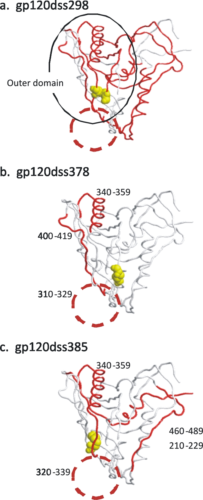
Sites of reduced epitope frequency in gp120 disulfide-bond variants compared to wild-type gp120, illustrated on the three-dimensional structure of gp120 (21). The view is of the side opposite the CD4 binding site. Disulfide bonds affected by the mutations are illustrated as space-filled atoms. Peptides that stimulated lower-frequency T-cell responses are highlighted in red. The V3 loop is illustrated as a red circle. (a) gp120dss298; (b) gp120dss378; (c) gp120dss385.
The T-cell response to gp120dss378 was shifted from the outer domain to the inner domain of gp120 (Fig. 6). This was primarily due to a significant reduction in the frequency of responses to three peptides in the outer domain: peptides 29 (residues 320 to 329) and 32 (residues 340 to 359), in the region of V3, and peptide 38 (residues 400 to 419), in the V4 loop (Fig. 7b). These three peptides were among the most immunogenic peptides for the group of mice immunized with wild-type protein. The V3 loop is in close proximity to the affected disulfide bond in the three-dimensional structure. We concluded that deletion of the C378-C441 disulfide bond destabilized the structure in this region of the protein. This destabilization increased the sensitivity of this region to proteolysis and reduced the presentation of the included epitopes to helper T cells.
The T-cell response to gp120dss385 produced a wild-type immunodominance pattern, although the overall frequency of response to this variant was reduced in comparison to the response to wild-type gp120 (Fig. 6). The frequencies of responses to five peptides were significantly reduced in comparison to those in wild-type-immunized mice (Fig. 7c). Of these peptides, four were immunodominant in the wild-type-immunized group, namely, peptides 19, 30, 32, and 44 (where immunodominant is defined as producing a response in a majority of immunized mice). We propose that this disulfide deletion caused a global destabilization of the protein structure (albeit less severe than that in gp120dss298), and the outcome was a shift of the protein's fate from presentation toward proteolytic destruction.
Serum antibody titers against wild-type gp120.
Serum antibody titers for reaction with wild-type gp120 protein were measured for each mouse by ELISA (Fig. 8a). The mean titer for sera raised against gp120dss378 was significantly greater than the mean titer for sera raised against wild-type gp120. Since the dependence of B-cell development on T-cell help has been well documented (8, 28, 29, 31, 32), the simultaneous decrease in helper T cells and increase in antibody were unexpected. There are at least three possible explanations for this observation. The first is that deletion of the disulfide bonds in the variant proteins exposed B-cell epitopes that are inaccessible in the wild-type protein. This hypothesis is supported by an increase in the antibody response to linear epitopes in mice immunized with the variant proteins (Fig. 9). A second possibility is that some helper T cells indirectly hinder B cells that produce gp120-specific antibody by providing help to polyclonal, non-antigen-specific B cells. This phenomenon was previously observed for lymphocytic choriomeningitis virus (LCMV) infection in mice (16, 25, 37). Recher et al. showed that partial depletion of CD4+ helper T cells resulted in an increase in anti-LCMV antibody (37). These “unproductive” CD4+ helper T cells may provide help to CTLs instead of B cells. A study by Kleen et al., using a similar peptide library, showed that the helper T-cell epitope map for humans infected with HIV correlates well with the CTL epitope map measured by the production of granzyme B (18). The third possible explanation for the increase of antigen-specific antibody in the context of a decreased helper T-cell response is that the increased antibody is due to helper-T-cell-independent B-cell responses, although we believe this to be highly unlikely, since all the antibodies tested were of the IgG isotype. Further studies will be necessary to distinguish which of these mechanisms is responsible for the increased antibody observed in the context of reduced T-cell responses.
FIG. 8.

Serum reactivity with gp120 (a) and serum inhibition of CD4 binding to gp120 (b) following immunization with wild-type or disulfide-bond-variant gp120. The mean value for sera from mice immunized with the indicated protein is indicated by a horizontal line. Binding of mouse antibodies was detected with HRP-conjugated goat anti-mouse IgG. Binding of sCD4 was detected as described in the legend for Fig. 4.
FIG. 9.

Profiles of serum reactivity with gp120 peptides following immunization with wild-type or disulfide-bond-variant gp120. Reactions achieved higher frequencies and involved more peptides with sera from mice immunized with the disulfide-bond variants.
Determination of serum antibody blocking of CD4 binding to wild-type gp120.
Antibodies that prevent the binding of CD4 to gp120 could potentially provide protection against HIV infection. In order to determine if immunization with the disulfide-bond variants altered the amounts of these antibodies in the serum, we performed a CD4 competition assay. Antibodies raised against gp120dss378 were able to block CD4 binding to wild-type gp120 (Fig. 8b). The inhibitory effect was seen at 1:100 and 1:400 dilutions of serum (mean inhibition equal to 17% and 14%, respectively). CD4 blocking was not observed for sera from mice immunized with any of the other variants or wild-type gp120. The result with gp120dss378 is the first demonstration, to our knowledge, of the production of significant amounts of CD4-blocking activity following immunization of mice with a protein antigen.
In light of the recent study showing that deletion of the C378-C441 disulfide bond did not impede protein folding, protein shedding, or viral infectivity (47), it seems possible that this disulfide bond is conserved in order to misdirect the immune response. The results presented here suggest that this disulfide bond stabilizes the bridging sheet, which not only favors CD4 binding but also prevents induction of CD4-blocking antibodies. While it is premature to conclude that deletion of the C378-C441 disulfide bond would increase the protectiveness of a vaccine, further studies with other animal models and using envelope glycoproteins from other HIV strains are warranted.
Supplementary Material
Acknowledgments
This work was supported by Public Health Service grant AI-080367 from the National Institute of Allergy and Infectious Diseases and by a cooperative pilot research grant from the Louisiana Vaccine Center.
Footnotes
Published ahead of print on 20 January 2010.
Supplemental material for this article may be found at http://jvi.asm.org/.
REFERENCES
- 1.Bevan, M. J. 2004. Helping the CD8(+) T-cell response. Nat. Rev. Immunol. 4:595-602. [DOI] [PubMed] [Google Scholar]
- 2.Borrow, P., H. Lewicki, B. H. Hahn, G. M. Shaw, and M. B. Oldstone. 1994. Virus-specific CD8+ cytotoxic T-lymphocyte activity associated with control of viremia in primary human immunodeficiency virus type 1 infection. J. Virol. 68:6103-6110. [DOI] [PMC free article] [PubMed] [Google Scholar]
- 3.Brown, S. A., T. D. Lockey, C. Slaughter, K. S. Slobod, S. Surman, A. Zirkel, A. Mishra, V. R. Pagala, C. Coleclough, P. C. Doherty, and J. L. Hurwitz. 2005. T cell epitope “hotspots” on the HIV type 1 gp120 envelope protein overlap with tryptic fragments displayed by mass spectrometry. AIDS Res. Hum. Retrovir. 21:165-170. [DOI] [PubMed] [Google Scholar]
- 4.Brown, S. A., J. Stambas, X. Zhan, K. S. Slobod, C. Coleclough, A. Zirkel, S. Surman, S. W. White, P. C. Doherty, and J. L. Hurwitz. 2003. Clustering of Th cell epitopes on exposed regions of HIV envelope despite defects in antibody activity. J. Immunol. 171:4140-4148. [DOI] [PubMed] [Google Scholar]
- 5.Carmicle, S., G. Dai, N. K. Steede, and S. J. Landry. 2002. Proteolytic sensitivity and helper T-cell epitope immunodominance associated with the mobile loop in Hsp10s. J. Biol. Chem. 277:155-160. [DOI] [PubMed] [Google Scholar]
- 6.Carmicle, S., N. K. Steede, and S. J. Landry. 2006. Antigen three-dimensional structure guides the processing and presentation of helper T-cell epitopes. Mol. Immunol. 44:1159-1168. [DOI] [PubMed] [Google Scholar]
- 7.Castellino, F., F. Zappacosta, J. E. Coligan, and R. N. Germain. 1998. Large protein fragments as substrates for endocytic antigen capture by MHC class II molecules. J. Immunol. 161:4048-4057. [PubMed] [Google Scholar]
- 8.Claman, H. N., E. A. Chaperon, and R. F. Triplett. 1966. Thymus-marrow cell combinations. Synergism in antibody production. Proc. Soc. Exp. Biol. Med. 122:1167-1171. [DOI] [PubMed] [Google Scholar]
- 9.Dai, G., S. Carmicle, N. K. Steede, and S. J. Landry. 2002. Structural basis for helper T-cell and antibody epitope immunodominance in bacteriophage T4 Hsp10: role of disordered loops. J. Biol. Chem. 277:161-168. [DOI] [PubMed] [Google Scholar]
- 10.Dai, G., N. K. Steede, and S. J. Landry. 2001. Allocation of helper T-cell epitope immunodominance according to three-dimensional structure in the human immunodeficiency virus type I envelope glycoprotein gp120. J. Biol. Chem. 276:41913-41920. [DOI] [PubMed] [Google Scholar]
- 11.Fontana, A., P. P. de Laureto, B. Spolaore, E. Frare, P. Picotti, and M. Zambonin. 2004. Probing protein structure by limited proteolysis. Acta Biochim. Pol. 51:299-321. [PubMed] [Google Scholar]
- 12.Fontana, A., G. Fassina, C. Vita, D. Dalzoppo, M. Zamai, and M. Zambonin. 1986. Correlation between sites of limited proteolysis and segmental mobility in thermolysin. Biochemistry 25:1847-1851. [DOI] [PubMed] [Google Scholar]
- 13.Hastings, K. T., R. L. Lackman, and P. Cresswell. 2006. Functional requirements for the lysosomal thiol reductase GILT in MHC class II-restricted antigen processing. J. Immunol. 177:8569-8577. [DOI] [PubMed] [Google Scholar]
- 14.Hubbard, S. J., R. J. Beynon, and J. M. Thornton. 1998. Assessment of conformational parameters as predictors of limited proteolysis sites in native protein structures. Protein Eng. 11:349-359. [DOI] [PubMed] [Google Scholar]
- 15.Hubbard, S. J., F. Eisenmenger, and J. M. Thornton. 1994. Modeling studies of the change in conformation required for cleavage of limited proteolytic sites. Protein Sci. 3:757-768. [DOI] [PMC free article] [PubMed] [Google Scholar]
- 16.Hunziker, L., M. Recher, A. J. Macpherson, A. Ciurea, S. Freigang, H. Hengartner, and R. M. Zinkernagel. 2003. Hypergammaglobulinemia and autoantibody induction mechanisms in viral infections. Nat. Immunol. 4:343-349. [DOI] [PubMed] [Google Scholar]
- 17.Kent, S. J., G. L. Ada, E. Hayes, and I. M. Lewis. 2001. Determining the immune mechanisms of protection from AIDS: correlates of immunity and the development of syngeneic macaques. Immunol. Rev. 183:94-108. [DOI] [PubMed] [Google Scholar]
- 18.Kleen, T. O., R. Asaad, S. J. Landry, B. O. Boehm, and M. Tary-Lehmann. 2004. Tc1 effector diversity shows dissociated expression of granzyme B and interferon-gamma in HIV infection. AIDS 18:383-392. [DOI] [PubMed] [Google Scholar]
- 19.Koup, R. A., J. T. Safrit, Y. Cao, C. A. Andrews, G. McLeod, W. Borkowsky, C. Farthing, and D. D. Ho. 1994. Temporal association of cellular immune responses with the initial control of viremia in primary human immunodeficiency virus type 1 syndrome. J. Virol. 68:4650-4655. [DOI] [PMC free article] [PubMed] [Google Scholar]
- 20.Kuroda, M. J., J. E. Schmitz, W. A. Charini, C. E. Nickerson, M. A. Lifton, C. I. Lord, M. A. Forman, and N. L. Letvin. 1999. Emergence of CTL coincides with clearance of virus during primary simian immunodeficiency virus infection in rhesus monkeys. J. Immunol. 162:5127-5133. [PubMed] [Google Scholar]
- 21.Kwong, P. D., R. Wyatt, S. Majeed, J. Robinson, R. W. Sweet, J. Sodroski, and W. A. Hendrickson. 2000. Structures of HIV-1 gp120 envelope glycoproteins from laboratory-adapted and primary isolates. Structure 8:1329-1339. [DOI] [PubMed] [Google Scholar]
- 22.Kwong, P. D., R. Wyatt, J. Robinson, R. W. Sweet, J. Sodroski, and W. A. Hendrickson. 1998. Structure of an HIV gp120 envelope glycoprotein in complex with the CD4 receptor and a neutralizing human antibody. Nature 393:648-659. [DOI] [PMC free article] [PubMed] [Google Scholar]
- 23.Landry, S. J. 1997. Local protein instability predictive of helper T-cell epitopes. Immunol. Today 18:527-532. [DOI] [PubMed] [Google Scholar]
- 24.Landry, S. J. 2008. Three-dimensional structure determines the pattern of CD4+ T-cell epitope dominance in influenza virus hemagglutinin. J. Virol. 82:1238-1248. [DOI] [PMC free article] [PubMed] [Google Scholar]
- 25.Lang, K. S., A. N. Hegazy, P. A. Lang, B. Eschli, M. Lohning, H. Hengartner, R. M. Zinkernagel, and M. Recher. 2007. “Negative vaccination” by specific CD4 T cell tolerisation enhances virus-specific protective antibody responses. PLoS One 2:e1162. [DOI] [PMC free article] [PubMed] [Google Scholar]
- 26.Li, P., M. A. Haque, and J. S. Blum. 2002. Role of disulfide bonds in regulating antigen processing and epitope selection. J. Immunol. 169:2444-2450. [DOI] [PubMed] [Google Scholar]
- 27.McHeyzer-Williams, M., L. McHeyzer-Williams, J. Panus, R. Pogue-Caley, G. Bikah, D. Driver, and M. Eisenbraun. 2003. Helper T-cell-regulated B-cell immunity. Microbes Infect. 5:205-212. [DOI] [PubMed] [Google Scholar]
- 28.Miller, J. F., P. M. De Burgh, and G. A. Grant. 1965. Thymus and the production of antibody-plaque-forming cells. Nature 208:1332-1334. [DOI] [PubMed] [Google Scholar]
- 29.Miller, J. F., and G. F. Mitchell. 1968. Cell to cell interaction in the immune response. I. Hemolysin-forming cells in neonatally thymectomized mice reconstituted with thymus or thoracic duct lymphocytes. J. Exp. Med. 128:801-820. [DOI] [PMC free article] [PubMed] [Google Scholar]
- 30.Mirano-Bascos, D., M. Tary-Lehmann, and S. J. Landry. 2008. Antigen structure influences helper T-cell epitope dominance in the human immune response to HIV envelope glycoprotein gp120. Eur. J. Immunol. 38:1231-1237. [DOI] [PMC free article] [PubMed] [Google Scholar]
- 31.Mitchell, G. F., and J. F. Miller. 1968. Cell to cell interaction in the immune response. II. The source of hemolysin-forming cells in irradiated mice given bone marrow and thymus or thoracic duct lymphocytes. J. Exp. Med. 128:821-837. [DOI] [PMC free article] [PubMed] [Google Scholar]
- 32.Mitchell, G. F., and J. F. Miller. 1968. Immunological activity of thymus and thoracic-duct lymphocytes. Proc. Natl. Acad. Sci. USA 59:296-303. [DOI] [PMC free article] [PubMed] [Google Scholar]
- 33.Moore, J. P., and J. Sodroski. 1996. Antibody cross-competition analysis of the human immunodeficiency virus type 1 gp120 exterior envelope glycoprotein. J. Virol. 70:1863-1872. [DOI] [PMC free article] [PubMed] [Google Scholar]
- 34.Pollard, S. R., W. Meier, P. Chow, J. J. Rosa, and D. C. Wiley. 1991. CD4-binding regions of human immunodeficiency virus envelope glycoprotein gp120 defined by proteolytic digestion. Proc. Natl. Acad. Sci. USA 88:11320-11324. [DOI] [PMC free article] [PubMed] [Google Scholar]
- 35.Polverino de Laureto, P., E. Scaramella, V. De Filippis, M. Bruix, M. Rico, and A. Fontana. 1997. Limited proteolysis of ribonuclease A with thermolysin in trifluoroethanol. Protein Sci. 6:860-872. [DOI] [PMC free article] [PubMed] [Google Scholar]
- 36.Rasmussen, R. A., R. Hofmann-Lehmann, P. L. Li, J. Vlasak, J. E. Schmitz, K. A. Reimann, M. J. Kuroda, N. L. Letvin, D. C. Montefiori, H. M. McClure, and R. M. Ruprecht. 2002. Neutralizing antibodies as a potential secondary protective mechanism during chronic SHIV infection in CD8+ T-cell-depleted macaques. AIDS 16:829-838. [DOI] [PubMed] [Google Scholar]
- 37.Recher, M., K. S. Lang, L. Hunziker, S. Freigang, B. Eschli, N. L. Harris, A. Navarini, B. M. Senn, K. Fink, M. Lotscher, L. Hangartner, R. Zellweger, M. Hersberger, A. Theocharides, H. Hengartner, and R. M. Zinkernagel. 2004. Deliberate removal of T cell help improves virus-neutralizing antibody production. Nat. Immunol. 5:934-942. [DOI] [PubMed] [Google Scholar]
- 38.Rinaldo, C., X. L. Huang, Z. F. Fan, M. Ding, L. Beltz, A. Logar, D. Panicali, G. Mazzara, J. Liebmann, M. Cottrill, et al. 1995. High levels of anti-human immunodeficiency virus type 1 (HIV-1) memory cytotoxic T-lymphocyte activity and low viral load are associated with lack of disease in HIV-1-infected long-term nonprogressors. J. Virol. 69:5838-5842. [DOI] [PMC free article] [PubMed] [Google Scholar]
- 39.Safrit, J. T., and R. A. Koup. 1995. The immunology of primary HIV infection: which immune responses control HIV replication? Curr. Opin. Immunol. 7:456-461. [DOI] [PubMed] [Google Scholar]
- 40.Schmitz, J. E., M. J. Kuroda, S. Santra, V. G. Sasseville, M. A. Simon, M. A. Lifton, P. Racz, K. Tenner-Racz, M. Dalesandro, B. J. Scallon, J. Ghrayeb, M. A. Forman, D. C. Montefiori, E. P. Rieber, N. L. Letvin, and K. A. Reimann. 1999. Control of viremia in simian immunodeficiency virus infection by CD8+ lymphocytes. Science 283:857-860. [DOI] [PubMed] [Google Scholar]
- 41.Schmitz, J. E., M. J. Kuroda, S. Santra, M. A. Simon, M. A. Lifton, W. Lin, R. Khunkhun, M. Piatak, J. D. Lifson, G. Grosschupff, R. S. Gelman, P. Racz, K. Tenner-Racz, K. A. Mansfield, N. L. Letvin, D. C. Montefiori, and K. A. Reimann. 2003. Effect of humoral immune responses on controlling viremia during primary infection of rhesus monkeys with simian immunodeficiency virus. J. Virol. 77:2165-2173. [DOI] [PMC free article] [PubMed] [Google Scholar]
- 42.Sun, Y., J. E. Schmitz, A. P. Buzby, B. R. Barker, S. S. Rao, L. Xu, Z. Y. Yang, J. R. Mascola, G. J. Nabel, and N. L. Letvin. 2006. Virus-specific cellular immune correlates of survival in vaccinated monkeys after simian immunodeficiency virus challenge. J. Virol. 80:10950-10956. [DOI] [PMC free article] [PubMed] [Google Scholar]
- 43.Surman, S., T. D. Lockey, K. S. Slobod, B. Jones, J. M. Riberdy, S. W. White, P. C. Doherty, and J. L. Hurwitz. 2001. Localization of CD4+ T cell epitope hotspots to exposed strands of HIV envelope glycoprotein suggests structural influences on antigen processing. Proc. Natl. Acad. Sci. USA 98:4587-4592. [DOI] [PMC free article] [PubMed] [Google Scholar]
- 44.Thali, M., C. Furman, D. D. Ho, J. Robinson, S. Tilley, A. Pinter, and J. Sodroski. 1992. Discontinuous, conserved neutralization epitopes overlapping the CD4-binding region of human immunodeficiency virus type 1 gp120 envelope glycoprotein. J. Virol. 66:5635-5641. [DOI] [PMC free article] [PubMed] [Google Scholar]
- 45.Thali, M., J. P. Moore, C. Furman, M. Charles, D. D. Ho, J. Robinson, and J. Sodroski. 1993. Characterization of conserved human immunodeficiency virus type 1 gp120 neutralization epitopes exposed upon gp120-CD4 binding. J. Virol. 67:3978-3988. [DOI] [PMC free article] [PubMed] [Google Scholar]
- 46.Tuen, M., M. L. Visciano, P. C. Chien, Jr., S. Cohen, P. D. Chen, J. Robinson, Y. He, A. Pinter, M. K. Gorny, and C. E. Hioe. 2005. Characterization of antibodies that inhibit HIV gp120 antigen processing and presentation. Eur. J. Immunol. 35:2541-2551. [DOI] [PubMed] [Google Scholar]
- 47.van Anken, E., R. W. Sanders, I. M. Liscaljet, A. Land, I. Bontjer, S. Tillemans, A. A. Nabatov, W. A. Paxton, B. Berkhout, and I. Braakman. 2008. Only five of 10 strictly conserved disulfide bonds are essential for folding and eight for function of the HIV-1 envelope glycoprotein. Mol. Biol. Cell 19:4298-4309. [DOI] [PMC free article] [PubMed] [Google Scholar]
- 48.Williams, M. A., B. J. Holmes, J. C. Sun, and M. J. Bevan. 2006. Developing and maintaining protective CD8+ memory T cells. Immunol. Rev. 211:146-153. [DOI] [PubMed] [Google Scholar]
- 49.Xiang, S. H., L. Wang, M. Abreu, C. C. Huang, P. D. Kwong, E. Rosenberg, J. E. Robinson, and J. Sodroski. 2003. Epitope mapping and characterization of a novel CD4-induced human monoclonal antibody capable of neutralizing primary HIV-1 strains. Virology 315:124-134. [DOI] [PubMed] [Google Scholar]
- 50.Yeh, J. C., J. R. Seals, C. I. Murphy, H. van Halbeek, and R. D. Cummings. 1993. Site-specific N-glycosylation and oligosaccharide structures of recombinant HIV-1 gp120 derived from a baculovirus expression system. Biochemistry 32:11087-11099. [DOI] [PubMed] [Google Scholar]
Associated Data
This section collects any data citations, data availability statements, or supplementary materials included in this article.