Abstract
We reported that complement cascade (CC) becomes activated in bone marrow (BM) during mobilization of hematopoietic stem/progenitor cells (HSPCs) induced by granulocyte-colony stimulating factor (G-CSF) and C5 cleavage plays an important role in optimal egress of HSPCs. In the current work, we explored whether CC is involved in mobilization of HSPCs induced by the CXCR4 antagonist, AMD3100. To address this question, we performed mobilization studies in mice that display a defect in the activation of the proximal steps of CC (Rag−/−, SCID, C2.Cfb−/−) as well as in mice that do not activate the distal steps of CC (C5−/−). We noticed that proximal CC activation-deficient mice (above C5 level), in contrast to distal step CC activation-deficient C5−/− ones mobilize normally in response to AMD3100 administration. We hypothesized that this discrepancy in mobilization could be explained by AMD3100 activating C5 in Rag−/−, SCID, C2.Cfb−/− animals in a non-canonical mechanism involving activated granulocytes. To support this granulocytes i) as first egress from BM and ii) secrete several proteases that cleave/activate C5 in response to AMD3100. We conclude that AMD3100-directed mobilization of HSPCs, similarly to G-CSF-induced mobilization, depends on activation of CC; however, in contrast to G-CSF, AMD3100 activates the distal steps of CC directly at the C5 level. Overall, these data support that C5 cleavage fragments and distal steps of CC activation are required for optimal mobilization of HSPCs.
Keywords: AMD3100, Complement, CXCR4, C5
Introduction
Hematopoietic stem/progenitor cells (HSPCs) circulate in the peripheral blood (PB) under steady state conditions at very low levels and their number increases in emergency situations such as infection and/or tissue damage.1–3 HSPCs could be also mobilized from bone marrow (BM) into PB after administration of some cytokines,4–7 growth factors,8–11 chemokines,12–14 and pharmacological agents.15–18 The cytokine granulocyte colony-stimulating factor (G-CSF) is currently the most frequently employed clinical drug that may efficiently mobilize HSPCs after a few consecutive daily injections. Some level of mobilization has also been achieved within one hour in experimental animals after injection of polysaccharide zymosan.19–21
Generally, all mobilizing agents induce a proteolytic environment in BM tissue.22–25 However, the molecular mechanisms controlling egress of HSPCs from BM into PB are still not well understood. Nevertheless, it is widely accepted that what is crucial for the BM egress of HSPCs is the attenuation of the stromal-derived growth factor-1 (SDF-1)-CXCR4 interaction between BM-secreted SDF-1 and HSPC-expressed CXCR4 and the adhesive interaction between Very Late Antigen-4 (VLA-4; α1β4 integrin) expressed on HSPCs and its ligand Vascular Adhesion Molecule-1 (VCAM-1; CD106), which is expressed in the BM microenvironment.26,27 Nevertheless, a significant number of patients, particularly those pretreated by chemotherapy, are resistant to G-CSF mobilization.28 This explains why new pro-mobilizing compounds are tested as being employed alone or in combination with G-CSF. One such compound is bicyclam AMD3100, which blocks the interaction between CXCR4 and SDF-1.29–31
On the other hand, augmenting evidence demonstrates that HSPC mobilization is regulated by elements of innate immunity, in particular by complement cascade (CC) protein cleavage fragments,32–37 neutrophils,38–41 and Toll receptors (TRs)42 that all play a pivotal and, until recently, underappreciated role in this process. Accordingly, we reported that CC becomes activated in BM during mobilization of HSPCs by the immunoglobulin (Ig)-dependent pathway and/or by the alternative Ig-independent pathway as seen, for example, during G-CSF- or zymosan-induced mobilization, respectively.33,34 To support this notion, we found that: i) Nonobese diabetic/severe combined immune deficient (NOD/SCID) and Rag−/− animals that do not activate the Ig-dependent CC classical pathway37; ii) C2 and Factor B-deficient (C2.Cfb−/−) mice that do not activate the classical and alternative CC pathways3; and iii) C5−/− mice that do not activate the distal pathway of CC are all poor G-CSF- and/or zymosan mobilizers.40,41
Moreover, our studies in C5-deficient mice revealed that C5 cleavage fragments (C5a and desArgC5a) are crucial for the egress of HSPCs and we postulated three levels at which they affect this process.41 First, stimulation of granulocytes directly in the BM microenvironment by C5a and desArgC5a enhances secretion of proteolytic enzymes, which perturb HSPCs retention signals (e.g., SDF-1-CXCR4 and VLA-4-VCAM-1 interactions). Second, the plasma desArgC5a strongly chemoattracts granulocytes. These granulocytes migrating from BM into PB highly express metalloproteinases (MPs) and “pave the way” through the endothelial barrier for HSPCs, which follow granulocytes (Ice Breaker Phenomenon). Finally, after egress from BM, granulocytes are stimulated in BM vessels by C5a and release several cationic peptides (e.g., cathelicidin and β2-defensin), some of which enhance/prime responsiveness of HSPCs to plasma SDF-1.41
In this study, we were interested in the novel mobilizing agent AMD3100. Because the primary target of this compound is CXCR4 expressed on BM-residing cells, we asked whether mobilization of HSPCs by this compound involves activation of CC as well. We found that AMD3100-directed mobilization, similarly to G-CSF-induced mobilization, depends on activation of CC; however, in contrast to G-CSF, AMD3100 activates CC at the distal stages directly at the C5 level by proteases released from activated granulocytes. Thus, our data further support the concept that C5 cleavage is crucial for optimal egress of HSPCs from BM.
Material and Methods
Animals
Pathogen-free, 4- to 6-week-old C5−/−, C5+/+, and Rag−/− mice were purchased from the Jackson Laboratory (Bar Harbor, ME; http://www.jax.org). C57BL/6, Balb/c, and SCID mice were purchased from the National Cancer Institute (Frederick, MD; http://www.cancer.gov). C2.Cfb−/− mice were a kind gift from Dr. M. Botto (Imperial College, London, UK). All mice were adopted by at least 2 weeks and used for experiments at age 6 to 8 weeks. Animal studies were approved by the Animal Care and Use Committee of the University of Louisville (Louisville, KY).
Mobilization
Mice were injected subcutaneously (s.c.) with 250 μg/kg of human G-CSF (Amgen, Thousand Oaks, CA; http://www.amgen.com) daily for 6 days. For zymosan mobilization, mice were injected intravenously (i.v.) with 0.5 mg/mouse. For AMD3100 mobilization, animals received this compound at a dose of 5 mg/kg s.c. At 6 h after the last G-CSF administration or at 1 h after zymosan and AMD3100 injection, mice were bled from the retro-orbital plexus for complete blood count (CBC) and 450 μl of PB was obtained from the vena cava with a 25-gauge needle and 1 ml syringe containing 50 μl of 100 mM ethylenediaminetetraacetic acid (EDTA).
CBC counts
Fifty microliters of PB was taken from the retro-orbital plexus of the mice and collected into microvette EDTA-coated tubes (Sarstedt Inc., Newton, NC; http://www.sarstedt.com/php/main.php). Samples were run within 2 h of collection on a HemaVet 950 (Drew Scientific Inc., Oxford, CT; http://www.drew-scientific.com).
Colony forming unit-granulocytes/macrophage (CFU-GM) assay
Red blood cells (RBCs) were lysed with BD Pharm Lyse buffer (BD Biosciences, San Jose, CA; http://www.bdbiosciences.com). Nucleated cells were subsequently washed twice and used for CFU-GM colonies as described elsewhere. Briefly, cells were resuspended in human methylcellulose base media provided by the manufacturer (R&D Systems, Inc., Minneapolis, MN; http://www.rndsystems.com) supplemented with 25 ng/ml recombinant murine granulocyte macrophage colony-stimulating factor (mGM-CSF) and 10 ng/ml recombinant murine interleukin-3 (mIL-3; Millipore, Billerica, MA; http://www.millipore.com). Cultures were incubated for 7 days, at which time they were scored for the number of CFU-GM colonies under an inverted microscope.
Evaluation of HSPC mobilization
The following formula was used for evaluation of circulating CFU-GM and Sca-1+/c-Kit+/Lin−/CD34− (SKL CD34−) cells: {[number of white blood cells (WBCs) x number of CFU-GM colonies]/number of WBCs plated = number of CFU-GM per microliter of PB} and {(number of WBCs x number of SKL CD34− cells)/number of gated WBCs = number of SKL cells per microliter of PB}, respectively.
Bone marrow nucleated cells (BMNCs)
BMNCs were prepared by flushing femurs and tibias of pathogen-free, 6- to 8-week-old mice without enzymatic digestion. BMNCs were lysed with BD Pharm Lyse buffer (BD Biosciences) to remove RBCs, washed, and resuspended in appropriate media for further analysis.
Fluorescence-activated cell sorting (FACS) analysis
BMNC staining was performed in medium containing 2% fetal bovine serum (FBS). All monoclonal antibodies (mAbs) were added at saturating concentrations and the cells were incubated for 30 min on ice, washed twice, resuspended in staining solution at a concentration of 5×10⁶ cells/ml, and subjected to analysis using an LSR II (Becton Dickinson, Mountainview, CA). The following anti-mouse Abs were used to detect fluorescein isothiocyanate (FITC)-anti-CD117 (c-Kit) (clone 2B8; BioLegend, San Diego, CA) and Phycoerythrin (PE)-Cy5 anti-mouse Ly-6A/E (Sca-1) (clone D7; eBioscience™, San Diego, CA). All anti-mouse lineage markers (Lin) were conjugated by PE and purchased from BD Biosciences: anti-CD45R/B220 (clone RA3-6B2); anti-Gr-1 (clone RB6-8C5); anti-t-cell receptor β (TCRβ; clone H57-597); anti-TCRγδ (clone GL3); anti-CD11b (clone M1/70); anti-Ter-119 (clone TER-119); and anti-CD34 (clone RAM34).
Sorting of BMNCs
Granulocytes (Gr-1+) were purified as described elsewhere. Briefly, BMNCs (1×10⁸ cells/ml) were resuspended in Roswell Park Memorial Institute (RPMI) medium containing 2% heat-inactivated FBS (GIBCO, Grand Island, NY), 10 mM N-2-hydroxyethylpiperazine-N′-2-ethanesulfonic acid (HEPES) buffer (GIBCO), and antibiotics (Mediatech Inc., Manassas, VA; http://www.cellgro.com). Next, BMNCs were incubated with saturating concentrations of directly labeled mAbs for 30 min on ice, washed twice, and sorted by multiparameter, live sterile cell sorting (MoFlo; Dako A/S, Fort Collins, CO).
Zymography
To evaluate matrix (M)MP-9 secretion by Gr-1+ cells, 2×10⁶ cells/ml were incubated (for 24 h at 37°C in 5% CO2) in the absence or presence of AMD3100 (1μM) and cell-conditioned media were analyzed immediately by zymography as described earlier by us.43
Phosphorylation of intracellular pathway proteins
Gr-1+ cells (5×10⁶/ml) were stimulated with 1 μg/ml of AMD3100 for 5, 10, and 15 min at 37°C and then lysed for 10 min on ice in M-Per lysing buffer (Pierce, Rockford, IL) containing protease and phosphatase inhibitors (Sigma, St. Louis, MO). The extracted proteins were separated and analyzed for phosphorylation of 44/42 mitogen-activating protein kinase (MAPK). Equal loading in the lanes was evaluated by stripping the blots and reprobing them with the appropriate mAb or polyclonal (p)Ab p42/44 anti-MAPK Ab clone 9102 (New England Biolabs, Ipswich, MA). The membranes were developed with an enhanced chemiluminescent (ECL) reagent (Amersham Life Sciences, Arlington Heights, IL), dried, and exposed to film (HyperFilm; Amersham).
Real-time quantitative polymerase chain reaction to evaluate the expression of proteolytic enzymes
Total mRNA was isolated using RNeasy Mini Kit (Qiagen Inc., Valencia, CA) from each 20,000 sorted cell populations. Messenger RNA was reverse-transcribed with 500U of Moloney murine leukemia virus reverse transcriptase (MoMLV-RT). The resulting cDNA fragments were amplified using 5 U of Thermus aquaticus (Taq) polymerase. Primer sequences for β-2 microglobulin (β2M) were forward primer: 5′-CAT ACG CCT GCA GAG TTA AGC-3′ and reverse primer: 5′-GAT CAC ATG TCT CGA TCC VAG TAG-3′. For MMP-2, primer sequences were forward primer: 5′-GGA CCC CGG TTT CCC TAA G-3′ and reverse primer: 5′-GTC CAC GAC GGC ATC CA-3′. For MMP-9, primer sequences were forward primer: 5′-CCT GGA ACT CAC ACG ACA TCT TC-3′ and reverse primer: 5′-TGG AAA CTC ACA CGC CAG AA-3′. For MMP-9, primer sequences were forward primer: 5′-CCT GGA ACT CAC ACG ACA TCT TC-3′ and reverse primer: 5′-TGG AAA CTC ACA CGC CAG AA-3′. For cathepsin G (CG), primer sequences were forward primer: 5′-CCA TCC GCC ATC CTG ATT AC-3′ and reverse primer: 5′-CCT CAG CTG CAG AAG CAT GA-3′. For neutrophil elastase (NE), primer sequences were forward primer: 5′-GGA TCT TCG AGA ATG GCT TTG A-3′ and reverse primer: 5′-TGG TAG CGG AGC CAT TGA G-3′. The relative quantitation value of the target, normalized to an endogenous control gene (β2M) and relative to a calibrator, is expressed as 2−ΔΔCt (fold difference), where ΔCt equals the Ct of the target genes minus the Ct of the endogenous control gene ((β2M), and ΔΔCt equals the ΔCt of the samples for the target gene minus the ΔCt of the calibrator for the target gene. To avoid the possibility of amplifying the contamination of DNA, a uniform amplification of the products was rechecked by analyzing the melting curves of the amplified products (dissociation graphs), the melting temperature (Tm) was 57–60°C, the product Tm was at least 10°C higher than the primer Tm.
Enzyme-Linked Immunosorbent Assay (ELISA) for complement cleavage fragment detection
Whole blood from C57BL/6 mice was obtained from the vena cava (1 ml syringe containing 50 μl of 100 mM EDTA) at various time points after PBS, AMD3100, or zymosan treatment. Plasma samples were prepared by taking the top fraction after centrifugation at 1,000xg for 10 min at 4°C. Some normal plasma from C57BL/6 mice were stimulated with AMD3100 in the presence or absence of purified Gr-1+ cells (105 cells) at 37°C for up to 30 min. Samples were stored at −80°C until use. All Abs, standard proteins, enzyme reagents, and substrate solutions were purchased from BD Biosciences. Assays were performed according to general instructions. Briefly, plasma samples were incubated in triplicate wells of microtiter plates, which had been pre-coated with antigen-specific capture Abs (C3a or C5a). Repeated washing and aspiration were conducted before each incubation step to remove unbound materials from the assay plates. This step was followed by incubation with specific biotin-labeled capture Abs and an enzyme reagent. Substrate solution was added to the wells of the microtiter plates and color developed in proportion to the amount of antigen bound in the initial step of the individual assay. The color development was stopped with the addition of an acid solution and the intensity of color was read using a microtiter plate spectrophotometer. The concentrations of complement C3a or C5a in plasma samples were determined by interpolation from individual standard curves composed of purified human C3a or C5a. Purified rat anti-mouse-C3a (clone I87-1162) or -C5a (clone I52-1486) Abs was used as the capture Ab, respectively (2 μg/ml, overnight at 4°C). Biotin-labeled rat anti-mouse-C3a (clone I87-419) or -C5a (clone I52-278) Abs was used as the detection Ab (2 μg/ml, 2 h at room temperature). Plasma samples were used at a 1:20 or 1:10 dilution for C3a or C5a detection, respectively.
Statistical analysis
Arithmetic means and standard deviations were calculated using Instat 1.14 software (Graphpad, San Diego, CA). Statistical significance was defined as P<0.05. Data were analyzed using Student’s t-test for unpaired samples.
Results
AMD3100 restores proper mobilization in Rag−/−, SCID, and C2.Cfb−/− mice that do not activate the proximal steps of CC, but is less effective in C5−/− animals
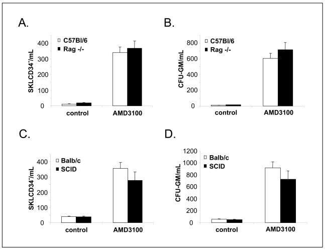
We reported that Ig-deficient mice that do not activate complement by the classical pathway are poor G-CSF-induced mobilizers.37 In a current study, we mobilized these mice by AMD3100 and to our surprise noticed that they mobilize a similar number of CD34−SKL− cells, CFU-GM progenitors, and leukocytes as their wild type littermates (Figure 1).
Figure 1. Normal AMD3100 mobilization of HSPCs in Ig-deficient mice.
Rag−/−, SCID, as well as age- and sex-matched control (C57BL/6 and Balb/c) mice were mobilized with AMD3100 (5 mg/kg s.c.) for 1 h. Number of circulating SKL CD34−/μL (A and C) and CFU-GM progenitors (B and D) per microliter of PB are shown. Data show representative results from three independent experiments with 6 animals/group.
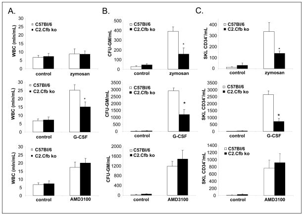
Next, we employed C2.Cfb−/− animals that possess defects at the upstream steps of CC activation by both classical (C2 deficiency) and alternative (factor B deficiency) pathways. As expected, we found that these mice are poor zymosan (Figure 2 upper panel) and G-CSF (middle panel) mobilizers; however, they are normal mobilizers after administration of AMD3100 (Figure 2 lower panel).
Figure 2. Impaired G-CSF- and zymosan- but normal AMD3100 mobilization of HSPCs in C2.Cfb-deficient mice.
C2.Cfb−/− mice as well as age- and sex-matched C57BL/6 mice were mobilized for 6 days with G-CSF (250 μg/kg s.c. per day; n=5 mice per group; upper panel) or 1 h with zymosan (0.5 mg/kg i.v.; n=5 mice per group; middle panel) or for 1 h with AMD3100 (5 mg/kg s.c.) n=5 mice per group (lower panel). Number of circulating leukocytes (A), CFU-GM progenitors (B), and SKL CD34− cells (C) per microliter of PB are shown. * P<.05 as compared with C5+/+ mice. Data show representative results from three independent experiments.
Finally, we performed mobilization in C5−/− animals that are not able to generate the distal components of CC activation, i.e., C5a and desArgC5a. In our previous work, we reported that these animals are poor G-CSF- and zymosan-induced mobilizers, which points to C5 cleavage fragments as being crucial for optimal HSPC egress.40,41 However, we report here that these animals are poor AMD3100 mobilizers (Figure 3), which is in contrast to mice with defects in activation of the proximal steps of CC activation. This indicates, C5 cleavage fragments are crucial for the optimal egress of HSPCs. We hypothesized that normal AMD3100 mobilization in animals with defects in activation of the proximal steps of CC suggests that C5 is activated/cleaved in these animals by some alternative mechanism.
Figure 3. Impaired AMD3100 mobilization of HSPCs in C5-deficient mice.
C5−/− as well as age- and sex-matched C5+/+ mice were mobilized with AMD3100 (5 mg/kg s.c.) for 1 h. Number of circulating leukocytes (A), CFU-GM progenitors (B), and SKL cells (C) per microliter of PB are shown. * P<.05 as compared with C5+/+ mice. Data show representative results from three independent experiments with 6 animals/group.
AMD3100 activates CC at the distal C5 level; potential involvement of granulocytes?
Next, we evaluated activation of CC in plasma at the proximal C3 and distal C5 level in normal animals mobilized by AMD3100 or zymosan. The concentration of C3 and C5 cleavage fragments, C3a/desArgC3a and C5a/desArgC5a, respectively, was measured by ELISA. Figure 4 panel A shows that AMD3100, in contrast to zymosan, does not activate CC at the C3 level. However, CC is activated at the C5 level both by AMD3100 and zymosan, but zymosan seems to be a much more potent C5 activator (Figure 4 panel B).
Figure 4. AMD3100 mobilization activates C5 cleavage and leukocytes precede egress on HSPCs during AMD3100 mobilization.
The concentration of C3 and C5 cleavage fragments-C3a/desArgC3a and C5a/desArgC5a, respectively, was measured by ELISA. Zymosan, but not AMD3100 or PBS administration activates CC at the C3 level (A) * P<.01. In contrast, CC is activated at the C5 level by both zymosan and AMD3100; however, zymosan seems to be a much more potent C5 activator (B) * P<.01. Panel C -Kinetics of mobilization of leukocytes (WBC) (a), neutrophils (NE) (b), monocytes (Mo) (c), and CFU-GM progenitors (d) in mice mobilized 0–90 min after administration of AMD3100 (5 mg/kg s.c.). Data are pooled from two independent experiments with 6 animals/experimental point. * P<.0001.
The activation of C5 by AMD3100 was an intriguing question. Based on data in the literature, we hypothesized that C5 can be alternatively cleaved in BM sinusoids by proteases secreted by mobilized neutrophils and monocytes.44–47 Thus, we became interested in the potential role of AMD3100 granulocyte-dependent activation of CC. Figure 4 panel C cell egress kinetic data show that granulocytes and monocytes are the first cells mobilized into PB by AMD3100 and that their egress precedes mobilization of HSPCs (Figure 4 panel C).
AMD3100 directly activates granulocytes that cleave/activate C5
Because AMD3100 perturbs the interaction of CXCR4 with SDF-1 in the BM microenvironment, we asked which additional steps of mobilization are affected by AMD3100. In particular, we were interested if mobilized phagocytic cells accumulating in PB before HSPCs (Figure 4 panel C) could be a source of C5-activating proteases.
First, we found that AMD3100 has no chemotactic activity against BM cells (data not shown); it activates phospohorylation of MAPKp42/44 in these cells (Figure 5 panel A). This observation is supported by a report that AMD3100 is a partial CXCR4 agonist.48 Next, we noticed by employing zymography that Gr-1+ phagocytic cells isolated from BM respond to stimulation of AMD3100 by secretion of metalloproteinases (MMPs) (Figure 5 panel B). Figure 5 panel C also shows that MMP-9 activity is elevated in PB from AMD3100-mobilized mice. Finally, real-time quantitative polymerase chain reaction (RQ-PCR) revealed that AMD3100 upregulates in BM-derived Gr-1+ cells expression of mRNA for MMP-9, cathepsin G (CG) and neutrophil elastaze (NE) (Figure 5 panel D).
Figure 5. AMD3100 directly activates Gr-1+ cells.
Gr-1+ cells were stimulated with AMD3100 for 5, 10, and 15 min. (A) The level of phosphorylation of MAPK42/44 was measured using Western blot. (B) Expression of MMP-9 in culture supernatants of cultured Gr-1+ cells stimulated with AMD3100 (1 μM) measured by zymography is shown. (C) Expression of MMP-9 in serum of control mice and mice mobilized by AMD3100 measured by zymography is shown. (D) Messenger RNA expression of poteolytic enzyme in Gr-1+ cells stimulated with AMD3100 (1 μM) or PBS a control. Experiments were repeated independently three times with similar results. * P<.0001. Representative data are shown.
Finally, to prove that phagocytic cells may secrete enzymes that activate/cleave C5, we sorted a population of Gr-1+ cells (Figure 6 panel A) from BM by FACS, stimulated them or not by AMD3100, and subsequently exposed normal murine plasma to these cells. By employing a sensitive ELISA assay, we noticed that these cells in fact activated/cleaved C5 (Figure 6 panel B).
Figure 6. BM-derived Gr-1+ cells activate/cleave C5.
(A) Gr-1+ cells sorted from murine BM. (B) The concentration of C5 cleavage fragments C5a/desArgC5a, respectively, was measured by ELISA. Gr-1+ cells and Gr-1+ cell stimulated by AMD3100 (indicated concentration) activate CC at the C5 level. * P<.001 compared to normal plasma, ** P<0.001 compared to plasma incubated only with Gr-1+ cells. Experiment was repeated independently two times with two sets of Gr-1+ sorted cells combined data are shown.
Discussion
The process of HSPC mobilization is still not fully understood and a significant number of patients, particularly those with previous histories of chemotherapy, are poor G-CSF mobilizers. Several host-related factors have been described to regulate mobilization, including: i) release of proteases in the BM microenvironment by activated myeloid cells, which perturb interactions of SDF-CXCR4,23,25 VLA-4-VCAM-1,26,27 and KL-c-KitR axes49–51; ii) release of neurotransmitters in the BM microenvironment by neural fibers that innervate BM tissue52; iii) down-regulation of SDF-1 expression in osteoblastic stem cell niches53,54; iv) activation of osteoclasts55; and v) up-regulation of serpin family protease inhibitors.56 Recent evidence from our laboratory demonstrates that mobilization of HSPCs is regulated by several components of innate immunity including CC and granulocytes.3,41
Accordingly, we reported that, e.g., G-CSF and cyclophosphamide strongly activate CC in BM by a classical pathway due to exposure during mobilization of neo-epitope on the surface of damaged BMNCs.36,37,57,58 This neo-epitope binds naturally occurring (N)Abs and subsequently neo-epitope-NAb complexes circulating in PB via C1q to trigger the classical Ig-dependent pathway of CC activation. In contrast, zymosan as a polysaccharide may directly activate CC by employing factor B and D involving the Ig-independent alternative CC activation pathway. The crucial role of CC in mobilization of HSPCs was demonstrated in experiments in mice with various defects of CC activation (e.g., Ig-deficient SCID and Rag−/−, C2.Cfb−/−, and C5−/− animals).Granulocytes as a source of proteolytic enzymes in the BM that perturb HSPC retention signals (SDF-CXCR4, VLA-4-VCAM-1, and KL-c-KitR axes) are another important component of innate immunity required for HSPC mobilization. To support this, it was reported that mobilization of HSPCs is severely reduced in neutropenic mice.13,14,39
The pivotal role of CC activation and granulocyte activation/egress was recently reported by us to be perturbed in C5-deficient mice. Accordingly, we described activation of CC in response to a mobilizing agent (G-CSF or zymosan) leads to C5 clevage and that C5 clevage fragments C5a and desArgC5a via BM-granulocytes orchestrate egress of HSPCs into PB. We noticed that C5a stimulates BM-granulocytes to secrete proteases, which contribute with other factors to establish a proteolytic environment on BM.40,41 Next, desArgC5a activated in BM sinusoids chemoattracts granulocytes that egress from BM as a first wave of cells to “pave the way” for HSPCs that follow. Finally, granulocytes mobilized into PB release several cationic peptides that increase responsiveness of HSPCs to SDF-1.
AMD3100 (plerixafor) is a bicyclam with two 1,4,8,11-tetraazacyklotetradecyne (cyclam) rings linked by 1,4-phenylenbis(methylene) moiety that is currently employed as an efficient mobilizing agent alone or in combination with other drugs (e.g., G-CSF). Based on its different molecular action as compared to G-CSF or zymosan, we asked whether optimal AMD3100-induced mobilization also depends on CC activation. To address this question, we employed mice that possess a defect in the activation of the proximal (SCID, Rag−/−, and C2.Cfb−/− mice) as well as distal (C5−/− mice) steps of CC. We also directly evaluated activation of CC at the proximal C3 and distal C5 levels by employing an ELISA assay to determine the presence of cleavage fragments in plasma of AMD3100-mobilized mice. We learned that mice with a defect in the activation of the proximal steps of CC mobilize normally; however, mobilization was still impaired in C5−/− mice. Furthermore, our ELISA studies revealed that after AMD3100 administration, C5, but not C3, is cleaved/activated in the plasma of these animals.
Based on these observations, we hypothesized that during AMD3100 mobilization, C5 could be cleaved in a cascade independent, non-canonical manner. We also paid close attention to Gr-1+ phagocytic cells (granulocytes and monocytes), which are able cleave/activate C5 as reported.44–47 In fact, we noticed that granulocytes and monocytes are the first cells that egress from BM during AMD3100 mobilization.
To address this issue, we studied the influence of AMD3100 stimulation on Gr-1+ cells sorted from BM that are enriched in granulocytes and monocytes and found that AMD3100 activates phosphorylation of MAPKp42/44 in these cells. This could be explained by AMD3100 being a partial agonist of CXCR4.48 Of importance, we identified for the first time that AMD3100 stimulates expression/secretion of several proteases (e.g., MMP-9, cathepsin, elstaze) in a Gr-1+ population. More importantly, in a direct set of experiments, we observed that C5 is cleaved/activated in plasma by Gr-1+ cells and that stimulation of these cells by AMD3100 enhances this effect. Altogether, these observations support the idea that mobilized/activated granulocytes activate the distal steps of CC in PB by inducing cleavage of C5. Thus, our data again confirm that C5 clevage fragments C5a and desArgC5a orchestrate optimal mobilization of HSPCs after AMD3100 administration.
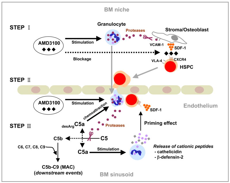
Based on these observations, we envision a flowing scenario in mobilization and CC activation induced by AMD3100 (Figure 7). First, at the BM microenvironment level, AMD3100 blocks the interaction of CXCR4+ granulocytes and CXCR4+ HSPCs with SDF-1 expressed in the BM microenvironment. At the same time, AMD3100 stimulates secretion in BM of MMPs and other proteolytic enzymes from activated granulocytes that help to perturb another axis crucial in HSPCs retention, i.e., VLA-4-VCAM-1. Next, granulocytes are the first cells to egress from BM and they “pave the way” for HSPCs to cross the BM-endothelial barrier. Finally, granulocytes activated/mobilized by AMD3100 into PB activate/cleave C5 and release C5a and desArgC5a that subsequently orchestrate/facilitate the final steps of HSPC egress.
Figure 7. AMD3100-directed HSPC mobilization depends on activation of CC by AMD3100 mobilized/activated granulocytes.
We envision three potential steps at which AMD3100 induces HSPC mobilization. First, AMD3100 blocks the interaction of CXCR4+ granulocytes and CXCR4+ HSPCs with SDF-1 expressed in the BM microenvironment. At the same time, however, AMD3100 stimulates secretion of metalloproteinases in BM from activated granulocytes, which helps to perturb/block another retention axis for HSPCs in BM, i.e., the VLA-4-VCAM-1 interaction (Step I). Next, granulocytes are the first cells to egress from BM and they “pave the way” for HSPCs to cross the BM-endothelial barrier (Ice Breaker Phenomenon; Step II). Finally, proteolytic enzymes released from AMD3100-activated granulocytes in BM sinusoids activate/cleave CC at the distal stages (i.e., C5) resulting in the release of C5a and desArgC5a, which facilitate the final steps of HSPC egress. Further studies however, require potential involvement of C5b-C9 - membrane attack complex (MAC) and our data indicate that some factors generated in plasma by activated MAC chemoattract HSPCs in a presence of AMD31000 in CXCR4-independent manner (manuscript in preparation) (Step III).
In conclusion, our data: i) better explain molecular mechanisms directing AMD3100-induced mobilization; ii) support a central role of innate immunity; and iii) provide evidence that C5 is activated/cleaved in plasma in a Gr-1+ cell-dependent manner. Granulocytes and C5 cleavage fragments orchestrate this process and, in light of our current data, mobilization of HSPCs could be envisioned as a part of the innate immunity response. However, further studies are needed to address if, in addition to C5a and desArgC5a cleavage fragments, other elements downstream from C5 of CC, e.g., C5b-C9 and/or membrane attack complex (MAC), are involved in the mobilization process.
Acknowledgments
Supported by NIH grant R01 CA106281, NIH R01 DK074720, and Stella and Henry Endowment to MZR.
References
- 1.Kyne L, Hausdorff JM, Knight E, Dukas L, Azhar G, Wei JY. Neutrophilia and congestive heart failure after acute myocardial infarction. Am Heart J. 2000;139:94–100. doi: 10.1016/s0002-8703(00)90314-4. [DOI] [PubMed] [Google Scholar]
- 2.Massberg S, Schaerli P, Knezevic-Maramica I, Köllnberger M, Tubo N, Moseman EA, et al. Immunosurveillance by hematopoietic progenitor cells trafficking through blood, lymph, and peripheral tissues. Cell. 2007;131:994–1008. doi: 10.1016/j.cell.2007.09.047. [DOI] [PMC free article] [PubMed] [Google Scholar]
- 3.Lee H, Ratajczak MZ. Innate immunity: a key player in the mobilization of hematopoietic stem/progenitor cells. Arch Immunol Ther Exp (Warsz) 2009;57:269–278. doi: 10.1007/s00005-009-0037-6. [DOI] [PubMed] [Google Scholar]
- 4.Andrews RG, Briddell RA, Knitter GH, Opie T, Bronsden M, Myerson D, et al. In vivo synergy between recombinant human stem cell factor and recombinant human granulocyte colony-stimulating factor in baboons enhanced circulation of progenitor cells. Blood. 1994;84:800–810. [PubMed] [Google Scholar]
- 5.Laterveer L, Lindley IJ, Hamilton MS, Willemze R, Fibbe WE. Interleukin-8 induces rapid mobilization of hematopoietic stem cells with radioprotective capacity and long-term myelolymphoid repopulating ability. Blood. 1995;85:2269–2275. [PubMed] [Google Scholar]
- 6.Stiff P, Gingrich R, Luger S, Wyres MR, Brown RA, LeMaistre CF, et al. A randomized phase 2 study of PBPC mobilization by stem cell factor and filgrastim in heavily pretreated patients with Hodgkin’s disease or non-Hodgkin’s lymphoma. Bone Marrow Transplant. 2000;26:471–481. doi: 10.1038/sj.bmt.1702531. [DOI] [PubMed] [Google Scholar]
- 7.To LB, Bashford J, Durrant S, MacMillan J, Schwarer AP, Prince HM, et al. Successful mobilization of peripheral blood stem cells after addition of ancestim (stem cell factor) in patients who had failed a prior mobilization with filgrastim (granulocyte colony-stimulating factor) alone or with chemotherapy plus filgrastim. Bone Marrow Transplant. 2003;31:371–378. doi: 10.1038/sj.bmt.1703860. [DOI] [PubMed] [Google Scholar]
- 8.Molineux G, Migdalska A, Szmitkowski M, Zsebo K, Dexter TM. The effects on hematopoiesis of recombinant stem cell factor (ligand for c-kit) administered in vivo to mice either alone or in combination with granulocyte colony-stimulating factor. Blood. 1991;78:961–966. [PubMed] [Google Scholar]
- 9.Glaspy JA, Shpall EJ, LeMaistre CF, Briddell RA, Menchaca DM, Turner SA, et al. Peripheral blood progenitor cell mobilization using stem cell factor in combination with filgrastim in breast cancer patients. Blood. 1997;90:2939–2951. [PubMed] [Google Scholar]
- 10.Molineux G, McCrea C, Yan XQ, Kerzic P, McNiece I. Flt-3 ligand synergizes with granulocyte colony-stimulating factor to increase neutrophil numbers and to mobilize peripheral blood stem cells with long-term repopulating potential. Blood. 1997;89:3998–4004. [PubMed] [Google Scholar]
- 11.Papayannopoulou T, Nakamoto B, Andrews RG, Lyman SD, Lee MY. In vivo effects of Flt3/Flk2 ligand on mobilization of hematopoietic progenitors in primates and potent synergistic enhancement with granulocyte colony-stimulating factor. Blood. 1997;90:620–629. [PubMed] [Google Scholar]
- 12.Pelus LM, Fukuda S. Chemokine-mobilized adult stem cells; defining a better hematopoietic graft. Leukemia. 2008;22:466–473. doi: 10.1038/sj.leu.2405021. [DOI] [PMC free article] [PubMed] [Google Scholar]
- 13.Pruijt JF, Fibbe WE, Laterveer L, Pieters RA, Lindley IJ, Paemen L, et al. Prevention of interleukin-8-induced mobilization of hematopoietic progenitor cells in rhesus monkeys by inhibitory antibodies against the metalloproteinase gelatinase B (MMP-9) Proc Natl Acad Sci USA. 1999;96:10863–10868. doi: 10.1073/pnas.96.19.10863. [DOI] [PMC free article] [PubMed] [Google Scholar]
- 14.King AG, Horowitz D, Dillon SB, Levin R, Farese AM, MacVittie TJ, et al. Rapid mobilization of murine hematopoietic stem cells with enhanced engraftment properties and evaluation of hematopoietic progenitor cell mobilization in rhesus monkeys by a single injection of SB-251353, a specific truncated form of the human CXC chemokine GRObeta. Blood. 2001;97:1534–1542. doi: 10.1182/blood.v97.6.1534. [DOI] [PubMed] [Google Scholar]
- 15.Papayannopoulou T. Current mechanistic scenarios in hematopoietic stem/progenitor cell mobilization. Blood. 2004;103:1580–1585. doi: 10.1182/blood-2003-05-1595. [DOI] [PubMed] [Google Scholar]
- 16.Fruehauf S, Seeger T, Topaly J. Innovative strategies for PBPC mobilization. Cytotherapy. 2005;7:438–446. doi: 10.1080/14653240500319135. [DOI] [PubMed] [Google Scholar]
- 17.Winkler IG, Levesque JP. Mechanisms of hematopoietic stem cell mobilization: When innate immunity assails the cells that make blood and bone. Exp Hematol. 2006;34:996–1009. doi: 10.1016/j.exphem.2006.04.005. [DOI] [PubMed] [Google Scholar]
- 18.Bensinger W, Dipersio JF, McCarty JM. Improving stem cell mobilization strategies: future directions. Bone Marrow Transplant. 2009;43:181–195. doi: 10.1038/bmt.2008.410. [DOI] [PubMed] [Google Scholar]
- 19.Vos O, Wilschut IJ. Further studies on mobilization of CFUs. Cell Tissue Kinet. 1979;12:257–267. doi: 10.1111/j.1365-2184.1979.tb00148.x. [DOI] [PubMed] [Google Scholar]
- 20.Sweeney EA, Priestley GV, Nakamoto B, Collins RG, Beaudet AL, Papayannopoulou T. Mobilization of stem/progenitor cells by sulfated polysaccharides does not require selectin presence. Proc Natl Acad Sci U S A. 2000;97:6544–6549. doi: 10.1073/pnas.97.12.6544. [DOI] [PMC free article] [PubMed] [Google Scholar]
- 21.Sweeney EA, Lortat-Jacob H, Priestley GV, Nakamoto B, Papayannopoulou T. Sulfated polysaccharides increase plasma levels of SDF-1 in monkeys and mice: involvement in mobilization of stem/progenitor cells. Blood. 2002;99:44–51. doi: 10.1182/blood.v99.1.44. [DOI] [PubMed] [Google Scholar]
- 22.Lévesque JP, Hendy J, Takamatsu Y, Williams B, Winkler IG, Simmons PJ. Mobilization by either cyclophosphamide or granulocyte colony-stimulating factor transforms the bone marrow into a highly proteolytic environment. Exp Hematol. 2002;30:440–449. doi: 10.1016/s0301-472x(02)00788-9. [DOI] [PubMed] [Google Scholar]
- 23.Petit I, Szyper-Kravitz M, Nagler A, Lahav M, Peled A, Habler L, et al. G-CSF induces stem cell mobilization by decreasing bone marrow SDF-1 and up-regulating CXCR4. Nat Immunol. 2002;3:687–694. doi: 10.1038/ni813. [DOI] [PubMed] [Google Scholar]
- 24.Pelus LM, Bian H, King AG, Fukuda S. Neutrophil-derived MMP-9 mediates synergistic mobilization of hematopoietic stem and progenitor cells by the combination of G-CSF and the chemokines GRObeta/CXCL2 and GRObetaT/CXCL2delta4. Blood. 2004;103:110–119. doi: 10.1182/blood-2003-04-1115. [DOI] [PubMed] [Google Scholar]
- 25.Lapidot T, Dar A, Kollet O. How do stem cells find their way home? Blood. 2005;106:1901–1910. doi: 10.1182/blood-2005-04-1417. [DOI] [PubMed] [Google Scholar]
- 26.Peled A, Grabovsky V, Habler L, Sandbank J, Arenzana-Seisdedos F, Petit I, et al. The chemokine SDF-1 stimulates integrin-mediated arrest of CD34+ cells on vascular endothelium under shear flow. J Clin Invest. 1999;104:1199–1211. doi: 10.1172/JCI7615. [DOI] [PMC free article] [PubMed] [Google Scholar]
- 27.Lévesque JP, Takamatsu Y, Nilsson SK, Haylock DN, Simmons PJ. Vascular cell adhesion molecule-1 (CD106) is cleaved by neutrophil proteases in the bone marrow following hematopoietic progenitor cell mobilization by granulocyte colony-stimulating factor. Blood. 2001;98:1289–1297. doi: 10.1182/blood.v98.5.1289. [DOI] [PubMed] [Google Scholar]
- 28.Sekhsaria S, Fleisher TA, Vowells S, Brown M, Miller J, Gordon I, et al. Granulocyte colony-stimulating factor recruitment of CD34+ progenitors to peripheral blood: Impaired mobilization in chronic granulomatous disease and adenosine deaminase deficient severe combined immunodeficiency disease patients. Blood. 1996;88:1104–1112. [PubMed] [Google Scholar]
- 29.Liles WC, Broxmeyer HE, Rodger E, Wood B, Hübel K, Cooper S, et al. Mobilization of hematopoietic progenitor cells in healthy volunteers by AMD3100, a CXCR4 antagonist. Blood. 2003;102:2728–2730. doi: 10.1182/blood-2003-02-0663. [DOI] [PubMed] [Google Scholar]
- 30.Broxmeyer HE, Orschell CM, Clapp DW, Hangoc G, Cooper S, Plett PA, et al. Rapid mobilization of murine and human hematopoietic stem and progenitor cells with AMD3100, a CXCR4 antagonist. J Exp Med. 2005;201:1307–1318. doi: 10.1084/jem.20041385. [DOI] [PMC free article] [PubMed] [Google Scholar]
- 31.Flomenberg N, Devine SM, Dipersio JF, Liesveld JL, McCarty JM, Rowley SD, et al. The use of AMD3100 plus G-CSF for autologous hematopoietic progenitor cell mobilization is superior to G-CSF alone. Blood. 2005;106:1867–1874. doi: 10.1182/blood-2005-02-0468. [DOI] [PubMed] [Google Scholar]
- 32.Molendijk WJ, van Oudenaren A, van Dijk H, Daha MR, Benner R. Complement split product C5a mediates the lipopolysaccharide-induced mobilization of CFU-s and haemopoietic progenitor cells, but not the mobilization induced by proteolytic enzymes. Cell Tissue Kinet. 1986;19:407–417. doi: 10.1111/j.1365-2184.1986.tb00738.x. [DOI] [PubMed] [Google Scholar]
- 33.Reca R, Mastellos D, Majka M, Marquez L, Ratajczak J, Franchini S, et al. Functional receptor for C3a anaphylatoxin is expressed by normal hematopoietic stem/progenitor cells, and C3a enhances their homing-related responses to SDF-1. Blood. 2003;101:3784–3793. doi: 10.1182/blood-2002-10-3233. [DOI] [PubMed] [Google Scholar]
- 34.Ratajczak J, Reca R, Kucia M, Majka M, Allendorf DJ, Baran JT, et al. Mobilization studies in mice deficient in either C3 or C3a receptor (C3aR) reveal a novel role for complement in retention of hematopoietic stem/progenitor cells in bone marrow. Blood. 2004;103:2071–2078. doi: 10.1182/blood-2003-06-2099. [DOI] [PubMed] [Google Scholar]
- 35.Ratajczak MZ, Reca R, Wysoczynski M, Kucia M, Baran JT, Allendorf DJ, et al. Transplantation studies in C3-deficient animals reveal a novel role of the third complement component (C3) in engraftment of bone marrow cells. Leukemia. 2004;18:1482–1490. doi: 10.1038/sj.leu.2403446. [DOI] [PubMed] [Google Scholar]
- 36.Ratajczak MZ, Reca R, Wysoczynski M, Yan J, Ratajczak J. Modulation of the SDF-1-CXCR4 axis by the third complement component (C3)--implications for trafficking of CXCR4+ stem cells. Exp Hematol. 2006;34:986–995. doi: 10.1016/j.exphem.2006.03.015. [DOI] [PubMed] [Google Scholar]
- 37.Reca R, Cramer D, Yan J, Laughlin MJ, Janowska-Wieczorek A, Ratajczak J, et al. A novel role of complement in mobilization: immunodeficient mice are poor granulocyte-colony stimulating factor mobilizers because they lack complement-activating immunoglobulins. Stem Cells. 2007;25:3093–3100. doi: 10.1634/stemcells.2007-0525. [DOI] [PubMed] [Google Scholar]
- 38.Liu F, Poursine-Laurent J, Link DC. Expression of the G-CSF receptor on hematopoietic progenitor cells is not required for their mobilization by G-CSF. Blood. 2000;95:3025–3031. [PubMed] [Google Scholar]
- 39.Pruijt JF, Verzaal P, van Os R, de Kruijf EJ, van Schie ML, Mantovani A, et al. Neutrophils are indispensable for hematopoietic stem cell mobilization induced by interleukin-8 in mice. Proc Natl Acad Sci USA. 2002;99:6228–6233. doi: 10.1073/pnas.092112999. [DOI] [PMC free article] [PubMed] [Google Scholar]
- 40.Wu W, Lee H, Wysoczynski M, Kucia M, Ratajczak J, Ratajczak MZ. Novel Observation That Mice Lacking the Fifth Complement Cascade Protein Component (C5) Are Very Poor Stem Cell Mobilizers Explained by Defective Egress of Granulocytes: A Novel Role for Bone Marrow Granulocytes to Act as “ice Breaker” Cells in Facilitating Egress of Hematopoietic Stem/Progenitor Cells. Blood. 2008;112:32. Abstract 67. [Google Scholar]
- 41.Lee HM, Wu W, Wysoczynski M, Liu R, Zuba-Surma EK, Kucia M, et al. Impaired mobilization of hematopoietic stem/progenitor cells in C5-deficient mice supports the pivotal involvement of innate immunity in this process and reveals novel promobilization effects of granulocytes. Leukemia. 2009;23:2052–2062. doi: 10.1038/leu.2009.158. [DOI] [PMC free article] [PubMed] [Google Scholar]
- 42.Nagai Y, Garrett KP, Ohta S, Bahrun U, Kouro T, Akira S, et al. Toll-like receptors on hematopoietic progenitor cells stimulate innate immune system replenishment. Immunity. 2006;24:801–812. doi: 10.1016/j.immuni.2006.04.008. [DOI] [PMC free article] [PubMed] [Google Scholar]
- 43.Wysoczynski M, Reca R, Lee H, Wu W, Ratajczak J, Ratajczak MZ. Defective engraftment of C3aR−/− hematopoietic stem progenitor cells shows a novel role of the C3a-C3aR axis in bone marrow homing. Leukemia. 2009;23:1455–1461. doi: 10.1038/leu.2009.73. [DOI] [PMC free article] [PubMed] [Google Scholar]
- 44.Huber-Lang M, Younkin EM, Sarma JV, Riedemann N, McGuire SR, Lu KT, et al. Generation of C5a by phagocytic cells. Am J Pathol. 2002;161:1849–1859. doi: 10.1016/S0002-9440(10)64461-6. [DOI] [PMC free article] [PubMed] [Google Scholar]
- 45.Ward PA. The dark side of C5a in sepsis. Nat Rev Immunol. 2004;4:133–142. doi: 10.1038/nri1269. [DOI] [PubMed] [Google Scholar]
- 46.Huber-Lang M, Sarma JV, Zetoune FS, Rittirsch D, Neff TA, McGuire SR, et al. Generation of C5a in the absence of C3: a new complement activation pathway. Nat Med. 2006;12:682–687. doi: 10.1038/nm1419. [DOI] [PubMed] [Google Scholar]
- 47.Asberg AE, Mollnes TE, Videm V. Complement activation by neutrophil granulocytes. Scand J Immunol. 2008;67:354–361. doi: 10.1111/j.1365-3083.2008.02077.x. [DOI] [PubMed] [Google Scholar]
- 48.Zhang WB, Navenot JM, Haribabu B, Tamamura H, Hiramatu K, Omagari A, et al. A point mutation that confers constitutive activity to CXCR4 reveals that T140 is an inverse agonist and that AMD3100 and ALX40–4C are weak partial agonists. J Biol Chem. 2002;277:24515–24521. doi: 10.1074/jbc.M200889200. [DOI] [PubMed] [Google Scholar]
- 49.Bellucci R, De Propris MS, Buccisano F, Lisci A, Leone G, Tabilio A, et al. Modulation of VLA-4 and L-selectin expression on normal CD34+ cells during mobilization with G-CSF. Bone Marrow Transplant. 1999;23:1–8. doi: 10.1038/sj.bmt.1701522. [DOI] [PubMed] [Google Scholar]
- 50.Lévesque JP, Hendy J, Takamatsu Y, Simmons PJ, Bendall LJ. Disruption of the CXCR4/CXCL12 chemotactic interaction during hematopoietic stem cell mobilization induced by GCSF or cyclophosphamide. J Clin Invest. 2003;111:187–196. doi: 10.1172/JCI15994. [DOI] [PMC free article] [PubMed] [Google Scholar]
- 51.Lévesque JP, Hendy J, Winkler IG, Takamatsu Y, Simmons PJ. Granulocyte colony-stimulating factor induces the release in the bone marrow of proteases that cleave c-KIT receptor (CD117) from the surface of hematopoietic progenitor cells. Exp Hematol. 2003;31:109–117. doi: 10.1016/s0301-472x(02)01028-7. [DOI] [PubMed] [Google Scholar]
- 52.Katayama Y, Battista M, Kao WM, Hidalgo A, Peired AJ, Thomas SA, et al. Signals from the sympathetic nervous system regulate hematopoietic stem cell egress from bone marrow. Cell. 2006;124:407–421. doi: 10.1016/j.cell.2005.10.041. [DOI] [PubMed] [Google Scholar]
- 53.McQuibban GA, Butler GS, Gong JH, Bendall L, Power C, Clark-Lewis I, et al. Matrix metalloproteinase activity inactivates the CXC chemokine stromal cell-derived factor-1. J Biol Chem. 2001;276:43503–43508. doi: 10.1074/jbc.M107736200. [DOI] [PubMed] [Google Scholar]
- 54.Semerad CL, Christopher MJ, Liu F, Short B, Simmons PJ, Winkler I, et al. G-CSF potently inhibits osteoblast activity and CXCL12 mRNA expression in the bone marrow. Blood. 2005;106:3020–3027. doi: 10.1182/blood-2004-01-0272. [DOI] [PMC free article] [PubMed] [Google Scholar]
- 55.Kollet O, Dar A, Lapidot T. The multiple roles of osteoclasts in host defense: bone remodeling and hematopoietic stem cell mobilization. Annu Rev Immunol. 2007;25:51–69. doi: 10.1146/annurev.immunol.25.022106.141631. [DOI] [PubMed] [Google Scholar]
- 56.van Pel M, van Os R, Velders GA, Hagoort H, Heegaard PM, Lindley IJ, et al. Serpina1 is a potent inhibitor of IL-8-induced hematopoietic stem cell mobilization. Proc Natl Acad Sci USA. 2006;103:1469–1474. doi: 10.1073/pnas.0510192103. [DOI] [PMC free article] [PubMed] [Google Scholar]
- 57.Zhang Zhang M, Austen WG, Jr, Chiu I, Alicot EM, Hung R, Ma M, et al. Identification of a specific self-reactive IgM antibody that initiates intestinal ischemia/reperfusion injury. Proc Natl Acad Sci USA. 2004;101:3886–3891. doi: 10.1073/pnas.0400347101. [DOI] [PMC free article] [PubMed] [Google Scholar]
- 58.Wysoczynski M, Kucia M, Ratajczak J, Ratajczak MZ. Cleavage fragments of third complement component (C3) enhance SDF-1 mediated platelet production during reactive thrombocytosis. Leukemia. 2007;21:973–982. doi: 10.1038/sj.leu.2404629. [DOI] [PubMed] [Google Scholar]


