
Fig. 4.
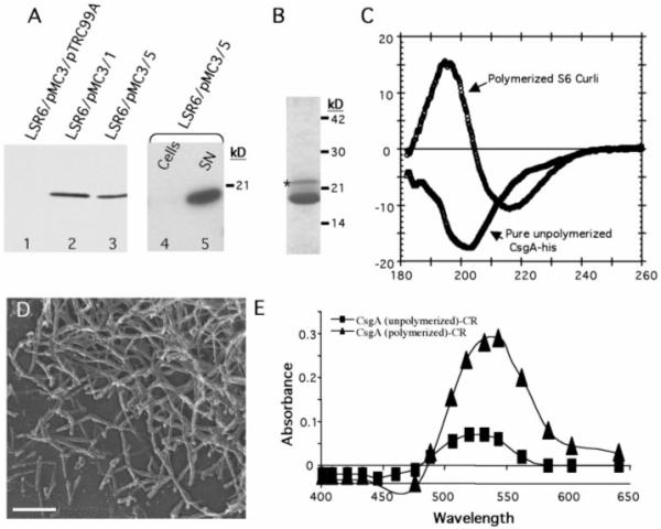
Purification and in vitro assembly of CsgA-his. (A) Western blot with anti-his (Covance, Richmond, California) to determine expression and cellular location of overexpressed CsgA-his. Log-phase cultures containing (lane 1) pMC3 (csgA-his) and pTrc99A (empty vector); (lane 2) pMC3 and pMC1 (csgG); or (lane 3) pMC3 and pMC5 (csgEFG) were induced with 0.5 mM IPTG for 1 hour; samples were removed and mixed with an equal amount of 2× SDS-PAGE dye and heated to 95°C before gel electrophoresis. CsgA-his expression with pTrc99A was detected only after overexposure of the blot. The cellular and supernatant fractions from cultures containing pMC3 and pMC5 were separated by centrifugation and loaded in lanes 4 and 5, respectively. (B) CsgA-his was purified from cleared LSR6/pMC3/pMC5 supernatants filtered through a 0.2-μm filter before loading on a disposable column packed with nickel NTA-agarose beads (Qiagen, Chatsworth, California). The column was washed with 10 column volumes of 10 mM tris (pH 7.4), 100 mM NaCl, and CsgA-his was eluted with 5 ml of wash buffer plus 100 mM imidazole and analyzed by Coomassie stain SDS-PAGE. Both the major band migrating at ~17.5 kD and the higher molecular weight, minor band (indicated by an asterisk) interacted with anti-CsgA (9). (C) The CD spectrum of wild-type S6 curli compared with that of 300 μg of soluble unpolymerized CsgA-his assayed immediately after purification. (D) High-resolution EM of pure CsgA-his preparations after a 1-week incubation at 4°C. Bar, 140 nm. (E) Absorbance of a 10 μM solution of CR with 100 μg of pure, unpolymerized CsgA-his (■) or pure polymerized CsgA-his (▲) after subtracting the absorbance of CR alone.