Abstract
Purpose
To determine putative alterations in the major lenticular proteins in Wistar rats of different ages and to compare these alterations with those occurring in rats with selenite-induced cataract.
Methods
Lenticular transparency was determined by morphological examination using slit-lamp biomicroscopy. Alterations in lenticular protein were determined by sodium dodecyl sulfate-PAGE (SDS–PAGE) and confirmed immunologically by western blot.
Results
Morphological examination did not reveal observable opacities in the lenses of the rats of different age groups; however, dense nuclear opacities were noted in lenses of rats in the selenite-cataract group. Western blot assays revealed age-related changes in soluble and urea-soluble lenticular proteins. Decreased αA- and βB1-crystallins in the soluble fraction and aggregation of αA-crystallin, in addition to the degraded fragment of βB1-crystallin, in the urea-soluble fraction appeared to occur in relation to increasing age of the rats from which the lenses were taken; similarly, cytoskeletal proteins appeared to decline with increasing age. The lenses from rats in the selenite-cataract group exhibited similar changes, except that there was also high molecular weight aggregation of αA-crystallin.
Conclusions
The results of this study suggest that there is loss, as well as aggregation, of αA-crystallin in the aging rat lens, although there is no accompanying loss of lenticular transparency.
Introduction
Over the last twenty years, the causes of blindness have changed in proportion and actual number. However, cataract has remained the major cause of blindness globally, particularly in Asia [1]. Cataract is a multifactorial disease associated with several risk factors such as aging, diabetes, exposure to sunlight, and hypertension; however, free radical-induced oxidative stress is postulated to be, perhaps, the major factor leading to senile cataract formation [2]. Generation of reactive oxygen species (ROS), resulting in degradation, crosslinking, and aggregation of lens proteins, is regarded as an important factor in cataractogenesis [3-5].
The mammalian lens consists mainly of proteins, which account for over 30% of its weight [6]. It is the architecture of the crystalline lattice that is ultimately responsible for lenticular transparency and for the proper focusing of light. The highly concentrated cellular proteins in mammalian lenses consist of two families of water-soluble lens crystallins, namely, α-crystallin and β/γ-crystallin [7], that are uniformly distributed in transparent lenticular cells. Dense opacification results when the proteins form large insoluble aggregates that approach or exceed the dimensions of the wavelength of light and produce large fluctuations in the index of refraction that result in increased light scattering [3,6,8].
Numerous changes in crystallin structure occur with age, mainly due to post-translational, non-enzymatic reactions [9]. Protein synthesis and turnover predominantly occur in the cortical region, and decrease with age in the nucleus. Previous experimental studies have shown the importance of cytoskeletal and membrane-associated proteins in the development and maintenance of the transparent structure of the cells of the lens [10-14]. Cytoskeletal proteins constitute 2%–4% of lenticular proteins, which include intermediate filaments, microfilaments, and microtubules [15,16]. Although the major proteins of the cytoskeleton of the lens are actin and vimentin, the tissue is also known to contain several similar proteins including tubulin, ankyrin, α-actinin, spectrin, myosin, tropomyosin, and beaded filaments [15-17]. Typically, the concentration of cytoskeletal proteins in the lens decreases with age, particularly in the nucleus. Loss of these proteins, including vimentin and α-actinin, is accelerated in the human lens during the development of senile cataract [12,18]. Therefore, studies of the protein patterns in the lens are of considerable importance with respect to an understanding of changes associated with aging and cataractogenesis.
Different animal models have been developed with a view to better understand the aging process and cataract formation [19-22]. None of these models exactly matches what occurs in human cataractogenesis. However, the model of selenite-induced cataractogenesis has many features to recommend it, namely: the morphological and biochemical characteristics have been extensively investigated; this model shows several general similarities to human cataract; the selenite cataract is rapidly induced; it is a useful rodent model for rapid screening of potential anticataract agents [23]. The aim of the present study was to determine the occurrence of possible alterations in certain crucial lenticular proteins (αA- and βB1-crystallins, and cytoskeletal proteins) during the process of aging, and to compare these changes with those occurring in selenite cataracts in Wistar rats.
Methods
Experimental animals
Rats of the Wistar strain were used in this study. The rats were housed with parents in large spacious cages, and the parents were given food and water ad libitum. The animal room was well ventilated and had a regular 12:12 h light/dark cycle throughout the experimental period. These animals were used in accordance with institutional guidelines and with the Association for Research in Vision and Ophthalmology Statement for the Use of Animals in Research. Rats in the following age groups were randomly selected (5 animals in each age group): 10-day-old, 16-day-old, 30-day-old, 90-day-old, 180-day-old, 365-day-old rats. Sixteen- day-old rats with selenite-induced cataract were also studied.
Selenite cataract
The cataractogenesis was induced by a single subcutaneous injection of sodium selenite (19 μmoles/kg bodyweight) on postpartum day 10.
Morphological observations
A slit-lamp biomicroscopic examination of each eye of 16-day-old and 365-day-old rats was performed to determine possible lenticular opacification (morphological assessment). Prior to performing the examination, mydriasis was achieved by using a topical ophthalmic solution containing tropicamide with phenylephrine (Maxdil Plus; Hi-Care Pharma, Chennai, India); one drop of the solution was instilled every 30 min for 2 h into each eye of each rat, with the animals being kept in a dark room, and, after 2 h, the eyes were viewed by the slit-lamp biomicroscope at 12× magnification.
The animals were sacrificed by cervical dislocation; the eyes of the different age groups were collected and the lenses were removed. Each lens was then homogenized with 10 times its mass of 20 mM phosphate buffer containing 1 mM ethylene glycol-bis(2-aminoethylether)-N,N,N’,N’-tetraacetic acid (EGTA; pH 7.2), and centrifuged at 14,000× g at 4 °C for 15 min. This process was repeated twice. The supernatant obtained was used for the determination of waster soluble protein (WS) while the pellet dissolved in 8 M urea was considered as urea-soluble (insoluble) protein. The protein concentration in each sample was estimated by the method of Bradford [24]. The estimated value of protein in each sample was used to calculate the ratio of urea-soluble to soluble protein. Both the fractions (soluble and urea-soluble) were aliquoted into portions and stored at −70 °C until analyzed by immunoblot.
Immunoblot
Immunoblot analysis was performed to determine the relative concentrations of the cytoplasmic proteins, αA-crystallin and βB1-crystallin, and of the cytoskeletal proteins, actin, vimentin, and spectrin, in samples of the various age groups. Proteins subjected to SDS–PAGE were electrophoretically transferred to a nitrocellulose (NC) sheet using a semidry blotting apparatus (Bio-Rad, Richmond, CA). Blotting was done at 25 V for 2 h. Blotted membranes were stained by Ponceau S solution to check for the efficiency of transfer; subsequently, blocking was done with 5% non-fat dry milk in Tris buffer saline (pH 7.5) with 0.1% (v/v) Tween-20 for 3 h. Antibodies (purchased from Sigma, St. Louis, MO) against αA-crystallin (1:1,000 dilution), βB1-crystallin (1:2,000 dilution), actin (1:100 dilution), vimentin (1:50 dilution) and spectrin (1:400 dilution) were used. Immunoreactivity was visualized with alkaline phosphatase conjugated to anti-mouse IgG or anti-rabbit IgG secondary antibodies and 5-bromo 4-chloro 3-indolyl phosphate/nitroblue tetrazolium chloride (BCIP/NBT; Genei, Bangalore, India). To analyze the small changes observed in the intensity of the bands, densitometry was conducted on the scanned images of the membrane. The program Quantity One SW (Bio-Rad) was used for the analysis of intensity of bands in each lane of the membrane.
Statistical analysis
Statistical analysis was performed with SPSS software (version 11.0) (SPSS Corporation, Chicago, IL). The results are expressed as the median (min–max), as the non-parametrical distribution requires nonparametrical analysis that compares medians instead of means. To test for correlations between two parameters, Spearman analysis was performed. P-values <0.05 were considered statistically significant.
Results
Morphological examination and protein ratio
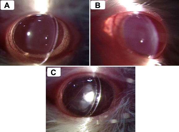
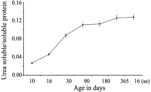
Morphological examination revealed clear transparent lenses in both eyes in all 10-day-old and 365-day-old rats. However, a complete nuclear opacity was observed in the lenses of each eye in 16-day-old rats that had been administered selenite (Figure 1). The ratio of urea-soluble to soluble lenticular protein was found to rise with increasing age from 0.027 (10-day-old rats) to 0.126 (365-day-old rats) and was found to be 0.127 in cataractous lenses (16-day-old, selenite-administered rats; Figure 2).
Figure 1.
Appearance of lenses in different groups of Wistar rats. Morphological assessment of cataract formation in Wistar rat pups by the slit-lamp biomicroscope. Clear lens observed in 16 day-old rats (A) and 365 day-old rats (B), and dense opacification of the lens observed in 16 day-old, selenite-administered rats (C).
Figure 2.
Effect of aging on the ratio of urea-soluble to soluble protein in lenses of rats of different age groups in comparison to rats with selenite cataractous lenses. The X-axis indicates the age in days and the Y-axis indicates the ratio of urea-soluble to soluble protein. 10 =10 day-old rat lenses; 16 = 16 day-old rat lenses; 30 = 30 day-old rat lenses; 90 = 90 day-old rat lenses; 180 = 180 day-old rat lenses; 365 = 365 day-old rat lenses; 16(se) = lenses of 16 day-old rats that had been administered selenite on post-partum day 10. The ratio of urea-soluble to soluble lenticular protein was found to rise with increasing age.
Immunoblot of αA- and βB1-crystallin
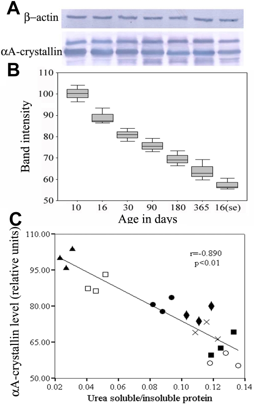
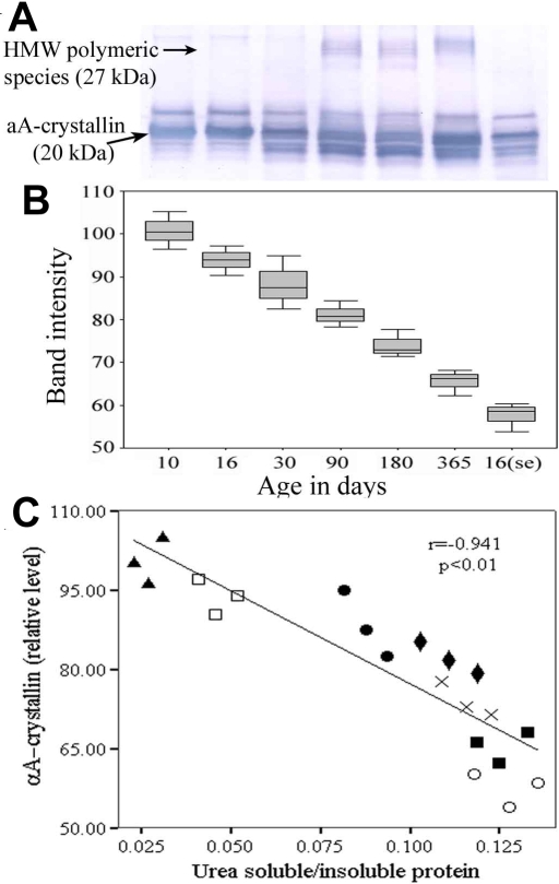
The relative amounts of αA- and βB1-crystallin in the soluble and urea-soluble fractions were further examined by immunoblotting with specific antibodies. Immunoblot analysis revealed that there was a decrease of αA-crystallin in the soluble fraction with increasing age, and this decrease was also seen in lenses with selenite cataract (Figure 3). There was also an obvious decrease of αA-crystallin in the urea-soluble fraction with increasing age; however, the anti-αA-crystallin antibody appeared to detect appreciable amounts of polymeric species of αA-crystallin with increasing age; such polymeric species of αA-crystallin were not detected in the soluble fraction or in lenses with selenite cataract (Figure 4). We also performed Spearman correlation analysis to examine whether intensity levels of αA-crystallin correlated well with the urea-soluble to soluble protein ratio. Figure 3C and Figure 4C demonstrate that there was a significant negative correlation (r=-0.890; p<0.01 and r=-0.941; p<0.01) between the band intensity levels of αA-crystallin and urea-soluble to soluble protein ratio in soluble and urea-soluble protein fractions, respectively.
Figure 3.
Occurrence of αA-crystallin in lenticular soluble protein fractions in different groups of Wistar rats. A typical western blot showing age-dependent relative concentrations of αA-crystallin in the soluble protein fractions of Wistar rat lenses. A: An equal amount of protein was loaded in each lane and subjected to electrophoresis. Blots were incubated with αA-crystallin antibody, with ß-actin as the loading control. B: band intensity of αA-crystallin levels relative to 10 day-old rat lenses. C: Linear regression analysis revealed a significant negative correlation between αA-crystallin and the urea-soluble to soluble protein ratio. (▲) Values in 10 day-old rat lenses; (□) Values in 16 day-old rat lenses; (●) Values in 30 day-old rat lenses; (♦) Values in 90 day-old rat lenses; (×) Values in 180 day-old rat lenses; (■) Values in 365 day-old rat lenses; (○) Values in the lenses of 16 day-old rats that had been administered selenite on post-partum day 10.
Figure 4.
Occurrence of αA-crystallin in lenticular urea-soluble protein fractions in different groups of Wistar rats. A typical western blot showing age-dependent relative concentrations of αA-crystallin in the urea-soluble protein fractions of Wistar rat lenses. A: An equal amount of protein was loaded in each lane and subjected to electrophoresis. Blots were incubated with αA-crystallin antibody. B: band intensity of αA-crystallin levels relative to 10 day-old rat lenses. C: Linear regression analysis revealed a significant negative correlation between αA-crystallin and the urea-soluble to soluble protein ratio. (▲) Values in 10 day-old rat lenses; (□) Values in 16 day-old rat lenses; (●) Values in 30 day-old rat lenses; (♦) Values in 90 day-old rat lenses; (×) Values in 180 day-old rat lenses; (■) Values in 365 day-old rat lenses; (○) Values in the lenses of 16 day-old rats that had been administered selenite on post-partum day 10.
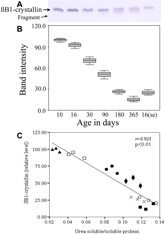
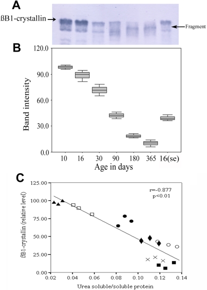
Similarly, the relative amount of βB1-crystallin in the soluble fractions was also found to decrease with increasing age (Figure 5). When the urea-soluble fraction was also examined for the presence of βB1-crystallin, decreased band intensity with a small fragmented band was observed in the lenses of aged rats, compared to the pattern in 10-day-old rats; in selenite-cataractous rat lenses, the fragment was seen at different places (Figure 6). Spearman analysis of correlation was also performed to examine whether there was any correlation between the intensity of the band corresponding to βB1-crystallin and the ratio of urea-soluble to soluble proteins. Figure 5C and Figure 6C demonstrate that there was a significant negative correlation between the intensity of the band corresponding to βB1-crystallin and the ratio of urea-soluble to soluble proteins in both the soluble (r=-0.923; p<0.01) and urea-soluble (r=-0.877; p<0.01) fractions.
Figure 5.
Occurrence of βB1-crystallin in lenticular soluble protein fractions in different groups of Wistar rats. A typical western blot showing age-dependent relative concentrations of βB1-crystallin in the soluble protein fractions of Wistar rat lenses. A: An equal amount of protein was loaded in each lane and subjected to electrophoresis. Blots were incubated with βB1-crystallin antibody. B: band intensity of βB1-crystallin levels relative to 10 day-old rat lenses. C: Linear regression analysis revealed a significant negative correlation between βB1-crystallin and the urea-soluble to soluble protein ratio. (▲) Values in 10 day-old rat lenses; (□) Values in 16 day-old rat lenses; (●) Values in 30 day-old rat lenses; (♦) Values in 90 day-old rat lenses; (×) Values in 180 day-old rat lenses; (■) Values in 365 day-old rat lenses; (○) Values in the lenses of 16 day-old rats that had been administered selenite on post-partum day 10.
Figure 6.
Occurrence of β1-crystallin in lenticular urea-soluble protein fractions in different groups of Wistar rats. A typical western blot showing age-dependent relative concentrations of β1-crystallin in the urea-soluble protein fractions of Wistar rat lenses. A: An equal amount of protein was loaded in each lane and subjected to electrophoresis. Blots were incubated with β1-crystallin antibody. B: Bnd intensity of β1-crystallin levels relative to 10 day-old rat lenses. C: Linear regression analysis revealed a significant negative correlation between βB1-crystallin and the urea-soluble to soluble protein ratio. (▲) Values in 10 day-old rat lenses; (□) Values in 16 day-old rat lenses; (●) Values in 30 day-old rat lenses; (♦) Values in 90 day-old rat lenses; (×) Values in 180 day-old rat lenses; (■) Values in 365 day-old rat lenses; (○) Values in the lenses of 16 day-old rats that had been administered selenite on post-partum day 10.
Immunoblot of cytoskeletal proteins
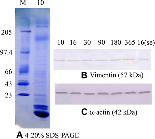
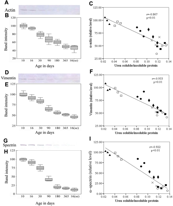
To identify the high molecular weight cytoskeletal proteins which were altered with increasing age, and in selenite cataract lenses, western blot analyses were performed on the soluble and urea-soluble protein fractions of the lenses of all groups. But no differences were found in the soluble fraction for high molecular weight proteins, actin and vimentin, in all the measured groups (Figure 7). In the urea-soluble fractions, the 42 kDa band reacted with anti-actin, the 57 kDa reacted with monoclonal anti-vimentin and the 235 kDa band reacted with polyclonal anti-spectrin (Figure 8).
Figure 7.
Occurrence of high molecular weight protein in lenticular soluble protein fractions in different groups of Wistar rats. Detection of changes in high molecular weight soluble protein by western blot analysis. An equal amount of the soluble protein fractions were loaded in each lane and subjected to 4%–20% SDS–PAGE. 10 = 10 day-old rat lens and the standard M = Molecular weight marker (A). Blots were subjected to immunoblot detection. Immunoblot analysis did not reveal any differences in the fraction for high molecular weight proteins, vimentin (B) and α-actin (C), in all the groups measured.
Figure 8.
Occurrence of high molecular weight proteins in lenticular urea-soluble protein fractions in different groups of Wistar rats. Detection of changes in high molecular weight insoluble protein by western blot analysis. Panels A, D, and G show immunoreactive bands of spectrin (240 kDa), vimentin (57 kDa), and α-actin (42 kDa), respectively. Box-plots show the band intensity of spectrin, vimentin, and α-actin, relative to the level in 10 day-old rat lenses (panels B, E, and H, respectively). Linear regression analysis revealed a significant negative correlation between the urea-soluble to soluble protein ratio and the levels of spectrin, vimentin, and alpha-actin (panels C, F, and I, respectively). (▲) Values in 10 day-old rat lenses; (□) Values in 16 day-old rat lenses; (●) Values in 30 day-old rat lenses; (♦) Values in 90 day-old rat lenses; (×) Values in 180 day-old rat lenses; (■) Values in 365 day-old rat lenses; (○) Values in the lenses of 16 day-old rats that had been administered selenite on post-partum day 10.
Quantitation of proteins was performed by densitometry. When compared to the values obtained in lenses of 10-day-old rats, a gradual decrease in the concentrations of these identified proteins was noted with increase in the ages of the test rats (10 days to 365 days) and this correlated with urea-soluble to soluble protein ratio for actin (r=-0.867, p<0.01), vimentin (r=-0.933, p<0.01) and spectrin (r=-0.922, p<0.01). Considering the levels of these proteins to be 100% on day 10, the levels of actin, vimentin and spectrin were found to be decreased to 53%, 51%, and 12%, respectively, in the lenses with selenite-induced cataract (Figure 8).
Discussion
The proteins that constitute the innermost core of the nucleus of the lens may be older than the postnatal life of the animal. Due to minimal or no turnover, lenticular proteins are subjected to considerable post-translational modifications which, in turn, disrupt lenticular architecture and alter the optical properties, leading to formation of a senile cataract [9]. We sought to determine the extent of age-related alterations in lenticular proteins in Wistar rats, and to compare these with the changes occurring in selenite-induced cataractous lenses.
In the present study, the ratio of urea-soluble to soluble lenticular proteins was found to increase with increasing age of the animals from which the lenses were taken (Figure 2), suggesting age-related increase in insolubilization of these proteins. Swamy and Abraham [25] observed that in rats, there was an increase in soluble proteins with age up to 20 months and a slow decrease in the later ages whereas urea-soluble proteins showed a continuous increase during the experimental period.
Age-related changes have been documented in the protein αA-crystallin, which is found abundantly in the lens; the molecular chaperone properties of this protein are believed to play a key role in maintaining transparency of the lens [26,27]. Changes in the αA-crystallin molecule possibly influence the lenticular changes that occur during normal development and aging [9,28-30]. With aging, αA-crystallin is believed to bind partially unfolded lenticular β- and γ-crystallins, thereby preventing their aggregation and precipitation and therein delaying lenticular opacification [31]. In the present investigation, an interesting observation made was the loss of αA-crystallin (20 kDa) in the soluble protein fraction and a gradual loss in the intensity of the band in the urea-soluble fraction with increasing age of the animals from which the lenses were taken (Figure 3 and Figure 4). Relative to αA-crystallin from 10 day-old rat lenses, anti-αA-crystallin antibody could detect an additional high molecular weight aggregate in the urea-soluble fraction (Figure 4).
It has been suggested that high molecular weight proteins might represent an intermediate stage in conversion of water-soluble to water-insoluble proteins in rabbit lenses [32]. Swamy and Abraham [25] showed that with increasing age, there was an increase of high molecular weight and insoluble proteins, with disappearance of γ-crystallins and sulfhydryl groups from the soluble fraction and an increase in the insoluble fraction and high molecular weight aggregates. Bindels et al. [33] proposed that in rodents, soluble high-molecular weight aggregates are formed by polymerization chiefly of α-crystallins.
Because of the possible function of βB1-crystallin in controlling the higher assembly of β-crystallins and the potential role of truncated versions of the protein in cataract formation [9], the present study also included an evaluation of the state of βB1-crystallin. In the present investigation, there appeared to be a loss of βB1-crystallin (30 kDa) in both soluble and urea-soluble fractions of lenticular protein with increasing age of the rats (Figure 5 and Figure 6). This finding is supported by previous studies that have shown that age-related proteolytic processing of human lenticular β-crystallins occurs mainly at the NH2-terminal extensions [34-36], and that the first crystallins modified are βB1- and βA1/A3-crystallin [34-37]. Here again, the appearance of an additional faint band in the soluble fraction, and a new band of molecular weight less than 30 kDa in the urea-soluble fractions may have been due to the cleavage of NH2-terminal extensions of βB1-crystallin. The presence of various NH2-terminally truncated forms of βB1-crystallin in the lower-molecular-weight fractions, together with the extensive degradation (15–41 residues) of the NH2-terminally truncated forms of βB1-crystallin with age as shown by mass spectroscopy, strengthens the idea that the NH2-terminal extension of βB1-crystallin is also involved in higher-order assembly [38].
Cytoskeletal proteins comprise 2%–4% of lenticular proteins and include actin, tubulin, vimentin, and spectrin. Cytoskeletal and membrane-associated proteins in the lens may have important functions in the development and maintenance of the transparent structure of the lens. In the present investigation, it was observed that with an increase in the ages of the rats from 10 days to 365 days, there was a 43% loss of vimentin, 68% loss of actin and 88% loss of spectrin in the lenses sampled. Immunoblot analysis did not reveal any variation in the relative quantity of actin and vimentin in the soluble fraction (Figure 7). Interestingly none of the changes was associated with loss of lenticular transparency as assessed morphologically (Figure 1). It has previously been reported that the age-related loss of vimentin, tubulin, and other cytoskeletal proteins in the nucleus of the human lens is not a direct initiator of nuclear cataract, since the same changes are evident even in old, clear (non-opaque) lenses [39-42].
When we examined the profile of lenticular proteins in selenite-induced cataractous lenses from 16-day-old rat pups, we observed changes that were very similar to the age-related changes occurring in rats ranging from 16-days-old to 365-days-old (Figure 2). There was an increase in the ratio of urea-soluble to soluble proteins (Figure 2) and the formation of insoluble protein aggregates of high molecular weight (Figure 2). Immunoblot analysis also revealed a decreased intensity of the 30 kDa βB1-crystallin in selenite-induced cataractous lenses (compared to 16-day-old [age-matched] lenses); such a decrease has been previously reported [43-45]. Immunoblot analysis also revealed a decrease of a αA-crystallin in the soluble fraction of lenses with selenite cataract (Figure 3), although polymeric species of a αA-crystallin were not detected in these lenses (Figure 4). With respect to cytoskeletal proteins, actin, vimentin, and spectrin were found to be decreased by as much as 50% to 88%, and proteins of molecular weight 42, 57, and 235 kDa were also markedly reduced. Matsushima et al. [45] also noted a similar reduction of these proteins to non-detectable levels in the nucleus and to 40% in the cortex of selenite cataracts. Loss of lenticular cytoskeletal proteins has been reported in oxidative stress-induced cataract models using buthionine sulfoxamine and selenite, with calcium activated proteolysis of calpain believed to be the major cause of the protein loss [45,46]. Hyperbaric oxygen-induced opacification of guinea pig lenses has also been reported to result in loss of cytoskeletal proteins due to oxidative damage resulting in disulfide cross-linkage, leading to high molecular weight protein aggregation [47]; loss of cytoskeletal protein due to high molecular weight protein aggregates have also been noted in human lenses [40,41].
Since similar changes occurred in the profile of lenticular proteins in rats of increasing age (up to 365 days) and in rats with selenite-induced cataract, the question arises as to why no lenticular opacification was noted in rats ranging in age from 10 days to 365 days, whereas dense nuclear opacification was only noted in rats that had been administered selenite. Possibly, an increased quantity of insoluble protein alone is not sufficient to cause lenticular opacification; it has been observed that some older, normal human lenses have a greater amount of water-insoluble protein than some younger, cataractous lenses [3]. Lenticular opacification also probably involves not simply quantitative, but also qualitative, changes related to the refractive index. Dense opacification results when the proteins form large insoluble aggregates that approach or exceed the dimensions of the wavelength of light and produce large fluctuations in the index of refraction that result in increased scattering of light [3,6,8]. In addition, there may be other, unique, factors that contribute to maintenance of transparency of the lens even when insolubilization of lenticular proteins has occurred. Elucidation of these factors may contribute to a better understanding of the mechanisms involved in cataractogenesis, and lead to improved methods of preventing or delaying the onset of cataract formation.
Acknowledgments
Instrumentation facility provided by University Grants Commission-Special Assistance Programme (UGC-SAP) of the Department of Animal Science, Bharathidasan University is acknowledged.
References
- 1.Foster A, Gilbert C, Johnson G. Changing patterns in global blindness: 1998–2008. Community Eye Health. 2008;21:37–9. [PMC free article] [PubMed] [Google Scholar]
- 2.Spector A. Oxidative stress induced cataract: mechanism of action. FASEB J. 1995;9:1173–82. [PubMed] [Google Scholar]
- 3.Spector A. The search for a solution to senile cataract. Invest Ophthalmol Vis Sci. 1984;25:130–46. [PubMed] [Google Scholar]
- 4.Taylor A, Nowell T. Oxidative stress and antioxidant function in relation to risk for cataract. Adv Pharmacol. 1997;38:515–36. doi: 10.1016/s1054-3589(08)60997-7. [DOI] [PubMed] [Google Scholar]
- 5.Truscott RJ. Age-related nuclear cataract: a lens transport problem. Ophthalmic Res. 2000;32:185–94. doi: 10.1159/000055612. [DOI] [PubMed] [Google Scholar]
- 6.Benedek GB. Theory of transparency of the eye. Appl Opt. 1971;10:459–73. doi: 10.1364/AO.10.000459. [DOI] [PubMed] [Google Scholar]
- 7.Andley UP. Crystallins in the eye: Function and pathology. Prog Retin Eye Res. 2007;26:78–98. doi: 10.1016/j.preteyeres.2006.10.003. [DOI] [PubMed] [Google Scholar]
- 8.Clark JI. Development and maintenance of lens transparency. In: Albert DA and Jacobiec FA, editors. Principles and practice of ophthalmology. Philadelphia: W.B. Saunders Company; 1994. p. 114–23. [Google Scholar]
- 9.Hanson SR, Hasan A, Smith DL, Smith JB. The major in vivo modifications of the human water-insoluble lens crystallins are disulfide bonds, deamidation, methionine oxidation and backbone cleavage. Exp Eye Res. 2000;71:195–207. doi: 10.1006/exer.2000.0868. [DOI] [PubMed] [Google Scholar]
- 10.Mousa GY, Trevithick JR. Actin in the lens: changes in actin during differentiation of lens epithelial cells in vivo. Exp Eye Res. 1979;29:71–81. doi: 10.1016/0014-4835(79)90167-2. [DOI] [PubMed] [Google Scholar]
- 11.Ellis M, Alousi S, Lawniczak J, Maisel H, Welsh M. Studies on lens vimentin. Exp Eye Res. 1984;38:195–202. doi: 10.1016/0014-4835(84)90103-9. [DOI] [PubMed] [Google Scholar]
- 12.Tagliavini J, Gandolfi SA, Maraini G. Cytoskeleton abnormalities in human senile cataract. Curr Eye Res. 1986;5:903–10. doi: 10.3109/02713688608995170. [DOI] [PubMed] [Google Scholar]
- 13.Bloemendal H. Disorganization of membranes and abnormal intermediate filament assembly lead to cataract. Invest Ophthalmol Vis Sci. 1991;32:445–55. [PubMed] [Google Scholar]
- 14.Carter JM, Hutcheson AM, Quinlan RA. In vitro studies on the assembly properties of the lens proteins CP49, CP115: coassembly with a-crystallin but not with vimentin. Exp Eye Res. 1995;60:181–92. doi: 10.1016/s0014-4835(95)80009-3. [DOI] [PubMed] [Google Scholar]
- 15.Alcala J, Maisel H. In: Dekker M, editor. Biochemistry of lens plasma membrane and cytoskeleton. In The ocular lens. New York: Marcel Dekker; 1985. p. 169–222. [Google Scholar]
- 16.Jaffe NS, Horwitz JH. Lens Biochemistry. In: Podos SM, Yaroff M, editors. Lens and cataract. New York: Gower Medical Publishing; 1992. p. 4.1–4.13. [Google Scholar]
- 17.Paterson CA, Delamere NA. In: Hart WM, Jr, editor. Adler’s physiology of the eye. St. Louis: Mosby-Year Book Inc; 1992. [Google Scholar]
- 18.Ozaki L, Jap P, Bloemendal H. Electron microscopic study of water-insoluble fractions in normal and cataractous human lens fibers. Ophthalmic Res. 1985;17:257–61. doi: 10.1159/000265382. [DOI] [PubMed] [Google Scholar]
- 19.Kador PF, Fukui HN, Fukushi S, Jernigan HM, Jr, Kinoshita JH. Philly mouse: a new model of hereditary cataract. Exp Eye Res. 1980;30:59–68. doi: 10.1016/0014-4835(80)90124-4. [DOI] [PubMed] [Google Scholar]
- 20.Kuck JF. Late onset hereditary cataract of the emory mouse. A model for human senile cataract. Exp Eye Res. 1990;50:659–64. doi: 10.1016/0014-4835(90)90110-g. [DOI] [PubMed] [Google Scholar]
- 21.Tripathi BJ, Tripathi RC, Borisuth NS, Dhaliwal R, Dhaliwal D. Rodent models of congenital and hereditary cataract in man. Lens Eye Toxic Res. 1991;8:373–413. [PubMed] [Google Scholar]
- 22.Okano T, Uga S, Ishikawa S, Shumiya S. Histopathological study of hereditary cataractous lenses in SCR strain rat. Exp Eye Res. 1993;57:567–76. doi: 10.1006/exer.1993.1161. [DOI] [PubMed] [Google Scholar]
- 23.Shearer TR, Ma H, Fukiage C, Azuma M. Selenite nuclear cataract: review of the model. Mol Vis. 1997;3:8. [PubMed] [Google Scholar]
- 24.Bradford MM. A rapid and sensitive method for the quantitation of microgram quantities of protein utilizing the principle of protein-dye binding. Anal Biochem. 1976;72:248–54. doi: 10.1016/0003-2697(76)90527-3. [DOI] [PubMed] [Google Scholar]
- 25.Swamy MS, Abraham EC. Lens protein composition, glycation and high molecular weight aggregation in ageing rats. Invest Ophthalmol Vis Sci. 1987;28:1693–701. [PubMed] [Google Scholar]
- 26.Takemoto L, Emmons T, Horwitz J. The C-terminal region of alpha-crystallin: involvement in protection against heat-induced denaturation. Biochem J. 1993;294:435–8. doi: 10.1042/bj2940435. [DOI] [PMC free article] [PubMed] [Google Scholar]
- 27.Boyle D, Takemoto L. Characterization of the alpha-gamma and alpha-beta complex: evidence for an in vivo functional role of alpha-crystallin as a molecular chaperone. Exp Eye Res. 1994;58:9–15. doi: 10.1006/exer.1994.1190. [DOI] [PubMed] [Google Scholar]
- 28.Lampi KJ, Ma Z, Hanson SR, Azuma M, Shih M, Shearer TR, Smith DL, Smith JB, David LL. Age-related changes in human lens crystallins identified by two-dimensional electrophoresis and mass spectrometry. Exp Eye Res. 1998;67:31–43. doi: 10.1006/exer.1998.0481. [DOI] [PubMed] [Google Scholar]
- 29.Han J, Schey KL. MALDI Tissue Imaging of Ocular Lens {alpha}-crystallin. Invest Ophthalmol Vis Sci. 2006;47:2990–6. doi: 10.1167/iovs.05-1529. [DOI] [PubMed] [Google Scholar]
- 30.Robinson NE, Lampi KJ, Speir JP, Kruppa G, Easterling M, Robinson AB. Quantitative measurement of young human eye lens crystallins by direct injection Fourier transform ion cyclotron resonance mass spectrometry. Mol Vis. 2006;12:704–11. [PubMed] [Google Scholar]
- 31.Horwitz J. Alpha-crystallin can function as a molecular chaperone. Proc Natl Acad Sci USA. 1992;89:10449–53. doi: 10.1073/pnas.89.21.10449. [DOI] [PMC free article] [PubMed] [Google Scholar]
- 32.Liem-The KN, Hoenders HJ. HM-crystallin as an intermediate in the conversion of water-soluble into water-insoluble rabbit lens proteins. Exp Eye Res. 1974;19:549–57. doi: 10.1016/0014-4835(74)90092-x. [DOI] [PubMed] [Google Scholar]
- 33.Bindels JG, Bours J, Hoenders HJ. Age-dependent variations in the distribution of rat lens water-soluble crystallins. Size fractionation and molecular weight determination. Mech Ageing Dev. 1983;21:1–13. doi: 10.1016/0047-6374(83)90011-8. [DOI] [PubMed] [Google Scholar]
- 34.David LL, Lampi KJ, Lund AL, Smith JB. The sequence of human βB1-crystallin cDNA allows mass spectrometric detection of βB1 protein missing portions of its N-terminal extension. J Biol Chem. 1996;271:4273–9. doi: 10.1074/jbc.271.8.4273. [DOI] [PubMed] [Google Scholar]
- 35.Lampi KJ, Ma Z, Shih M, Shearer TR, Smith JB, Smith DL, David LL. Sequence analysis of βA3, βB3, and βA4 crystallins completes the identification of the major proteins in young human lens. J Biol Chem. 1997;272:2268–75. doi: 10.1074/jbc.272.4.2268. [DOI] [PubMed] [Google Scholar]
- 36.Ma Z, Hanson SRA, Lampi KJ, David LL, Smith DL, Smith JB. Age-related changes in human lens crystallins identified by HPLC and mass spectrometry. Exp Eye Res. 1998;67:21–30. doi: 10.1006/exer.1998.0482. [DOI] [PubMed] [Google Scholar]
- 37.Gupta R, Srivastava K, Srivastava OP. Truncation of motifs III and IV in human lens βA3-crystallin destabilizes the structure. Biochemistry. 2006;45:9964–78. doi: 10.1021/bi060499v. [DOI] [PubMed] [Google Scholar]
- 38.Ajaz MS, Ma Z, Smith DL, Smith JB. Size of human lens –crystallin aggregates are distinguished by N-terminal truncation of B1. J Biol Chem. 1997;272:11250–5. doi: 10.1074/jbc.272.17.11250. [DOI] [PubMed] [Google Scholar]
- 39.Kuwabara T. Microtubules in the lens. Arch Ophthalmol. 1968;79:189–95. doi: 10.1001/archopht.1968.03850040191017. [DOI] [PubMed] [Google Scholar]
- 40.Bradley RH, Ireland ME, Maisel H. Age changes in the skeleton of the human lens. Acta Ophthalmol (Copenh) 1979;57:461–9. doi: 10.1111/j.1755-3768.1979.tb01830.x. [DOI] [PubMed] [Google Scholar]
- 41.Ringens PJ, Hoenders HJ, Bloemendal H. Effect of aging on the water-soluble and water-insoluble protein pattern in normal human lens. Exp Eye Res. 1982;34:201–7. doi: 10.1016/0014-4835(82)90054-9. [DOI] [PubMed] [Google Scholar]
- 42.Maisel H, Ellis M. Cytoskeletal proteins of the aging human lens. Curr Eye Res. 1984;3:369–81. doi: 10.3109/02713688408997222. [DOI] [PubMed] [Google Scholar]
- 43.David LL, Shearer TR. Beta-crystallins insolubilized by calpain II in vitro contain cleavage sites similar to beta-crystallins insolubilized during cataract. FEBS Lett. 1993;324:265–70. doi: 10.1016/0014-5793(93)80131-d. [DOI] [PubMed] [Google Scholar]
- 44.Shearer TR, Shih M, Azuma M, David LL. Precipitation of crystallins from young rat lens by endogenous calpain. Exp Eye Res. 1995;61:141–50. doi: 10.1016/s0014-4835(05)80033-8. [DOI] [PubMed] [Google Scholar]
- 45.Matsushima H, David LL, Hiraoka T, Clark JI. Loss of cytoskeletal proteins and lens cell opacification in the selenite cataract model. Exp Eye Res. 1997;64:387–95. doi: 10.1006/exer.1996.0220. [DOI] [PubMed] [Google Scholar]
- 46.Calvin HI, Patel SA, Zhang JP, Li MY, Fu SC. Progressive modifications of mouse lens crystallins in cataracts induced by buthionine sulfoximine. Exp Eye Res. 1992;54:611–9. doi: 10.1016/0014-4835(92)90140-n. [DOI] [PubMed] [Google Scholar]
- 47.Padgaonkar VA, Lin LR, Leverenz VR, Rinke A, Reddy VN, Giblin FJ. Hyperbaric oxygen in vivo accelerates the loss of cytoskeletal proteins and MIP26 in guinea pig lens nucleus. Exp Eye Res. 1999;68:493–504. doi: 10.1006/exer.1998.0630. [DOI] [PubMed] [Google Scholar]


