Abstract
Purpose
There are published studies of outcomes in the use of ESIN that raise concerns about serious complications: the aim of this study is to report our experiences over 17 years of use of this technique, which shows that complications and failures are insignificant when the method is applied correctly.
Method
We present a retrospective analysis of 553 children with forearm shaft fractures treated with elastic stable intramedullary nailing over a period of 17 years. The 354 boys (64%) and 199 girls (36%) had an average age of 9.1 years. A total of 61% of the fractures were located in the midshaft, 21% in the distal diaphyseal and 18% in the proximal third. Continuous documentation of treatment, postoperative course and follow-up after an average time of 37 months formed the basis of this study. The analysis included all kinds of failures and complications.
Results
The following complications and problems were encountered: 5 children with wound infections and disturbed wound healing, 1 case of osteomyelitis, 7 children with ulnar non-unions, 14 children with delayed unions, 6 cases of loss of correction, 15 children with lesions of the superficial radial nerve, one case of malplacement of a nail, 5 children with skin perforations caused by the ends of implants and 27 children with refractures.
Conclusion
The analysis of the failures and complications shows that a differentiated approach to the data has to be taken. Most complications occur because of incorrect use of the method with neglect of biomechanical principles. The usage of the ESIN method is extended to more problematic regions, such as the distal diaphyseal portion of the forearm, and therefore, an increase in complications is likely. Despite this risk, ESIN should still be the standard treatment for forearm shaft fractures in children, and no change in therapeutical strategy is necessary. However, it is of special importance to follow the right indication and to pay attention to biomechanical principles and to correct technical procedure.
Keywords: Elastic stable intramedullary nailing, Children, Complications, Forearm fracture
Introduction
With approximately 6% of all fractures in children forearm shaft fractures are quite a frequent fracture until the mid 1990s, the conservative therapy of infantile and adolescent forearm fractures with an upper arm cast was regarded as “gold standard”. In children younger than 10 years, perfect anatomic reduction is not necessary because remodeling may correct residual deformity [1, 2]. Different studies [3–5] have shown that angulation of the forearm greater than 10° should be treated because remodeling is unpredictable. Fuller [6] concluded that the loss of supination and pronation is proportional to the reduction of rotational malunion. He noted that in malunion no spontaneous correction of deformity occurs in girls older than 8 years and in boys older than 10 years of age.
A near-anatomic reduction is necessary in children older than 10 years to preserve full range of motion [2–4]. Frequent problems after conservative treatment, such as loss of correction with the necessity of repeated reduction as well as consolidation in inadequate alignment, caused impaired function. This led to a change in the treatment of unstable forearm fractures in children [6–11].
Many surgical management strategies have been described for the treatment for unstable forearm shaft fractures in children: plate fixation, external fixations, pins with plaster as well as intramedullary nailing [8, 12–15].
Elastic stable intramedullary nailing (ESIN) was rapidly established as “state-of-the-art” treatment for the unstable displaced forearm fractures because of the good results and the fact that the method is easy to learn [15–19].
In addition, ESIN is a minimally invasive technique, and the anatomic alignment is generally easy to reach and to stabilize until bony consolidation, while it provides easy and functional postoperative care modalities.
In spite of this, there are an increasing number of reports of complications such as pseudarthrosis, delayed union, infections as well as injuries of peripheral nerves and tendons after the use of ESIN [15–17, 20].
To prevent uncritical use, it is necessary to evaluate complications and failures of ESIN. The aim of the study was to report our experiences with complications and technical problems seen during 17 years of using the ESIN method.
Patients and methods
The study uses data from 537 children and adolescents who had a forearm shaft fracture and were subsequently treated with ESIN at our institution in the period between January 1992 and July 2008. The average time until follow-up was 37 months. In addition, we included 55 patients who were treated with ESIN elsewhere but were seen in the follow-ups in our clinic. In total, the study comprises 592 cases.
Until 2001, the records and radiographs of the patients were retrospectively evaluated, and since 2001, the data were acquired in a prospective manner. A total of 39 patients had to be excluded from the study due to insufficient patient records or availability of the data.
Of 553 patients, 354 were boys (64%) and 199 girls (36%). In 59%, the left arm was injured and in 41% the right arm. The age at injury ranged from 4 to 16 years with an average age of 9.1 years.
A total of 387 children (70%) sustained the fracture outdoors while playing or during sports activities, 83 (15%) at home, 44 (8%) were involved in an accident and 39 (7%) had other accidents.
Of the forearm fractures, 61% were located in the midshaft, 21% in the distal diaphyseal and 18% in the proximal third. Most of the fractures that had to be treated with ESIN were complete fractures (91%). The main indication for intervention was an intolerable initial axial deviation (86%), e.g., both-bone fractures in the same height with dislocation or angulation of more than 40 degrees. In another 6%, a closed reduction without surgery was initially found to be satisfactory, but it came to a second loss of correction that led to stabilization with ESIN. In the remaining 8%, the extent of the soft tissue lesions was the reason for a treatment with ESIN.
Most of the fractures (91%) were closed injuries and 9% were open.
The fracture line was transverse in 67%, oblique in 27%, and there were wedges in 6%. In all forearms, both bones were stabilized by ESIN. In our institution, we used stainless steel implants (K-Wire) in all cases. The diameter of the nails was 2/3 of the medullary canal, measured in the midshaft region (1.6–2.8 mm). In all children, a distal lateral radial approach and a descending splinting of the ulna were done. In 91%, closed reduction and nailing were performed, and open reductions were necessary in 9%.
Finally, 553 children were included, and information about the patients’ gender, age, type of fracture, open or closed reduction, postoperative wound healing and bone consolidation, intra- and postoperative complications as well as functional outcome was recorded.
Results
The following complications and problems were encountered (Table 1):
Table 1.
Complications’ overview
| Wound infections | n = 5 |
| Osteomyelitis | n = 1 |
| Non-union of the ulna | n = 7 |
| Delayed union | n = 14 |
| Secondary dislocation of the fracture | n = 6 |
| Lesion of the superficial radial nerve | n = 15 |
| Malplacement | n = 1 |
| Perforation of the nail | n = 5 |
| Refracture | n = 27 |
Wound infections and disturbed wound healing
In 5 cases, wound infections were seen, all of which had been treated primarily at our institution (5/553). Two wound infections were located at the distal forearm. These skin contusions occurred due to an excessively minimized surgical approach. In two cases, irritations at the proximal ulna in terms of a painful bursa were seen, which led to early removal of the osteosynthesis material, while the fracture consolidation was already sufficient. In one child, a wound infection in the area of the complication wound after first-degree open fracture took place, which made a revision with debridement necessary.
Osteomyelitis
Severe and early infection with secretion of pus 5 days after trauma was found in one 9-year-old girl with an unstable forearm shaft fracture in the middle third with a first-degree open fracture of the ulna (Fig. 1), which was treated with ESIN elsewhere. Revision surgery with repeated (2 times) vacuum sealing was necessary to cope with the infection while the ESIN were left in place (Fig. 1). Subsequently, a bone sequestration in the ulna developed. Since the patient had no pain, the forearm rotation function gradually improved, and the clinical-chemical parameters were tolerable, and no further revision was necessary. After 14 months, the sequestration of the ulna was absorbed, and the girl showed a free range of forearm turning motion and had no residual discomfort.
Fig. 1.
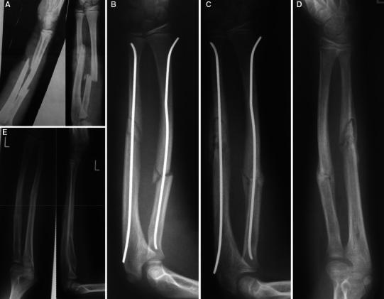
a 9-year-old girl with first-degree open forearm fracture, the complication wound is located at the ulna. b Debridement of the wound, closed reduction and ESIN. c 5 Days after trauma, an early infection appears with spontaneous purulent discharge from the complication wound. Premature removal of the ESIN 10 weeks after trauma. d Sequestration in the ulna 4 months after trauma. e 14 Months after trauma, complete adsorption of the sequestration accompanied by clinically free function
Pseudarthrosis and delayed union
All fractures of the children and adolescents that showed no bony consolidation after 6 months were classified as pseudarthrosis (Table 2). A case was described as delayed union if no bony consolidation was achieved by 12 weeks.
Table 2.
Demographic and fracture data of patients with non-unions of the ulna
| Patient | Age/sex | Primary or refracture | Soft tissue damage | Fracture localization | Reduction | Non-union type | Complications | Indication for surgery |
|---|---|---|---|---|---|---|---|---|
| 1 | 9/m | Primary | Closed | Mid-diaphysis | Ulna open; radius open | Hyperthrophic | Technical error, wound infection | Increasing axial deviation |
| 2 | 9/m | Primary | Closed | Distal diaphysis | Ulna open; radius closed | Hypothrophic | None | Increasing axial deviation |
| 3 | 12/m | Refracture | Closed | Mid-diaphysis | Ulna open; radius closed | Hyperthrophic | Wound infection, early removal of Ulna nail | Increasing axial deviation |
| 4 | 12/m | Refracture | Closed | Mid-diaphysis | Ulna open; radius closed | Hyperthrophic | None | No surgery |
| 5 | 12/m | Refracture | Closed | Mid-diaphysis | Ulna closed; radius closed | Hyperthrophic | None | No surgery |
| 6 | 13/m | Refracture | Closed | Mid-diaphysis | Ulna open; radius closed | Hyperthrophic | None | No surgery |
| 7 | 15/w | Primary | I° open ulna | Mid-diaphysis | Ulna open; radius closed | Hypothrophic | Different sizes of nails | Broken nail |
In our study, 7 patients fulfilled the criteria for pseudarthrosis, four of which were primarily treated elsewhere and the other three from the beginning at our institution. All of the pseudarthroses became manifest only in the ulna (Fig. 2), never in the radius. In six children, the pseudarthrosis was located in the middle third of the shaft and in one child in the distal third.
Fig. 2.
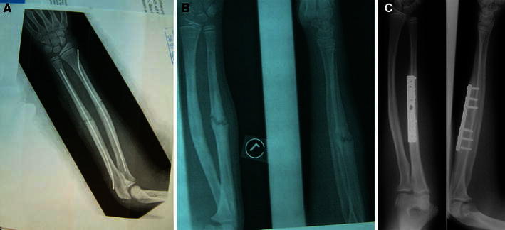
a 12-year-old boy with ESIN in forearm shaft fracture of the middle third portion. The ulna had to be reduced openly to allow intramedullary stabilization. The postoperative radiograph shows no signs of technical errors. b 15 Months after injury, there was a hypertrophic pseudarthrosis. c 10 Months after debridement of the pseudarthrosis and implantation of cancellous bone as well as plate osteosynthesis, there was complete consolidation without any functional deficits
In six cases, closed fractures were treated and in one case a first-degree open fracture. In six cases, an open reduction of the ulna had to be performed, since the closed maneuver with ESIN did not succeed. The radius was reduced closed in six cases and in one case openly with following intramedullary stabilization.
Four cases were refractures, and in one of these, it was the second refracture.
In one child, a technical mistake caused the development of the pseudarthrosis. In this patient, nails with two different diameters had to be used, because the medullary cavity was inaccessible after refracture. In four children, revision surgery was necessary; all of these had plating osteosynthesis. In three patients, spontaneous healing of the pseudarthrosis occurred.
Five cases showed a hypertrophic form; in two cases, a hypotrophic pseudarthrosis was found.
In three cases, an increasing malunion led to revision surgery, and in one of these, breaking of the Kirschner wire preceded the pseudarthrosis.
In 14 children, bony consolidation did not take place by 12 weeks, and these were classified as delayed unions. All of these fractures healed between the 11th and the 16th postoperative week. In 13 patients, the healing of the shaft of the ulna was delayed and in two patients that of the radius. In 5 cases, the ulna had to be openly reduced, and in a further case, a first-degree open fracture of the ulna had to be treated. A delayed union was noted 14 times in the middle third of the shaft and only one time in the distal third.
In our series of 537 patients, 31 fractures of the ulna and 18 of the radius were reduced in an open way. Of the 31 openly reduced ulna-fractures, 3 showed a pseudarthrosis and 5 a delayed union. We did not see any pseudarthroses of the radius.
Secondary loss of correction
In six cases, a loss of correction while the ESIN being in place was seen, and four cases of these took place in the distal diaphyseal third of the forearm, where the loss of correction did not lead to functional deficits. In two cases, a loss of correction due to a technical error was seen. The technical error was the choice of implants of too small diameter used in fractures of the middle third (Fig. 3).
Fig. 3.
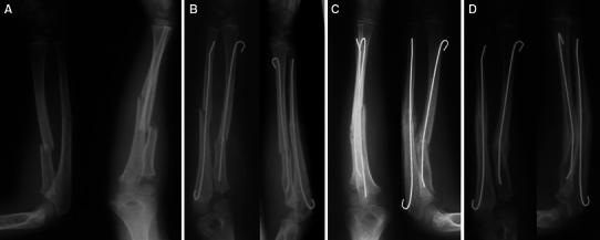
a 8-year-old girl with proximal forearm shaft fracture. b Open reduction of the ulna fracture and closed reduction of the radius with ESIN stabilization. The intramedullary nail is of an insufficient diameter (less than 2/3 of the diameter of the medullary cavity), providing no 3-point stabilization of the radius. c Loss of correction in the radius. d Partial correction through remodeling
Lesion of the superficial radial nerve
In 13 children treated in our clinic and 2 children who had been treated elsewhere, hypaesthesias in the area of the superficial radial nerve were found.
In 8 cases, the lesion originated from the primary operation, and in 7 cases, it occurred at the time of material removal. In 13 children, the hypaesthesia was temporary, and in two cases treated in our own institution, it diminished but persisted after all.
Malplacement of ESIN
In one child with refracture, a paraosseous location of the nail became obvious in the documentation x-ray after finishing the operation, and it was changed during the same anesthetic (Fig. 4).
Fig. 4.
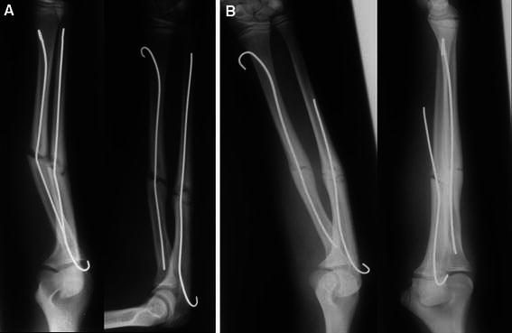
a 13-year-old boy with refracture of the forearm after fracture consolidation. b Intraoperative problems while inserting the ESIN with two technical mistakes: paraosseous malplacement of the ulna nail, that is too short as well, even if it were properly placed
Perforation of osteosynthesis material
In five children, the ESIN had to be removed prematurely, since the skin was perforated by the osteosynthesis material (Fig. 5). In three cases, the patients had been treated at our institution and in the other two cases elsewhere.
Fig. 5.
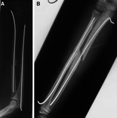
a ESIN with skin perforation due to sharp and long nail end in the ulna. b ESIN with Kirschner wires without sufficient bending at the ulnar end and consecutive perforation of the skin
In all cases, the reasons for the perforations had been technical mistakes. When K-wires were used, the ESIN-endpieces were not adequately bent and thus led to the perforation. When titanium elastic nails were used, long and sharp edges resulting from the cutoff were the reason for the described problem. In 4 children, the ESIN was removed from radius and ulna simultaneously, and in one case, the non-perforating nail was removed after complete fracture consolidation.
A bursitis had developed in 14 children at the nail ends. These lesions were not clinically relevant and were first described at the time of examination. These minor appearances were not evaluated as a complication.
Refractures
In 14 cases (11 treated at our institution/3 treated elsewhere), refractures while ESIN in place were found. All children suffered another adequate trauma. No technical problems could be identified as causes for the refractures. There was no high-grade soft tissue damage at the primary fracture.
In 5 cases, the second trauma happened in the first 6 weeks after the first fracture resulting in loss of correction, while the fracture had not healed yet. In these cases, a bending of the ESIN nails had occurred, and thus, they were not refractures in the common sense. In 8 cases, the fracture was already fully consolidated, and a refracture due to a second trauma resulted. One of these cases showed a delayed union. We recommend to remove the metal after 6 months; the average time until metal removal was 6.5 (5–15) months, and only in 5% of the patients, metal removal was performed earlier.
A refracture after implant removal was found in 13 children. In all of these children, implant removal was performed between 6 and 8 months after trauma, and we did not observe earlier implant removals.
Of the 13 refractures, 6 happened within the first 10 weeks, another 5 within 1 year and another 2 within 2 years after implant removal.
Discussion
Since the first publications of the French and Spanish research groups about elastic stable intramedullary nailing (ESIN) of forearm shaft fractures in the early 1980s [7, 8, 10], the technique developed to be the standard treatment of unstable forearm fractures in childhood and adolescence [9]. The reasons for this are the easy and minimally invasive instrumentation, the uncomplicated postoperative care and the safe and stable internal fixation. The technique is easy to learn and has a low rate of complications. In general, in most studies, positive results were reported, just few authors report about problems and complications, sometimes regarding the entire spectrum of ESIN applications. The aim of the study was explicitly to report on problems and complications of ESIN in the forearm in a very big study group over a long observation period.
Disturbance of wound healing
Although disturbances of wound healing or superficial wound infections following surgery in children are very rare, there are reports in a few publications [11, 16, 21, 22]. Mann [21] reported on 7 superficial infections in 54 patients with forearm fractures and Griffet [22] on 7 infections in 80 patients.
In our study, we saw 5 superficial infections in 553 children. The reason for these seems to be soft tissue stress during opening the cortical bone with the reamer or during insertion of the nail. The nail is usually inserted via a minimally invasive approach. A contusion of the soft tissues may occur due to the necessary angulation of the reamer or the nail while insertion. As prevention, we recommend to use (in the radius) a more distal skin incision regarding the planned entry point in the bone and to avoid excessive minimal approaches. The incision should be about 1.5 cm on the radial side. If contusions of the skin are already apparent, the traumatized skin should be resected before the suture.
Osteomyelitis
In patients primarily treated in our institution, no case of osteomyelitis was found, though there is in our study one case that had been primarily treated elsewhere with consequent development of osteomyelitis. Still this is generally a rare problem that is also very sparsely reported. Most authors reporting on ESIN in forearm fractures also did not find osteomyelitis as a complication [8, 16, 18, 19, 21]. Two authors, Cullen [23] and Schmittenbecher [15], report on single cases of osteomyelitis. In these cases as well as in the one case of our study, the fractures were primarily of the open type. Cullen and Schmittenbecher removed the ESIN, whereas in our case, the ESIN was left in place after thorough debridement and vacuum sealing. A sequestration developed, but with a persistent good forearm rotation function and normal clinical-chemical parameters, spontaneous healing and consolidation could be observed without further intervention.
Pseudarthrosis and delayed union
There are only a few reports [23–25] on delayed union or even pseudarthrosis after ESIN treatment in the forearm.
Schmittenbecher [26] reported in a multi-center study with 532 patients on 10 patients with delayed union, 7 cases in the ulna and 3 cases in the radius. A pseudarthrosis had not been described. Ogonda [27] mentions 2 patients with delayed union and one with a pseudarthrosis, in all cases the ulna was concerned in its middle third portion. Lieber [16] documented in a multi-center study with 400 patients two cases of delayed unions, but no pseudarthrosis.
As Schmittenbecher [26] and Oganda [27] reported, we also observed that the middle third portion of the ulna seems to be especially susceptible to develop a pseudarthrosis. In our 7 cases of ulna pseudarthrosis, the middle third portion was affected in 6.
Wright and Glowczweskie [28] described a relative “watershed-zone” regarding the intraossary blood circulation in the middle third portion of the ulna. This may be of significance regarding the bone healing, especially when the periosteal blood flow is damaged by open fracture or by open reduction maneuvers. Another problem may be the loss of the fracture hematoma [29] in these cases.
In our study, 6 out of 7 children with pseudarthrosis (one case had an open fracture) and 5 of 14 patients with delayed union initially had an open reduction. In the study of delayed unions described by Schmittenbecher [26], 30% open fractures were seen, and in 60%, open reduction had been performed. Ogonda [27] reported on 3 closed fractures, and in 2, the ulna had to be openly reduced. This should not mean though that first- and second-degree open fractures and open reduction are contraindicated in stabilization of forearm shaft fractures with ESIN. In any case, it should be a principle to limit damage of periosteal perfusion to a minimum [29].
Ogonda [27] made the point that the retrograde nailing of the ulna causes a distraction of the fracture and thus leads to a disturbance of the bone healing process. In our study, we could not see support for this theory, just as Schmittenbecher interpreted his data.
In the cases of our study, which developed pseudarthrosis, 3 out of 6 children had had a refracture, and in the study of Schmittenbecher [26], there was one child with a refracture. There may be a correlation between prior damage of the periosteal blood supply due to the first fracture, which could lead to an additional deficit in the healing process of a refracture.
A premature removal of osteosynthesis material was performed in our patients in 2 cases. Lieber [16] described this in one of two cases and Ogonda [27] in all 3 cases of his group that developed a pseudarthrosis. The most common reason for early removal was a deep infection.
The type of pseudarthrosis that is found is of utmost importance. There is a significant prognostic difference between a hypertrophic and a hypotrophic pseudarthrosis. In our patients, we saw 5 hypertrophic pseudarthroses. In the study described by Oganda [27], also 2 cases of hypertrophic pseudarthrosis were found, all of which consolidated after a period of 10 months spontaneously. In the absence of functional deficits, it is a well-established option to await the natural course of a hypertrophic pseudarthrosis in the juvenile age. In some cases, a creeping axial deviation may occur; in these cases, a revision with proper correction and stabilization should be performed, since permanent loss of function has to be feared even in cases with secondary remodeling of the prior deformity.
Secondary loss of correction
Loss of correction is most commonly seen in the distal third. Anyhow this zone is not ideal for treatment with ESIN. The distal radial fragment is too short and may not be sufficiently held by the nail. Due to this instability in the distal fragment, a loss of correction may occur. Therefore, the external fixateur is described as a favorable solution in this problem by some authors. In our opinion, a molded forearm cast that is applied in addition to the ESIN in the first 2–4 weeks is sufficient to prevent or at least significantly reduce loss of correction without too much discomfort for the patient. Some authors [7, 9, 20] describe it as a false indication to treat fractures in the distal forearm shaft third/quarter with ESIN. Alternatives might be plating, percutaneous K-wire osteosynthesis or the external fixateur. Our experience with ESIN though suggests that even suboptimal ESIN treatment in this difficult zone beats the other beforehand named methods. However, it is of special importance to use sufficiently stable or thick nails to prevent loss of correction, which happened in one case in our study. In two cases of our series, a loss of correction in the proximal third due to a technical error was seen. The error was the choice of implants of a too small diameter (Fig. 3). The diameter of the nails has to be 2/3 of the medullary canal, measured in the midshaft region.
Lesion of the superficial radial nerve
Lesions of the superficial radial nerve are a common complication in forearm shaft fractures treated with ESIN [8, 11, 18, 23, 30].
The lesion occurs in the primary fracture treatment as well as at the time of material removal at a similar rate. There is a controversy whether the intraoperative identification of the superficial radial nerve is to be advised. On one hand, there are reports about unproblematic and also mandatory identification of the nerve, and on the other hand, many authors do not see the necessity to do so. Since the nerve splits into diverse sensory branches in the region of the surgical approach, we are of the opinion that with careful blunt subcutaneous preparation and a sufficient approach (ca. 2 cm), there is minimal risk of causing a nerve lesion. At the time of material removal, there is usually some scarring that also obstructs easy identification of the nerve, which may lead to damage while trying to do so.
Many of the operations to remove the osteosynthesis material are widely considered as surgery for ‘starters’, i.e., inexperienced surgeons. In these cases, a slightly enlarged approach should be considered to maintain a good exposure of the endangered structures.
Due to the described problems with the superficial radial nerve, the need for better alternatives had been seen, and thus, for example, the approach dorsal of the Lister’s tubercle had been propagated [9]. This approach though has its own morbidity regarding the complexity of the extensor tendons. It may result in the need to elaborately reconstruct complete lesions of the extensor pollicis longus tendon and the extensor carpi radialis brevis tendon [8, 9].
Malplacement of ESIN
The paraosseous malplacement of an ESIN nail is a severe technical mistake. The problem may occur, if under fluoroscopy only one radiographic plane is taken and then trusted, that the nail is safely intramedullarily placed. To avoid this, fluoroscopy in 2 planes differing by 90° has to be performed as a routine safety control.
Perforation of the nail ends
Irritation induced by the implanted material is a well-known problem [7, 16, 18, 23], and in some cases, even perforation of the skin may occur. This complication usually is the result of technical mistakes.
After the fracture reduction and the positioning of the nails, great care should be diverted to the correct cutting and placing of the nail ends, to avoid possible soft tissue problems.
In our institution, Kirschner wires are used that are bent by 180° to be placed smoothly next to the bone after the final placement. Since the remaining prominence is then round and without edges, we very rarely encounter soft tissue irritations or perforations.
Migration of the nails may eventually occur and thus lead to irritation or even skin perforation. The reason may be an insufficiently pre-bent and/or too thin nail.
Refractures
Refractures after forearm fractures are the most common extremity shaft refractures in childhood and adolescence [30]. The risk of a forearm shaft refracture is reportedly approximately 3–8%, and in most cases, the primary fracture having been of the greenstick type [8, 9, 31–33]. In these fracture types, a delayed consolidation of the bone and an increased rate of refractures are observed. Accordingly, 3 children of our study had greenstick fractures as primary fracture.
Many authors report on refractures after material removal [15–17, 23, 34]; therefore, the recommendation should be to remove the osteosynthesis material after complete consolidation of the fracture, i.e., not before 16 weeks after trauma. Some authors are of the opinion that early metal removal leads to a significantly higher risk of refractures [8, 15, 16, 20]. We do not have to cope with this problem because we perform metal removal later, generally after a period of 6 months.
Conclusions
The analysis of the complications of elastic stable intramedullary nailing in children’s forearm fractures shows that a differentiated approach has to be taken to the assessment of the data. Most of the complications are caused by incorrect use of the method, thus being the surgeon himself the reason for the failure of the method. Though the usage of ESIN is easy to learn, it still has to be learned meticulously. If the use of the ESIN method is extended to more problematic regions, such as the distal diaphyseal portion of the forearm, an increase in complications is likely. It has to be added that in a minority of patients, these complications should be considered unavoidable.
The surgeon must know about possible complications in order to safely prevent them.
In our opinion, the ESIN method for treating forearm shaft fractures is a safe and well-suited method for children and adolescents, since it is functionally and cosmetically appealing and leads to very good results with an overall small complication rate.
References
- 1.Hugstone JC. Fractures of the forearm in children. J Bone Joint Surg Am. 1982;64-A:14–17. [Google Scholar]
- 2.Price CT, Scott DS, Kurzner ME, Flynn JC. Malunited forearm fractures in children. J Pediatr Orthop. 1990;10:705–712. doi: 10.1097/01241398-199011000-00001. [DOI] [PubMed] [Google Scholar]
- 3.Sarmiento A, Ebramzadeh E, Brys D, Tarr R. Angular deformities and forearm function. J Orthop Res. 1992;10:121–133. doi: 10.1002/jor.1100100115. [DOI] [PubMed] [Google Scholar]
- 4.Tarr R, Garfinkel AI, Sarmiento A. The effects of angular and rotational deformities of both bones of the forearm: an in vitro study. J Bone Joint Surg Am. 1984;66-A:65–70. [PubMed] [Google Scholar]
- 5.Daruwalla JS. A study of radioulnar movements following fractures of the forearm in children. Clin Orthop. 1979;139:114–120. [PubMed] [Google Scholar]
- 6.Fuller DJ, McCullought CJ. Malunited fractures of the forearm in children. J Bone Joint Surg B. 1982;64:364–367. doi: 10.1302/0301-620X.64B3.7096406. [DOI] [PubMed] [Google Scholar]
- 7.Perez Sicilia JE, Morote Jurado JL, Corbacho Girones JM, Hernandez Cabrera JA, Gonzales Buendia R. Osteosinthesis pecutanea en fracturas diafisarias de antebrazo en ninos y adolescentes. Rev Esp Cir Osteoartic. 1977;12:321–334. [Google Scholar]
- 8.Lascombes P, Prevot J, Ligier N, Metaizeau JP, Poncelet T. Elastic stable intramedullary nailing in forearm shaft fractures in children: 85 cases. J Pediatr Orthop. 1990;10:167–171. doi: 10.1097/01241398-199003000-00005. [DOI] [PubMed] [Google Scholar]
- 9.Schmittenbecher PP. State of the art treatment of the forearm fractures. Inj Int J Care Inj. 2005;36:25–34. doi: 10.1016/j.injury.2004.12.010. [DOI] [PubMed] [Google Scholar]
- 10.Metaizeau JP. L’osteosynthese de l’enfant: techniques et indications. Rev Chir Orthop. 1983;69:495–511. [PubMed] [Google Scholar]
- 11.Schoemaker SD, Comstock CP, Mubarak SJ, Wenger DR, Chambers HG. Intramedullary Kirschner wire fixation of open or unstable forearm fractures in children. J Pediatr Orthop. 1999;19:329–337. [PubMed] [Google Scholar]
- 12.Ortega R, Loder RT, Louis DS. Open reduction and internal fixation of forearm fractures in children. J Pediatr Orthop. 1996;16:651–654. doi: 10.1097/01241398-199609000-00019. [DOI] [PubMed] [Google Scholar]
- 13.Kay S, Smith C, Oppenheim WL. Both Bone midshaft forearm fractures in children. J Pediatr Orthop. 1986;6:306–310. doi: 10.1097/01241398-198605000-00009. [DOI] [PubMed] [Google Scholar]
- 14.Nielsen AB, Simson O. Displaced forearm fractures in children treated with AO plates. Injury. 1984;15:393–396. doi: 10.1016/0020-1383(84)90204-3. [DOI] [PubMed] [Google Scholar]
- 15.Schmittenbecher PP, Dietz HG, Linhart WE, Slongo T. Complications and problems in intramedullary nailing of childrens’ fractures. Eur J Trauma. 2000;26:287–293. doi: 10.1007/PL00002453. [DOI] [Google Scholar]
- 16.Lieber J, Joeris A, Knorr P, Schalomon J, Schmittenbecher PP. ESIN in forearm fractures. Eur J Trauma. 2005;31:3–11. doi: 10.1007/s00068-005-1071-7. [DOI] [Google Scholar]
- 17.Yung SH, Lam CYA, Choi KY, Ng KW, Maffulli N, Cheng JCY. Percutaneous intramedullary Kirschner wiring for displaced diaphyseal forearm fractures in children. J Bone Joint Surg B. 1998;80:91–94. doi: 10.1302/0301-620X.80B1.8110. [DOI] [PubMed] [Google Scholar]
- 18.Jubel A, Andermahr J, Isenberg J, Issavand A, Prokop A, Rehm KE. Outcomes and complications of elastic stable intramedullary nailing for forearm fractures in children. J Pedia Orthop B. 2005;14:375–380. doi: 10.1097/01202412-200509000-00012. [DOI] [PubMed] [Google Scholar]
- 19.Garg NK, Ballal MS, Malek IA, Webster RA, Bruce CE. Use of elastic stable intramedullary nailing for treating unstable forearm fractures in children. J Trauma. 2008;65:109–115. doi: 10.1097/TA.0b013e3181623309. [DOI] [PubMed] [Google Scholar]
- 20.Slongo T. Complications and failures of the ESIN technique. Injury. 2005;36:78–85. doi: 10.1016/j.injury.2004.12.017. [DOI] [PubMed] [Google Scholar]
- 21.Mann D, Schnabel M, Baacke M, Gotzen L. Ergebnisse der elastischen stabilen intramedullären Nagelung (ESIN) bei Unterarmschaftfrakturen im Kindesalter. Unfallchirurg. 2003;106:102–109. doi: 10.1007/s00113-002-0474-8. [DOI] [PubMed] [Google Scholar]
- 22.Griffet J, El Hayek T, Baby M. Intramedullary nailing of forearm fractures in children. J Pediatr Orthop B. 1990;8:88–89. [PubMed] [Google Scholar]
- 23.Cullen MC, Dennis RR, Giza E, Crawford AH. Complications of intramedullary fixation of pediatric forearm fractures. J Pediatr Orthop. 1998;18:14–21. [PubMed] [Google Scholar]
- 24.Luhmann SJ, Gordon JE, Schoenecker PL. Intramedullary fixation of unstable both-bone forearm fractures in children. J Pediatr Orthop. 1998;18:451–456. [PubMed] [Google Scholar]
- 25.Lascombes P, Haumont T, Journeau P. Use and abuse of flexible intramedullary nailing in children and adolescents. J Pediatr Orthop. 2006;26:827–834. doi: 10.1097/01.bpo.0000235397.64783.d6. [DOI] [PubMed] [Google Scholar]
- 26.Schmittenbecher PP, Fitze G, Gödecke J, Kraus R, Schneidmüller D. Delayed healing of the forearm shaft fractures in children after intramedullary nailing. J Pediatr Orthop. 2008;28:303–306. doi: 10.1097/BPO.0b013e3181684cd6. [DOI] [PubMed] [Google Scholar]
- 27.Ogonda L, Wong-Chung J, Wray R, Canavan B. Delayed union and non-union of the ulna following intramedullary nailing in children. J Pediatr Orthop B. 2004;13:330–333. doi: 10.1097/01202412-200409000-00009. [DOI] [PubMed] [Google Scholar]
- 28.Wright TW, Glowczewskie F. Vascular anatomy of the ulna. J Hand Surg (Am) 1998;23:800–804. doi: 10.1016/S0363-5023(98)80153-6. [DOI] [PubMed] [Google Scholar]
- 29.Greenbaum B, Zionts LE, Ebramzadeh E. Open fractures of the forearm in children. J Orthop Trauma. 2001;15:111–118. doi: 10.1097/00005131-200102000-00007. [DOI] [PubMed] [Google Scholar]
- 30.Landin LA. Epidemiology of children’s fractures. J Pediatr Orthop B. 1997;6:79–83. doi: 10.1097/01202412-199704000-00002. [DOI] [PubMed] [Google Scholar]
- 31.Schwarz AF, Höcker K, Schwarz N, Jelen M, Styhler W, Mayr J, Brass D, Jansky W, Poigenfürst J, Straub G. Die Refraktur des kindlichen Unterarmes. Unfallchirurg. 1996;99:175–182. [PubMed] [Google Scholar]
- 32.Fiala M, Carey TP. Paediatric forearm fractures: an analysis of refracture rates. Orthop Trans. 1994;18:1265–1266. [Google Scholar]
- 33.Litton LO, Adler F. Refracture of the forearm in children: a frequent complication. J Trauma. 1963;3:41–51. doi: 10.1097/00005373-196301000-00004. [DOI] [PubMed] [Google Scholar]
- 34.Mittal R, Hafez MA, Templeton PA. Failure of forearm intramedullary elastic nails. Injury. 2004;35:1319–1321. doi: 10.1016/j.injury.2003.10.029. [DOI] [PubMed] [Google Scholar]


