Abstract
A variable number of tandem repeats (short (S) vs long (L)) in the promoter region of the serotonin transporter gene (5-HTTLPR) and a functional variant of a single-nucleotide polymorphism (rs25531) in 5-HTTLPR have been recently associated with increased risk for major depressive disorder (MDD). In particular, relative to L/L or LA homozygotes (hereafter referred to as L′ participants), S carriers or Lg-allele carriers (S′ participants) have been found to have a higher probability of developing depression after stressful life events, although inconsistencies abound. Previous research indicates that patients with MDD are characterized by executive dysfunction and abnormal activation within the anterior cingulate cortex (ACC), particularly in situations requiring adaptive behavioral adjustments following errors and response conflict (action monitoring). The goal of this study was to test whether psychiatrically healthy S′ participants would show abnormalities similar to those of MDD subjects. To this end, 19 S′ and 14 L′ participants performed a modified Flanker task known to induce errors, response conflict, and activations in various ACC subdivisions during functional magnetic resonance imaging. As hypothesized, relative to L′ participants, S′ participants showed (1) impaired post-error and post-conflict behavioral adjustments; (2) larger error-related rostral ACC activation; and (3) lower conflict-related dorsal ACC activation. As similar behavioral and neural dysfunctions have been recently described in MDD patient samples, the current results raise the possibility that impaired action monitoring and associated ACC dysregulation may represent risk factors increased vulnerability to depression.
Keywords: serotonin transporter promoter polymorphism (5-HTTLPR), anterior cingulate cortex, action monitoring, conflict monitoring, executive functioning, depression
INTRODUCTION
Both genetic and environmental factors are implicated in the etiology of major depressive disorder (MDD; Wong and Licinio, 2001). However, despite evidence indicating that depression is heritable (Sullivan et al, 2000), few genes have been consistently linked to MDD, and inconsistencies abound. Given the relatively small effects of individual genes and the heterogeneous nature of MDD, it is unlikely that a one-to-one relationship between specific genes and MDD exists (Sanders et al, 2004). Pinpointing possible neurobiological abnormalities associated with individual symptoms or core disease dysfunctions might provide a useful platform for improving our understanding of the etiology of MDD. Accordingly, reducing heterogeneity through the study of less complex and more clearly delineated aspects of MDD could enhance our ability to isolate ‘endophenotypes,' ie, phenotypes lying within the causal chain between gene and disorder (Hasler et al, 2004).
Impaired executive functioning is a hallmark feature of depression (Austin et al, 2001). In particular, individuals with MDD are characterized by ‘action monitoring' deficits, including reduced accuracy in trials after mistakes (Beats et al, 1996; Elliott et al, 1996; Holmes and Pizzagalli, 2008b) or involving conflict between competing responses (Paradiso et al, 1997; Siegle et al, 2004). Fitting conceptualizations that impaired action monitoring may reflect an important depressive endophenotype (Olvet and Hajcak, 2008), such deficits have also been described in individuals at increased genetic risk for MDD (Althaus et al, 2009; Fallgatter et al, 2004) or in those in remission (Vanderhasselt and De Raedt, 2009).
Parallel cognitive neuroscience research has greatly advanced our understanding of action monitoring. The anterior cingulate cortex (ACC), in particular, has a key role in the generation of adaptive behaviors after errors and fluctuating task difficulty (Botvinick et al, 1999). Highlighting functional specialization, dorsal ACC (dACC) and rostral ACC (rACC) regions are preferentially recruited during conflict monitoring and error processing, respectively (Ridderinkhof et al, 2004). Recently, data indicating that MDD is characterized by region-specific ACC dysfunctions have emerged. Thus, in response to errors, MDD subjects show potentiated rACC activation (Holmes and Pizzagalli, 2008b), consistent with ERP studies highlighting increased error sensitivity in depression (Chiu and Deldin, 2007; but see Schrijvers et al, 2008, 2009). Conversely, during high-response conflict trials, MDD subjects show reduced dACC activation (George et al, 1997; Holmes and Pizzagalli, 2008a). Whether impaired action monitoring and associated ACC dysregulation represent correlates of, or vulnerability factors for, MDD is largely unknown.
A variable number of tandem repeats (VNTRs; short (S) and long (L)) in the promoter region of the serotonin transporter (5-HTTLPR) gene might confer increased MDD risk, particularly following stress (Brown and Harris, 2008; Caspi et al, 2003; cf. Risch et al, 2009). Critically, healthy S-allele carriers have heightened ERN amplitude (Althaus et al, 2009; Fallgatter et al, 2004), suggesting that the action monitoring dysfunction may be a mechanism contributing to the increased vulnerability to depression. Of relevance, a functional variant of a single-nucleotide polymorphism (SNP; rs25531, A/G) in 5-HTTLPR has also been found to impact the function of the serotonin transporter (5-HTT). Specifically, when combined with the L VNTR allele, the G allele results in 5-HTT mRNA levels and clinical outcomes similar to the S allele (Hu et al, 2005; Murphy et al, 2008; Zalsman et al, 2006). Thus, lack of consideration of this SNP might explain some of the inconsistencies in the literature.
The goal of this study was to test whether the S and/or LG allele might confer an increased risk for depression through action monitoring deficits and dysregulated ACC functioning. To this end, we collected functional magnetic resonance imaging (fMRI) data during performance of a task known to induce errors and response conflict from psychiatrically healthy participants genotyped for both the 5-HTTLPR VNTR and the SNP. Given previous evidence of (1) increased risk for depression in S-allele and LG-allele carriers (Caspi et al, 2003; Zalsman et al, 2006), (2) increased ERN amplitudes in S-allele carriers (Althaus et al, 2009; Fallgatter et al, 2004), and (3) heightened rACC response to errors and impaired post-error behavioral adjustments in MDD (Chiu and Deldin, 2007; Holmes and Pizzagalli, 2008b), we hypothesized that S- and LG-allele carriers (hereafter referred to as S′), relative to LA/LA homozygotes (hereafter referred to as L′), would exhibit increased rACC activation to errors and decreased post-error performance. In addition, given conflict-monitoring deficits and associated decreased dACC activation in MDD (Holmes and Pizzagalli, 2008a), we expected reduced behavioral performance and decreased dACC activity during high-response conflict trials in the S′ group, relative to the L′ group.
MATERIALS AND METHODS
Participants
Between November 2006 and May 2008, 38 right-handed participants were recruited from the Boston area. Exclusion criteria included current/past Axis I or neurological disorder, current/past psychotropic medication, acute physical illness, and loss of consciousness. To prevent population stratification confounds, Caucasians of European ancestry were recruited. Participants provided written informed consent to a protocol approved by the Committee on the Use of Human Subjects in Research (Harvard University) and Partners Health Care System Human Subjects Committee.
The Structured Clinical Interview for the DSM-IV (SCID; First et al, 2007) was administered by masters-level clinicians to assess eligibility. Eligible participants completed the Beck Depression Inventory-II (BDI-II; Beck et al, 1996), the Mood and Anxiety Symptom Questionnaire (MASQ; Watson et al, 1995), and the Perceived Stress Scale (PSS; Cohen et al, 1983) to assess anxiety, depression symptoms, general distress, and perception of ongoing stressors. Genotyping was accomplished using a saliva sample (Oragene, DNA Genotek, Ottawa, ON, Canada). Data pertaining to fMRI were collected within 1 week of the SCID. Participants were debriefed after study completion and compensated with $10 per hour and $60 for the interview and fMRI session, respectively. Data obtained from five participants were lost because of chance performance (n=1), technical issues (n=3), or excessive head movement (n=1). The final sample consisted of 33 participants (L′: n=14, S′: n=19). Genotype groups did not differ in any demographic or self-reported variable (Table 1).
Table 1. Summary of Demographic and Self-Report Data.
| Variable |
L′ subjects (n=14) |
S′ subjects (n=19) |
χ2/t-value | p-value | ||
|---|---|---|---|---|---|---|
| Mean | SD | Mean | SD | |||
| Age | 22.92 | 4.07 | 23.88 | 3.01 | 0.74 | >0.46 |
| Years of education | 15.81 | 3.73 | 16.47 | 2.42 | 0.60 | >0.55 |
| Percentage female | 64.3 | N/A | 63.2 | N/A | 0.004 | >0.94 |
| BDI-II | 1.11 | 1.56 | 1.00 | 1.24 | 0.22 | >0.83 |
| MASQ AA | 18.29 | 1.38 | 17.95 | 2.63 | 0.44 | >0.67 |
| MASQ AD | 47.86 | 10.88 | 45.42 | 9.80 | 0.51 | >0.51 |
| MASQ GDA | 14.71 | 4.39 | 13.47 | 2.37 | 0.88 | >0.38 |
| MASQ GDD | 15.74 | 7.23 | 14.36 | 2.44 | 0.68 | >0.50 |
Abbreviations: AA, anxious arousal; AD, anhedonic depression; BDI-II, Beck Depression Inventory (Beck et al, 1996); GDA, general distress-anxiety; GDD, general distress-depression; MASQ, Mood and Anxiety Symptom Questionnaire (Watson et al, 1995).
Flanker Task
Trials began with a probe consisting of five arrows presented in the center of the screen. Participants were instructed to respond as quickly and accurately as possible with the index finger of their hand corresponding to the direction the center arrow was pointing. Trials were either congruent (‘<<<<<,' ‘>>>>>') or incongruent (‘<<><<,' ‘>><>>'). To familiarize participants with the task, and to generate reaction-time (RT) data required to compute individually titrated response windows (see below), a practice block was presented before data collection (46 congruent, 24 incongruent trials).
After the presentation of the practice block, participants completed 5 blocks during fMRI data collection, each with 46 congruent and 24 incongruent trials. Trials consisted of a probe presented for 200 ms, followed by a variable inter-trial interval (ITI; 2250–7250 ms). During experimental blocks, feedback was presented for 300 ms, followed by a variable ITI (2250–7250 ms). Positive feedback (a schematic smiling face) followed correct responses made within the individually titrated response window (85th percentile of each participant's practice RT). Negative feedback (a schematic frowning face) followed incorrect responses, or responses exceeding the response window. To accommodate performance shifts, the response window was updated at the midpoint and endpoint of each block.
The number of congruent trials preceding each incongruent trial was fully randomized using optseq2 (http://surfer.nmr.mgh.harvard.edu/optseq/). To maximize statistical orthogonality among conditions, ISIs and ITIs were determined using a genetic algorithm (Wager and Nichols, 2003).
Genetic Analyses
DNA was purified, extracted, hydrated, and stored at −80°C. 5-HTTLPR VNTR and SNP (rs25531) genotyping was performed following established procedures (Wendland et al, 2006). Briefly, in a 20 μl solution, genomic DNA (25 ng) was amplified through a PCR in the presence of 1 × multiplex master mix (Qiagen, Valencia, CA) and primers (forward: 5′-TCCTCCGCTTTGGCGCCTCTTCC-3′ reverse: 5′-TGGGGGTTGCAGGGGAGATCCTG-3′ Integrated DNA Technologies, Coralville, IA). Next, 7 μl of PCR product was digested by HpaII (13 μl; New England BioLabs, Ipswich, MA) in a reaction assay with 1 × NEBuffer1 and 1 × BSA (Ambion, Foster City, CA). Finally, 4 μl of the remaining PCR product and 18 μl of the restriction enzyme assay solution were loaded onto a 2.0% agarose gel (E-Gel, Invitrogen, Carlsbad, CA) and visualized after 15, 25, and 45 min. Participants were grouped as LA homozygotes (LA/LA, n=14) and S or LG carriers (S/S homozygotes, n=4; S/L or S/LA, n=15). This choice was motivated by previous research highlighting the functional dominance of the S allele (Hariri et al, 2005; Brown and Hariri, 2006), and by the observation that pairing of the G allele and L VNTR allele results in functional (Hu et al, 2005) and clinical (Zalsman et al, 2006) outcomes similar to those of the S allele. Neither the VNTR nor SNP deviated significantly from the Hardy–Weinberg equilibrium, both p's>0.81.
fMRI Acquisition
fMRI images were acquired on a 1.5-T Symphony/Sonata scanner (Siemens Medical Systems; Iselin, NJ). The protocol included a T1-weighted MPRAGE volume (TR/TE: 2730/3.39 ms; FOV: 256 mm; voxel dimensions: 1 × 1 × 1.33 mm3; 128 slices), and 5 functional gradient echo T2*-weighted echoplanar runs (TR/TE: 2500/35 ms; FOV: 200 mm; voxel dimensions: 3.125 × 3.125 × 3 mm3; 35 slices). Tilted slice acquisition (30° to AC-PC line) and z-shimming were used to minimize susceptibility artifacts (Deichmann et al, 2003).
Data Reduction and Statistical Analyses
Behavioral data
Only trials with a response were examined, and RT analyses were restricted to correct responses. To minimize the influence of outliers, RTs exceeding mean±3 SDs (after log transformation) were excluded (3.17±3.97%). Primary analyses focused on behavioral adjustments occurring with response conflict and error commission. Flanker interference effects were calculated as [RTIncongruent trials−RTCongruent trials] and (AccuracyCongruent trials−AccuracyIncongruent trials), with higher scores indicating increased interference. The Gratton effect, a measure of post-conflict behavioral adjustments (Gratton et al, 1992), was calculated as [RTIncongruent trials following congruent trials−RTIncongruent trials following incongruent trials] and [AccuracyIncongruent trials following incongruent trials−AccuracyIncongruent trials following congruent trials], with higher scores reflecting increased cognitive control. Post-error adjustments (Rabbitt, 1966; Laming, 1979) were operationalized as [RTAfter incorrect trials−RTAfter correct trials] and [AccuracyAfter incorrect trials−AccuracyAfter correct trials], with higher scores indicating more adaptive behavioral adjustments. Finally, following previous studies (eg, Kerns et al, 2004), a ‘post-conflict RT adjustment score' was computed. This effect examines whether participants show faster RT on incongruent trials preceded by incongruent trials (iI) relative to incongruent trials preceded by congruent trials (cI), as well as decreased RT on congruent trials preceded by congruent trials (cC) relative to congruent trials preceded by incongruent trials (iC). Post-conflict RT adjustment scores were calculated as (iC−cC)+(cI−iI). For the sake of brevity, only effects involving Group are reported for post-conflict RT adjustment scores. Overall, to disentangle adjustment effects, analyses assessing post-error adjustments were restricted to trials following incongruent trials, whereas Flanker–Gratton post-conflict adjustment effects were restricted to post-correct trials.
Imaging data
Data were analyzed using FS-FAST and FreeSurfer (http://surfer.nmr.mgh.harvard.edu/). After slice time and motion correction, fMRI data were detrended and spatially smoothed (Gaussian filter, FWHM: 6 mm3). Before group analyses, data were resampled into MNI305 space (2 mm3 voxels).
Functional data were analyzed using a general linear model (GLM), with motion parameters included as nuisance regressors. The hemodynamic response was modeled as a γ-function (2.25-s delay, 1.25-s dispersion) and convolved with stimulus/response onsets. A temporal whitening filter was used to account for autocorrelation in the noise. A random-effects model was for population-based inferences. For each participant, one mean image was generated per condition and then combined in a series of linear contrasts. Mimicking the behavioral analysis, only trials with a response were considered.
Statistical Analyses
Behavioral data
Exploratory analyses revealed no effects of demographics. As Shapiro–Wilk tests showed that the RT and accuracy data were not normally distributed, all statistical tests were conducted on log-transformed data (for ease of interpretation, non-normalized means and SDs are presented in text and figures). Separate mixed 2 × 2 analyses of variance (ANOVAs) with Group (L′, S′) and Condition (eg, incongruent RT, congruent RT) as factors were conducted. To examine post-conflict adjustments, Group × Current Trial (incongruent trials vs congruent trials) × Previous Trial (post-incongruent trials vs post-congruent trials) ANOVAs were conducted. Post hoc Newman–Keuls tests were conducted in case of significant ANOVA findings.
fMRI data
Between-group whole-brain random effects comparisons were computed for the contrasts of interest: Flanker effects (incongruent correct responses>congruent correct responses) and errors (incongruent error responses>incongruent correct responses). Given previous findings of ACC abnormalities (Pezawas et al, 2005) and increased ERN amplitudes (Althaus et al, 2009; Fallgatter et al, 2004) in S-allele carriers, primary analyses targeted cingulate regions. The left and right ACC (Brodmann areas (BAs) 24/32, and spanning into the posterior and subgenual cingulate cortex, BAs 31/23/25) were defined through an automated parcellation system (Fischl et al, 2004), which was visually inspected for accuracy. To correct for multiple comparisons, 10 000 Monte Carlo permutations were run (AlphaSim), yielding a combination of p<0.005 and 14-voxel cluster extent to achieve a corrected p<0.05 within the ACC. In case of significant findings, β-weights were extracted from the cluster exceeding the statistical threshold, averaged across voxels, and entered into Group × Condition ANOVAs. As regions surviving the permutation reflect, by design, significant Group × Condition interactions, follow-up tests are directly reported.
Finally, secondary whole-brain analyses were performed. On the basis of permutations considering the entire brain volume, these analyses were thresholded at p<0.005 with a minimum cluster extent of 79 voxels, yielding a corrected p<0.05. Owing to the limited number of errors, analyses of trials after an initial mistake were not possible.
RESULTS
Behavioral Data
Flanker effects
As expected, a main effect of Condition (incongruent vs congruent) emerged for both log-transformed accuracy (F(1, 31)=75.19, p<0.001; partial η2=0.71) and RT (F(1, 31)=234.44, p<0.001; partial η2=0.88) scores. Participants were more accurate (0.99±0.02) and faster (432.08±55.75 ms) for congruent relative to incongruent trials (accuracy: 0.83±0.09; RT: 517.89±84.57 ms). No effects involving Group emerged (F's<0.64, p's>0.43).
Post-error adjustment effects
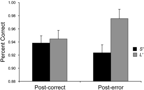
When examining post-error accuracy, the main effect of Condition (post-error vs post-correct trials) was not significant (F(1, 31)=0.45, p>0.51), whereas the main effect of Group was trending (F(1, 31)=4.29, p<0.056; partial η2=0.12). Critically, the Group × Condition interaction (F(1, 31)=4.29, p<0.047; partial η2=0.12) was significant (Figure 1). Post hoc tests showed that the interaction was due to the expected increase in post-error (0.98±0.03) relative to post-correct (0.95±0.06) accuracy for the L′ group (p<0.046), but not for the S′ participants (post-error: 0.92±0.06 vs post-correct: 0.94±0.04; p>0.35). In addition, as hypothesized, relative to the L′ group, S′ participants were significantly less accurate after incorrect (p<0.005) but not correct (p>0.77) responses. No significant effects emerged when considering RT (F's<1.91, p's>0.18). Overall, these results indicate significantly reduced post-error behavioral adjustments (Laming effects) in the S′ group relative to the L′ participants.
Figure 1.
Mean (and SE) accuracy in trials immediately after an incorrect vs correct trial for the L′ (n=14) and S′ (n=19) participants. To disentangle error and congruency effects, only incongruent trials were considered. Error bars represent SE.
Post-conflict adjustment effects
When considering accuracy, the main effect of Condition (post-congruent incongruent trials vs post-incongruent incongruent trials) was significant (F(1, 31)=4.57, p<0.041; partial η2=0.13), due to increased post-incongruent (0.84±0.12) relative to post-congruent (0.81±0.10) accuracy. Contrary to our hypotheses, neither the main effect of Group nor the Group × Condition interaction were significant (F's<0.27, p's>0.61).
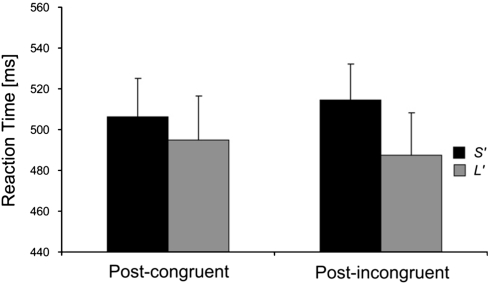
For RT, a Group × Condition interaction emerged (F(1, 31)=4.84, p<0.035; partial η2=0.14; Figure 2). Post hoc tests showed that this effect was due to slower post-incongruent (514.50±75.99) relative to post-congruent (506.38±70.70 ms) RT for the S′ group (p<0.08), a pattern absent in L′ participants (post-incongruent: 487.50±79.89 ms vs post-congruent 494.82±94.01; p>0.21). However, no significant group differences emerged (p's>0.28). The main effects of Condition (F(1, 31)=0.08, p>0.79) and Group (F(1, 31)=0.67, p>0.42) were not significant.
Figure 2.
Mean (and SE) reaction times in trials immediately after an incongruent vs congruent trial for the L′ (n=14) and S′ (n=19) participants. To disentangle error and congruency effects, only correct trials were considered. Error bars represent SE.
When considering post-conflict adjustment accuracy scores (Kerns et al, 2004), the Group × Current Trial (incongruent trials vs congruent trials) × Previous Trial (post-incongruent trials vs post-congruent trials) ANOVA revealed no effects involving Group (F's<0.46, p's>0.49), in line with analyses on the traditional Gratton accuracy effect. When considering adjustment RT scores, the only significant effect involving Group was the Group × Previous Trial interaction (F(1, 31)=7.48, p<0.01; partial η2=0.19). Post hoc tests revealed that, for the S′ group, post-incongruent RTs were significantly slower than post-congruent RTs (481.63±60.60 vs 470.62±57.54 ms; p<0.001); for L′ participants, no differences emerged between the two conditions (post-incongruent: 458.86±70.27 ms, post-congruent: 457.69±72.02; p>0.58).
fMRI Data
Flanker effect (incongruent correct responses>congruent correct responses)
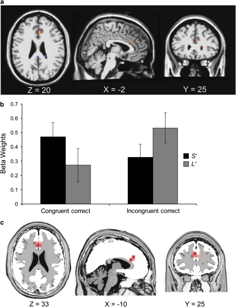
As hypothesized, compared with the S′ group, L′ participants exhibited significantly larger activation during high- vs low-conflict trials in a left dACC cluster falling into the cognitive subdivision of BA 24 (Ridderinkhof et al, 2004; Vogt et al, 1995; Figure 3a; Table 2a). Follow-up tests revealed that L′ participants showed significantly higher dACC activation during incongruent relative to congruent trials (incongruent: 0.533±0.424; congruent: 0.274±0.367; p<0.016); interestingly, S′ participants showed a reverse pattern (incongruent: 0.327±0.374, congruent: 0.471±0.476; p<0.031). In spite of these within-group differences, the L′ and S′ groups did not differ in their activation for congruent (p>0.21) or incongruent (p>0.15) trials.
Figure 3.
Conflict-related activity for the L′ (n=14) vs S′ (n=19) participants. (a) Axial, saggital, and coronal slices showing the dACC cluster (incongruent vs congruent) thresholded at p<0.005 (x=−2, y=25, z=20). (b) Mean (and SE) β-weights extracted from the dACC cluster exceeding the statistical threshold (p<0.05, corrected). (c) dACC cluster reported in a previous ERP study, in which MDD subjects exhibited decreased current density relative to control participants 620 ms after the presentation of an incongruent trial in a Stroop task (peak voxel Talairach coordinates: x=−10, y=25, z=33; Holmes and Pizzagalli, 2008a). The Euclidian distance between the fMRI and ERP voxel is 15.8 mm.
Table 2. Summary of Primary (Regions-of-Interest) Analyses Contrasting L′ and S′ Participants with ACC Regions.
| Peak voxel location | Volume (mm3) | X | Y | Z | Z-score | BA |
|---|---|---|---|---|---|---|
| (a) Response conflict (incongruent correct vs congruent correct) | ||||||
| L dACC | 176 | −2 | 25 | 20 | 3.70 | 24 |
| R Posterior ACC | 192 | 2 | −33 | 25 | −4.74 | 23 |
| (b) Error commission (incongruent error vs incongruent correct) | ||||||
| L rACC | 288 | −12 | 35 | 14 | −3.59 | 32 |
| L Subgenual ACC | 128 | −6 | 18 | −10 | 2.92 | 32 |
Abbreviations: ACC, anterior cingulate cortex; BA, Brodmann area; L, left; R, right.
Coordinates are in Talairach space. All regions meet p<0.05, corrected (based on ACC volume: p<0.005, uncorrected, cluster >14 voxels). Reported coordinates and Z-scores are for peak voxels. Z-scores >0 indicate greater activation in the L′ than in the S′ group; Z-scores <0 indicate greater activation in the S′ than in the L′ group.
Significant group differences also emerged in a cluster within the right posterior cingulate cortex (BA 23). Compared with the L′ group, S′ participants showed significantly higher activation during high- vs low-conflict trials (Table 2). However, follow-up tests indicated that genotype groups did not significantly differ in their posterior cingulate activation for congruent (p>0.29) or incongruent trials (p>0.32). Nevertheless, in the S′ group, posterior cingulate activation was significantly higher for incongruent (0.540±0.388) than for congruent (0.345±0.405) trials (p<0.015); for L′ participants, a trend in the opposite direction emerged (incongruent: 0.362±0.569; congruent: 0.515±0.503; p<0.077).
Error responses (incongruent error responses>incongruent correct responses)
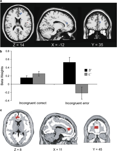
As hypothesized, relative to L′ participants, the S′ group showed significantly increased activation after errors in a more rostral region of the ACC (at the border between the affective and cognitive subdivision of BA 24; Ridderinkhof et al, 2004; Vogt et al, 1995; Figure 4a; Table 2b). Follow-up tests indicated that, relative to L′ participants, S′ participants displayed greater rACC activation for incorrect (p<0.001), but not correct (p>0.20), responses. Moreover, within-group tests showed that S′ participants activated the rACC after incorrect rather than correct responses more strongly (0.525±0.517 vs 0.153±0.207; p<0.012), whereas the L′ group exhibited the reverse pattern (incorrect: −0.217±0.584; correct: 0.253±0.225; p<0.013).
Figure 4.
Error-related activity for the L′ (n=14) vs S′ (n=19) participants. (a) Axial, saggital, and coronal slices showing the rACC cluster (error vs correct) thresholded at p<0.005 (x=−12, y=35, z=14). (b) Mean (and SE) β-weights extracted from the rACC cluster exceeding the statistical threshold (p<0.05, corrected). (c) rACC cluster emerging from a previous ERP study, in which MDD subjects exhibited increased current density relative to control participants 80 ms after committing an error in a Stroop task (peak voxel Talairach coordinates: x=10, y=41, z=7; Holmes and Pizzagalli, 2008b). The Euclidian distance between the fMRI and ERP voxel is 21.4 mm
In addition to the rACC, a second ACC region survived the statistical threshold. This region encompassed the subgenual ACC (BA 32) and was characterized by significantly higher activation in L′ relative to S′ participants (Table 2b). Follow-up analyses confirmed that groups differed for incorrect (p<0.007), but not correct (p>0.87) responses. Moreover, within-group analyses indicated that, in S′ participants, errors were associated with significantly reduced subgenual activation relative to correct responses (errors: −0.604±0.680; correct response: −0.109±0.449; p<0.004). In L′ participants, a trend in the opposite direction was observed (errors: 0.356±1.038; correct response: −0.085±0.350; p=0.087).
Whole-Brain Analyses
Flanker effect (incongruent correct responses>congruent correct responses)
No additional significant group differences emerged. Regions characterized by significant Condition effects irrespective of genotype are summarized in Table 3a. Among other regions, increased activation to incongruent trials was seen in the left insula (x=−30, y=22, z=−2) and a medial region encompassing the dACC (x=4, y=26, z=35), two regions previously associated with incongruency effects in Flanker tasks (Wager et al, 2005). However, the dACC cluster (74 voxels) missed the cluster extent (79 voxels) and should thus be interpreted cautiously.
Table 3. Summary of Secondary (Whole-Brain) Analyses in the Entire Sample (Irrespective of Genotype).
| Peak voxel location | Volume (mm3) | X | Y | Z | Z-score |
|---|---|---|---|---|---|
| (a) Response conflict (incongruent correct vs congruent correct) | |||||
| L. Middle frontal gyrus | 2680 | −28 | 22 | 33 | −4.74 |
| L. Superior frontal gyrus | 2088 | −10 | 61 | 27 | −4.57 |
| L. Lingual gyrus | 672 | −10 | −59 | 0 | −4.62 |
| L. Insula | 1408 | −30 | 22 | −2 | 6.28 |
| L. Lingual gyrus | 3128 | −24 | −71 | −4 | −5.75 |
| R. dACCa | 592 | 4 | 26 | 35 | 3.84 |
| R. Insula | 736 | 52 | −29 | 19 | −4.84 |
| R. Lingual gyrus | 2912 | 22 | −67 | 1 | −5.04 |
| R. Middle temporal gyrus | 848 | 55 | −11 | −10 | −4.75 |
| (b) Error commission (incongruent error vs incongruent correct) | |||||
| L. Precentral gyrus | 3200 | −38 | −17 | 37 | 5.50 |
| L. Caudate | 1440 | −16 | −17 | 20 | 3.92 |
| L. Superior temporal gyrus | 1376 | −52 | −49 | 14 | 5.74 |
| L. Precentral gyrus | 4144 | −50 | −1 | 6 | 4.66 |
| L. Insula | 1736 | −38 | 21 | 5 | 5.49 |
| L. Putamen | 752 | −18 | 9 | −1 | 4.69 |
| R. Middle frontal gyrus | 4504 | 52 | 1 | 40 | 7.20 |
| R. dACC | 13 328 | 8 | 13 | 39 | 7.81 |
| R. Superior frontal gyrus | 1152 | 24 | 43 | 28 | 4.08 |
| R. Caudate | 832 | 12 | 0 | 18 | 3.90 |
| R. Insula | 5048 | 50 | −39 | 20 | 5.20 |
| R. Posterior cingulate | 10 408 | 18 | −59 | 9 | 5.18 |
| R. Inferior frontal gyrus | 1464 | 52 | 32 | 7 | 4.18 |
| R. Thalamus | 2448 | 6 | −19 | −3 | 4.46 |
| R. Brain stem | 664 | 4 | −31 | −16 | 5.72 |
Abbreviations: ACC, anterior cingulate cortex; L, left; R, right.
Coordinates are in Talairach space.
Does not meet cluster extent threshold (74 voxels). All other regions meet p<0.05, corrected (whole-brain: p<0.005, uncorrected, cluster >79 voxels).
Error responses (incongruent error responses>incongruent correct responses)
As above, no additional group differences outside the ACC emerged (p<0.005; cluster size >79 voxels). Regions with significant differences between error and correct responses are summarized in Table 3b. Briefly, relative to correct responses, errors elicited higher activation in several prefrontal and limbic regions, including the left and right insula (x=−38, y=21, z=5; x=50, y=−39, z=20), putamen (x=−18, y=9, z=−1), and inferior frontal gyrus (x=52, y=32, z=7).
DISCUSSION
The main goal of this study was to investigate putative action monitoring dysfunctions in 5-HTTLPR S- or LG-allele carriers. Relative to a group of demographically matched L (VNTR) or LA (SNP) homozygotes, S′ participants were characterized by (1) reduced post-error and post-conflict behavioral adjustments, (2) decreased conflict-related dACC activation, and (3) increased error-related rACC activation. It is noteworthy that these findings emerged in the absence of any discernable differences in self-report of mood, and raise the possibility that dysregulated ACC functioning—specifically rACC hyperactivity to errors and dACC hypoactivity to response conflict—may represent mechanisms through which the 5-HTTLPR genotype confers an increased risk for MDD or emotional disorders.
Echoing previous findings of increased ERN and rACC activation in response to errors in 5-HTT short carriers (Althaus et al, 2009; Fallgatter et al, 2004) and MDD subjects (Holmes and Pizzagalli, 2008b), the S′ group exhibited potentiated error-related rACC activation. In addition, consistent with the role of the ACC in the generation of adaptive behavioral responses (Kerns et al, 2004), decreased conflict-related dACC activity and less adaptive post-conflict shifts in RT were observed in the S′ group, relative to the L′ group. These findings mirror previous results in MDD of reduced cognitive control (Paradiso et al, 1997; Siegle et al, 2004) and dACC activation during response conflict (Holmes and Pizzagalli, 2008a). Of note, the peak fMRI voxel showing group differences in conflict monitoring was 15.8 mm from the peak voxel associated with decreased conflict-related dACC activation in a MDD sample tested with a Stroop paradigm and ERP source localization techniques (Figure 3c; Holmes and Pizzagalli, 2008a).
In addition to dACC hypoactivation during high-conflict trials, relative to L′ participants, S′ participants showed relatively higher activation in the posterior cingulate cortex (BA 23) during incongruent trials. Interestingly, posterior cingulate activation has been reported during tasks involving threat-related stimuli (Maddock and Buonocore, 1997), arousing facial expressions (Critchley et al, 2000a), and somatic arousal (Critchley et al, 2000b), raising the possibility that posterior cingulate hyperactivation might reflect increased autonomic/somatic arousal in S′ participants during high-conflict trials. Alternatively, it is important to emphasize the fact that the posterior cingulate constitutes a core component of the default network, a distributed system of regions active at rest (for a review see, Buckner et al, 2008). Accordingly, the present findings might reflect impaired de-activation in S′ participants, in line with recent observations that reduced task-elicited deactivation of the posterior cingulate cortex is associated with subsequent error responses (Eichele et al, 2008). As these findings were not hypothesized a priori, their interpretation should, however, be considered tentative.
The observation of reduced conflict monitoring, as well as dysregulated dACC and subgenual ACC activation in individuals with an increased genetic vulnerability to MDD is particularly intriguing, as findings in the emotion regulation literature suggest that emotion regulation and reappraisal depend on an interplay between prefrontal and ACC regions and regions implicated in emotional reactivity, including the amygdala and insula (for a review, see Phillips et al, 2008; Ochsner and Gross, 2005, 2008). Accordingly, impairments in basic mechanisms implicated in cognitive control may contribute to the development of more complex emotion dysregulation observed in MDD, including amplification of the significance of failure (eg, Wenzlaff and Grozier, 1988) and difficulty in suppressing failure-related thoughts (eg, Conway et al, 1991), just to name few examples. This speculation is supported by evidence that individuals with more adaptive cognitive control (as assessed through ERN amplitudes and post-error behavioral adjustments) are less affected by daily life stress, a finding hypothesized to result from shared processes recruited in situations of increased cognitive conflict and regulation of negative reactions to stressful life events (Compton et al, 2008; see also Ochsner et al, 2009 for a recent demonstration that affective and cognitive conflict depends on a partially overlapping neural network). Future studies will be required to directly test the hypothesis that deficits in core cognitive processes (eg, action monitoring) may contribute to the generation of more complex impairments observed in MDD, including self-referential processing (Lemogne et al, 2009) and emotion regulation (Phillips et al, 2008), as well as increased risk for emotional disorders.
Although groups did not differ in their overall accuracy or RTs, or in accuracy after correct responses, S′ carriers were significantly less accurate after committing a mistake and showed significantly higher rACC activation to errors relative to L or LA homozygotes. These data are consistent with evidence of larger ERN in S-allele carriers (Althaus et al, 2009; Fallgatter et al, 2004), particularly as ACC regions have been implicated in the generation of the ERN (eg, van Veen and Carter, 2002). In addition, the present findings closely mirror recent ERP evidence of impaired post-error behavioral adjustments and potentiated error-related rACC activation in MDD patients (Figure 4c; Holmes and Pizzagalli, 2008b). Interestingly, the rACC peak voxel emerging from the current fMRI study was 21.4 mm away from the peak reported in our previous ERP study in MDD, a difference that is within the spatial resolution of the source localization technique used in our ERP study (Holmes and Pizzagalli, 2008b). As heightened reactivity to performance mistakes has been associated with increased negative affect (Hajcak et al, 2004) and punishment sensitivity (Boksem et al, 2006), the present findings suggest that, in S and LG SNP carriers, enhanced rACC response to errors and a failure to adaptively adjust behaviors after mistakes may constitute a basic cognitive mechanism associated with increased vulnerability to emotional disorders.
Consistent with previous findings in MDD (eg, George et al, 1997; Holmes and Pizzagalli, 2008a, 2008b), evidence of error-related rACC hyperactivity and conflict-related dACC hypoactivity in the S′ group reveals the presence of a multifaceted dysfunction of action monitoring system in individuals at increased genetic risk for depression when challenged by life stressors (Caspi et al, 2003; Kendler et al, 2001; but see Risch et al, 2009). Given the hypothesized role of the ACC in (1) the recruitment of prefrontal control mechanisms after errors and shifts in task difficulty (Holmes and Pizzagalli, 2008b; Kerns et al, 2004), and (2) the downregulation of limbic system activity after the presentation of self-relevant negative stimuli (Siegle et al, 2007), the presence of action monitoring deficits and associated ACC dysregulation in S and LG carriers may contribute to deficits in behavioral and/or emotional regulation in the context of adverse life events. Providing support for this assumption, S-allele carriers are characterized by heightened amygdala activity after the presentation of fearful/threatening facial expressions (Hariri et al, 2002, 2005) and reduced functional coupling between the perigenual ACC and amygdala (Pezawas et al, 2005). These findings have been hypothesized to reflect a failure of ACC-driven downregulation of activity in S-allele carriers rather than a primary abnormality in the amygdala (Hariri et al, 2006). Along similar lines, in patients with MDD, a decreased functional relationship between the amygdala and ACC activity has been observed during periods of rest (Anand et al, 2005) and after the presentation of self-relevant negative words (Siegle et al, 2007). In addition, disrupted functional connectivity has been observed in MDD subjects between dorsolateral prefrontal cortex and rACC, as well as dACC regions implicated in the recruitment of cognitive control after errors and response conflict (eg, Dannlowski et al, 2009; Holmes and Pizzagalli, 2008b; for a review, see Savitz and Drevets, 2009).
It should be noted that there have been inconsistent findings regarding the modulatory role of the 5-HTTLPR genotype on ACC responses to other stimuli, such as emotional faces. For example, Dannlowski et al (2008), recently reported increased responses in S-allele carriers to masked facial emotions in a region encompassing the supragenual and perigenual ACC. In contrast, Shah et al (2009) observed reduced ventral ACC activation to fearful and happy face in S-allele carriers. In light of methodological differences between these studies (eg, the use of subliminal vs supraliminal presentation; consideration of possible conjoint effects of 5-HTTLPR and rs25531), it is unclear whether these data highlight region-specific abnormalities in S-allele carriers. Accordingly, in the context of these data and the present findings, it is unlikely that a uniform relationship exists between ACC functioning and the 5-HTTLPR genotype. Given the dissociable roles of specific ACC regions (Ridderinkhof et al, 2004), future research examining the role of genetic variants affecting 5-HT (and other neuromodulators) on putative links between disrupted functional connectivity within frontocingulate pathways and action monitoring deficits is clearly warranted.
Interestingly, robust group differences in behavioral and fMRI data emerged in the absence of observable differences in self-reported affect. The present data replicate previous findings that failed to identify relationships between the 5-HTTLPR genotype and self-reported affect/personality (eg, Ball et al, 1997; Deary et al, 1999; Flory et al, 1999; Hariri et al, 2002; Katsuragi et al, 1999). Thus, the 5-HTTLPR genotype might affect physiological responses subserving specific cognitive processes without yielding an observable difference in self-reported measures (Hariri et al, 2006). Overall, the present pattern of findings highlights the utility of coupling molecular genetic and neuroimaging techniques in the search for psychiatric endophenotypes.
It should be noted that, in addition to enhanced risk for MDD after stressful life events (eg, Caspi et al, 2003), 5-HTTLPR S-allele carriers are at increased risk for other psychiatric illnesses, including PTSD (Broekman et al, 2007), ADHD (Beitchman et al, 2003), and alcoholism (Hu et al, 2005), among others. Interestingly, behavioral and neuroimaging evidence of action monitoring dysfunction have been observed in these disorders (eg, Endrass et al, 2008; Falconer et al, 2008; Wiersema et al, 2009), providing additional support for links between the 5-HTTLPR genotype and ACC functioning. Given these findings, further research will be necessary to establish whether the relationship between 5-HTT polymorphisms and action monitoring is specific to depression or rather represents a general risk factor for psychiatric illnesses with an affective component.
The limitations of this study should be acknowledged. First, our sample size was limited, which might have led to type I errors (Munafo et al, 2008), most prominently the absence of group differences in the amygdala. Second, individual differences in action monitoring, as with other complex behavioral traits, are most likely generated through the complex interactions of various environmental factors and a multitude of genes (Brown and Hariri, 2006; Prathikanti and Weinberger, 2005). Accordingly, although the present findings provide important insight into possible psychological and neurobiological factors linking 5-HTTLPR to increased vulnerability to psychopathology, the focus on a single candidate gene is an important limitation. Along similar lines, because of the relatively small sample size, analyses investigating the interactions among different genes were not possible. Given the hypothesized role of the mesencephalic dopamine system in the physiological correlates of action monitoring (Holroyd and Coles, 2002), future studies should be conducted on a scale allowing for the examination of interactions between multiple genes. Third, the limited size of the stimulus and response sets in the current version of the flanker task prevented analyses disentangling the potential overlap between response-conflict and repetition/negative priming effects (eg, Mayr et al, 2003; Ullsperger et al, 2005), which may mask the specific contribution of conflict adaptation (Bugg, 2008). Although the present findings are consistent with previous data (Holmes and Pizzagalli, 2008a, 2008b) stemming from paradigms in which conflict adaptation has been observed irrespective of priming (eg, Kerns et al, 2004), future studies using flanker tasks with larger stimulus and response sets are required to examine the unique contributions of stimulus repetition and conflict effects.
Despite these limitations, the present data suggest that action monitoring dysfunctions (and associated post-error rACC hyperactivity and post-conflict dACC hypoactivity) might constitute basic cognitive mechanisms through which 5-HTTLPR polymorphisms confer an increased vulnerability to emotional disorders, particularly when facing environmental stressors. Longitudinal studies in additional samples at increased risk for MDD (eg, unaffected offspring of depressed parents, remitted depressed samples) will be required to evaluate the predictive validity of these mechanisms vis-à-vis onset of psychopathology.
Acknowledgments
This study was funded by a Sackler Scholar in Psychobiology Research Grant (RB), a National Institute of Health Training Grant 1 F31MH078346 (AJH), and NIMH Research Grants R01MH68376 and R21MH078979 (DAP). We thank Nancy Brooks, Alison Brown, Daniel G Dillon, Jesen Fagerness, Miles Nugent, Roy H Perlis, Sara Rubenstein, and Patrice Vamivakas for their contributions and assistance with various aspects of this research.
The authors declare that over the past 3 years, Dr Pizzagalli has received research support from GlaxoSmithKline, Merck and ANT North America (Advanced Neurotechnology) for projects unrelated to the current study; consulting fees from ANT and AstraZeneca, and honoraria from AstraZeneca. Dr Holmes and Mr Bogdan declare no competing interests.
References
- Althaus M, Groen Y, Wijers AA, Mulder LJ, Minderaa RB, Kema IP, et al. Differential effects of 5-HTTLPR and DRD2/ANKK1 polymorphisms on electrocortical measures of error and feedback processing in children. Clin Neurophysiol. 2009;120:93–107. doi: 10.1016/j.clinph.2008.10.012. [DOI] [PubMed] [Google Scholar]
- Anand A, Li Y, Wang Y, Wu J, Gao S, Bukhari L, et al. Activity and connectivity of brain mood regulating circuit in depression: a functional magnetic resonance study. Biol Psychiatry. 2005;57:1079–1088. doi: 10.1016/j.biopsych.2005.02.021. [DOI] [PubMed] [Google Scholar]
- Austin MP, Mitchell P, Goodwin GM. Cognitive deficits in depression: possible implications for functional neuropathology. Br J Psychiatry. 2001;178:200–206. doi: 10.1192/bjp.178.3.200. [DOI] [PubMed] [Google Scholar]
- Ball D, Hill L, Freeman B, Eley TC, Strelau J, Riemann R, et al. The serotonin transporter gene and peer-rated neuroticism. Neuroreport. 1997;8:1301–1304. doi: 10.1097/00001756-199703240-00048. [DOI] [PubMed] [Google Scholar]
- Beats BC, Sahakian BJ, Levy R. Cognitive performance in tests sensitive to frontal lobe dysfunction in the elderly depressed. Psychol Med. 1996;26:591–603. doi: 10.1017/s0033291700035662. [DOI] [PubMed] [Google Scholar]
- Beck AT, Steer RA, Brown GK.1996Beck Depression Inventory Manual2nd ed. The Psychological Corportation, San Antonio, TX [Google Scholar]
- Beitchman JH, Davidge KM, Kennedy JL, Atkinson L, Lee V, Shapiro S, et al. The serotonin transporter gene in aggressive children with and without ADHD and nonaggressive matched controls. Ann N Y Acad Sci. 2003;1008:248–251. doi: 10.1196/annals.1301.025. [DOI] [PubMed] [Google Scholar]
- Boksem MA, Tops M, Wester AE, Meijman TF, Lorist MM. Error-related ERP components and individual differences in punishment and reward sensitivity. Brain Res. 2006;1101:92–101. doi: 10.1016/j.brainres.2006.05.004. [DOI] [PubMed] [Google Scholar]
- Botvinick M, Nystrom LE, Fissell K, Carter CS, Cohen JD. Conflict monitoring versus selection-for-action in anterior cingulate cortex. Nature. 1999;402:179–181. doi: 10.1038/46035. [DOI] [PubMed] [Google Scholar]
- Broekman BFP, Olff M, Boer F. The genetic background of PTSD. Neurosci Biobehav Rev. 2007;31:348–362. doi: 10.1016/j.neubiorev.2006.10.001. [DOI] [PubMed] [Google Scholar]
- Brown GW, Harris TO. Depression and the serotonin transporter 5-HTTLPR polymorphism: a review and a hypothesis concerning gene-environment interaction. J Affect Disord. 2008;111:1–12. doi: 10.1016/j.jad.2008.04.009. [DOI] [PubMed] [Google Scholar]
- Brown SM, Hariri AR. Neuroimaging studies of serotonin gene polymorphisms: exploring the interplay of genes, brain, and behavior. Cogn Affect Behav Neurosci. 2006;6:44–52. doi: 10.3758/cabn.6.1.44. [DOI] [PubMed] [Google Scholar]
- Buckner RL, Andrews-Hanna JR, Schacter DL. The brain's default network: anatomy, function, and relevance to disease. Ann NY Acad Sci. 2008;1124:1–38. doi: 10.1196/annals.1440.011. [DOI] [PubMed] [Google Scholar]
- Bugg JM. Opposing influences on conflict-driven adaptation in the Eriksen flanker task. Mem Cogn. 2008;36:1217–1227. doi: 10.3758/MC.36.7.1217. [DOI] [PMC free article] [PubMed] [Google Scholar]
- Caspi A, Sugden K, Moffitt TE, Taylor A, Craig IW, Harrington H, et al. Influence of life stress on depression: moderation by a polymorphism in the 5-HTT gene. Science. 2003;301:386–389. doi: 10.1126/science.1083968. [DOI] [PubMed] [Google Scholar]
- Chiu PH, Deldin PJ. Neural evidence for enhanced error detection in major depressive disorder. Am J Psychiatry. 2007;164:608–616. doi: 10.1176/ajp.2007.164.4.608. [DOI] [PubMed] [Google Scholar]
- Cohen S, Kamarck T, Mermelstein R. A global measure of perceived stress. J Health Soc Behav. 1983;24:385–396. [PubMed] [Google Scholar]
- Compton RJ, Robinson MD, Ode S, Quandt LC, Fineman SL, Carp J. Error monitoring ability predicts daily stress regulation. Psych Science. 2008;19:702–708. doi: 10.1111/j.1467-9280.2008.02145.x. [DOI] [PubMed] [Google Scholar]
- Conway M, Howell A, Giannopoulos C. Dysphoria and thought suppression. Cogn Ther Res. 1991;15:153–166. [Google Scholar]
- Critchley HD, Daly E, Phillips M, Brammer M, Bullmore E, Williams S. Explicit and implicit neural mechanisms for processing of social information from facial expressions: a functional magnetic resonance imaging study. Hum Brain Mapp. 2000a;9:93–105. doi: 10.1002/(SICI)1097-0193(200002)9:2<93::AID-HBM4>3.0.CO;2-Z. [DOI] [PMC free article] [PubMed] [Google Scholar]
- Critchley HD, Elliott R, Mathias CJ, Dolan RJ. Neural activity relating to generation and representation of galvanic skin conductance responses: a functional magnetic resonance imaging study. J Neurosci. 2000b;8:3033–3040. doi: 10.1523/JNEUROSCI.20-08-03033.2000. [DOI] [PMC free article] [PubMed] [Google Scholar]
- Dannlowski U, Ohrmann P, Bauer J, Deckert J, Hohoff C, Kugel H, et al. 5-HTTLPR biases amygdala activity in response to masked facial expressions in major depression. Neuropharmacol. 2008;33:418–424. doi: 10.1038/sj.npp.1301411. [DOI] [PubMed] [Google Scholar]
- Dannlowski U, Ohrmann P, Konrad C, Domschke K, Bauer J, Kugel H, et al. Reduced amygdala-prefrontal coupling in major depression: association with MAOA genotype and illness severity. Int J Neuropsychopharmacol. 2009;12:11–22. doi: 10.1017/S1461145708008973. [DOI] [PubMed] [Google Scholar]
- Deary IJ, Battersby S, Whiteman MC, Connor JM, Fowkes FG, Harmar A. Neuroticism and polymorphisms in the serotonin transporter gene. Psychol Med. 1999;29:735–739. doi: 10.1017/s0033291798007557. [DOI] [PubMed] [Google Scholar]
- Deichmann R, Gottfried JA, Hutton C, Turner R. Optimized EPI for fMRI studies of the orbitofrontal cortex. Neuroimage. 2003;19:430–441. doi: 10.1016/s1053-8119(03)00073-9. [DOI] [PubMed] [Google Scholar]
- Eichele T, Debener S, Calhoun V, Specht K, Hugdahl K, von Cramon DY, et al. Prediction of human errors by maladaptive changes in event-related brain networks. Proc Natl Acad Sci USA. 2008;105:6173–6178. doi: 10.1073/pnas.0708965105. [DOI] [PMC free article] [PubMed] [Google Scholar]
- Elliott R, Sahakian BJ, McKay AP, Herrod JJ, Robbins TW, Paykel ES. Neuropsychological impairments in unipolar depression: the influence of perceived failure on subsequent performance. Psychol Med. 1996;26:975–989. doi: 10.1017/s0033291700035303. [DOI] [PubMed] [Google Scholar]
- Endrass T, Klawohn J, Schuster F, Kathmann N. Overactive performance monitoring in obsessive-compulsive disorder: ERP evidence from correct and erroneous reactions. Neuropsychologia. 2008;46:1877–1887. doi: 10.1016/j.neuropsychologia.2007.12.001. [DOI] [PubMed] [Google Scholar]
- Falconer E, Bryant R, Felmingham KL, Kemp AH, Gordon E, Peduto A, et al. The neural networks of inhibitory control in posttraumatic stress disorder. J Psychiatry Neurosci. 2008;33:413–422. [PMC free article] [PubMed] [Google Scholar]
- Fallgatter AJ, Herrmann MJ, Roemmler J, Ehlis AC, Wagener A, Heidrich A, et al. Allelic variation of serotonin transporter function modulates the brain electrical response for error processing. Neuropsychopharmacology. 2004;29:1506–1511. doi: 10.1038/sj.npp.1300409. [DOI] [PubMed] [Google Scholar]
- First MB, Spitzer RL, Gibbon M, Williams J. Structured Clinical Interview for DSM-IV-TR Axis I Disorders—Patient Edition (SCID-I/P, 1/2007 revision) Biometrics Research, New York State Psychiatric Institute: New York; 2007. [Google Scholar]
- Fischl B, van der KA, Destrieux C, Halgren E, Segonne F, Salat DH, et al. Automatically parcellating the human cerebral cortex. Cereb Cortex. 2004;14:11–22. doi: 10.1093/cercor/bhg087. [DOI] [PubMed] [Google Scholar]
- Flory JD, Manuck SB, Ferrell RE, Dent KM, Peters DG, Muldoon MF. Neuroticism is not associated with the serotonin transporter (5-HTTLPR) polymorphism. Mol Psychiatry. 1999;4:93–96. doi: 10.1038/sj.mp.4000466. [DOI] [PubMed] [Google Scholar]
- George MS, Ketter TA, Parekh PI, Rosinsky N, Ring HA, Pazzaglia PJ, et al. Blunted left cingulate activation in mood disorder subjects during a response interference task (the Stroop) J Neuropsychiatry Clin Neurosci. 1997;9:55–63. doi: 10.1176/jnp.9.1.55. [DOI] [PubMed] [Google Scholar]
- Gratton G, Coles MG, Donchin E. Optimizing the use of information: strategic control of activation of responses. J Exp Psychol Gen. 1992;121:480–506. doi: 10.1037//0096-3445.121.4.480. [DOI] [PubMed] [Google Scholar]
- Hajcak G, McDonald N, Simons RF. Error-related psychophysiology and negative affect. Brain Cogn. 2004;56:189–197. doi: 10.1016/j.bandc.2003.11.001. [DOI] [PubMed] [Google Scholar]
- Hariri AR, Drabant EM, Munoz KE, Kolachana BS, Mattay VS, Egan MF, et al. A susceptibility gene for affective disorders and the response of the human amygdala. Arch Gen Psych. 2005;62:146–152. doi: 10.1001/archpsyc.62.2.146. [DOI] [PubMed] [Google Scholar]
- Hariri AR, Drabant EM, Weinberger DR. Imaging genetics: perspectives from studies of genetically driven variation in serotonin function and corticolimbic affective processing. Biol Psychiatry. 2006;59:888–897. doi: 10.1016/j.biopsych.2005.11.005. [DOI] [PubMed] [Google Scholar]
- Hariri AR, Mattay VS, Tessitore A, Kolachana B, Fera F, Goldman D, et al. Serotonin transporter genetic variation and the response of the human amygdala. Science. 2002;297:400–403. doi: 10.1126/science.1071829. [DOI] [PubMed] [Google Scholar]
- Hasler G, Drevets WC, Manji HK, Charney DS. Discovering endophenotypes for major depression. Neuropsychopharmacology. 2004;29:1765–1781. doi: 10.1038/sj.npp.1300506. [DOI] [PubMed] [Google Scholar]
- Holmes AJ, Pizzagalli DA. Response conflict and frontocingulate dysfunction in unmedicated participants with major depression. Neuropsychologia. 2008a;46:2904–2913. doi: 10.1016/j.neuropsychologia.2008.05.028. [DOI] [PMC free article] [PubMed] [Google Scholar]
- Holmes AJ, Pizzagalli DA. Spatiotemporal dynamics of error processing dysfunctions in major depressive disorder. Arch Gen Psychiatry. 2008b;65:179–188. doi: 10.1001/archgenpsychiatry.2007.19. [DOI] [PMC free article] [PubMed] [Google Scholar]
- Holroyd CB, Coles MG. The neural basis of human error processing: reinforcement learning, dopamine, and the error-related negativity. Psychol Rev. 2002;109:679–709. doi: 10.1037/0033-295X.109.4.679. [DOI] [PubMed] [Google Scholar]
- Hu X, Oroszi G, Chun J, Smith TL, Goldman D, Schuckit MA. An expanded evaluation of the relationship of four alleles to the level of response to alcohol and the alcoholism risk. Alcohol Clin Exp Res. 2005;29:8–16. doi: 10.1097/01.alc.0000150008.68473.62. [DOI] [PubMed] [Google Scholar]
- Katsuragi S, Kunugi H, Sano A, Tsutsumi T, Isogawa K, Nanko S, et al. Association between serotonin transporter gene polymorphism and anxiety-related traits. Biol Psychiatry. 1999;45:368–370. doi: 10.1016/s0006-3223(98)00090-0. [DOI] [PubMed] [Google Scholar]
- Kendler KS, Thornton LM, Gardner CO. Genetic risk, number of previous depressive episodes, and stressful life events in predicting onset of major depression. Am J Psychiatry. 2001;158:582–586. doi: 10.1176/appi.ajp.158.4.582. [DOI] [PubMed] [Google Scholar]
- Kerns JG, Cohen JD, MacDonald AW, III, Cho RY, Stenger VA, Carter CS. Anterior cingulate conflict monitoring and adjustments in control. Science. 2004;303:1023–1026. doi: 10.1126/science.1089910. [DOI] [PubMed] [Google Scholar]
- Laming D. Autocorrelation of choice-reaction times. Acta Psychol (Amst) 1979;43:381–412. doi: 10.1016/0001-6918(79)90032-5. [DOI] [PubMed] [Google Scholar]
- Lemogne C, Bastard G, Mayberg H, Volle E, Bergouignan L, Lehericy S. In search of the depressive self: extended medial prefrontal network during self-referential processing in major depression. Soc Cogn Affect Neur. 2009;4:305–312. doi: 10.1093/scan/nsp008. [DOI] [PMC free article] [PubMed] [Google Scholar]
- Maddock RJ, Buonocore MH. Activation of left posterior cingulate gyrus by the auditory presentation of threat-related words: an fMRI study. Psychiatry Res. 1997;75:1–14. doi: 10.1016/s0925-4927(97)00018-8. [DOI] [PubMed] [Google Scholar]
- Mayr U, Awh L, Laurey P. Conflict adaptation effects in the absence of executive control. Nat Neurosci. 2003;6:450–452. doi: 10.1038/nn1051. [DOI] [PubMed] [Google Scholar]
- Munafo M, Brown S, Hariri AR. Serotonin transporter (5-HTTLPR) genotype and amygdala activation: a meta-analysis. Biol Psychiatry. 2008;63:852–857. doi: 10.1016/j.biopsych.2007.08.016. [DOI] [PMC free article] [PubMed] [Google Scholar]
- Murphy DL, Fox MA, Timpano KR, Moya PR, Ren-Patterson R, Andrews AA, et al. How the serotonin story is being rewritten by new gene-based discoveries principally related to SLC6A4, the serotonin transporter gene, which functions to influence all cellular serotonin systems. Neuropharmacol. 2008;55:932–960. doi: 10.1016/j.neuropharm.2008.08.034. [DOI] [PMC free article] [PubMed] [Google Scholar]
- Ochsner KN, Gross JJ. The cognitive control of emotion. Trends Cogn Sci. 2005;9:242–249. doi: 10.1016/j.tics.2005.03.010. [DOI] [PubMed] [Google Scholar]
- Ochsner KN, Gross JJ. Cognitive emotion regulation: insights from social cognitive and affective neuroscience. Curr Dir Psychol Sci. 2008;17:153–158. doi: 10.1111/j.1467-8721.2008.00566.x. [DOI] [PMC free article] [PubMed] [Google Scholar]
- Ochsner KN, Hughes B, Robertson ER, Cooper JC, Gabrieli JDE. Neural systems supporting the control of affective and cognitive conflicts. J Cogn Neurosci. 2009;21:1841–1854. doi: 10.1162/jocn.2009.21129. [DOI] [PMC free article] [PubMed] [Google Scholar]
- Olvet DM, Hajcak G. The error-related negativity (ERN) and psychopathology: toward an endophenotype. Clin Psychol Rev. 2008;28:1343–1354. doi: 10.1016/j.cpr.2008.07.003. [DOI] [PMC free article] [PubMed] [Google Scholar]
- Paradiso S, Lamberty GJ, Garvey MJ, Robinson RG. Cognitive impairment in the euthymic phase of chronic unipolar depression. J Nerv Ment Dis. 1997;185:748–754. doi: 10.1097/00005053-199712000-00005. [DOI] [PubMed] [Google Scholar]
- Pezawas L, Meyer-Lindenberg A, Drabant EM, Verchinski BA, Munoz KE, Kolachana BS, et al. 5-HTTLPR polymorphism impacts human cingulate-amygdala interactions: a genetic susceptibility mechanism for depression. Nat Neurosci. 2005;8:828–834. doi: 10.1038/nn1463. [DOI] [PubMed] [Google Scholar]
- Phillips ML, Ladouceur CD, Drevets WC. A neural model of voluntary and automatic emotion regulation: implications for understanding the pathophysiology and neurodevelopment of bipolar disorder. Mol Psychiatry. 2008;13:833–857. doi: 10.1038/mp.2008.65. [DOI] [PMC free article] [PubMed] [Google Scholar]
- Prathikanti S, Weinberger DR. Psychiatric genetics—the new era: genetic research and some clinical implications. Br Med Bull. 2005;73–74:107–122. doi: 10.1093/bmb/ldh055. [DOI] [PubMed] [Google Scholar]
- Rabbitt PM. Errors and error correction in choice-response tasks. J Exp Psychol. 1966;71:264–272. doi: 10.1037/h0022853. [DOI] [PubMed] [Google Scholar]
- Ridderinkhof KR, Ullsperger M, Crone EA, Nieuwenhuis S. The role of the medial frontal cortex in cognitive control. Science. 2004;306:443–447. doi: 10.1126/science.1100301. [DOI] [PubMed] [Google Scholar]
- Risch N, Herrell R, Lehner T, Liang KY, Eaves L, Hoh J, et al. Interaction between the serotonin transporter gene (5-HTTLPR), stressful life events, and risk of depression: a meta-analysis. JAMA. 2009;301:2462–2471. doi: 10.1001/jama.2009.878. [DOI] [PMC free article] [PubMed] [Google Scholar]
- Sanders AR, Duan J, Gejman PV. Complexities in psychiatric genetics. Int Rev Psychiatry. 2004;16:284–293. doi: 10.1080/09540260400014393. [DOI] [PubMed] [Google Scholar]
- Savitz JB, Drevets WC. Imaging phenotypes of major depressive disorder: genetic correlates. Neuroscience. 2009;164:300–330. doi: 10.1016/j.neuroscience.2009.03.082. [DOI] [PMC free article] [PubMed] [Google Scholar]
- Schrijvers D, de Bruijn ER, Maas Y, De Grave C, Sabbe BG, Hulstijn W. Action monitoring in major depressive disorder with psychomotor retardation. Cortex. 2008;44:569–579. doi: 10.1016/j.cortex.2007.08.014. [DOI] [PubMed] [Google Scholar]
- Schrijvers D, de Bruijn ER, Maas YJ, Vancoillie P, Hulstijn W, Sabbe BG. Action monitoring and depressive symptom reduction in major depressive disorder. Int J Psychophysiol. 2009;71:218–224. doi: 10.1016/j.ijpsycho.2008.09.005. [DOI] [PubMed] [Google Scholar]
- Shah MP, Wang F, Kalmar JH, Chepenik LG, Tie K, Pittman B, et al. Role of variation in the serotonin transporter protein gene (SLC6A4) in trait disturbances in the ventral anterior cingulate in bipolar disorder. Neuropharmacol. 2009;34:1301–1310. doi: 10.1038/npp.2008.204. [DOI] [PMC free article] [PubMed] [Google Scholar]
- Siegle GJ, Steinhauer SR, Thase ME. Pupillary assessment and computational modeling of the Stroop task in depression. Int J Psychophysiol. 2004;52:63–76. doi: 10.1016/j.ijpsycho.2003.12.010. [DOI] [PubMed] [Google Scholar]
- Siegle GJ, Thompson W, Carter CS, Steinhauer SR, Thase ME. Increased amygdala and decreased dorsolateral prefrontal BOLD responses in unipolar depression: related and independent features. Biol Psychiatry. 2007;61:198–209. doi: 10.1016/j.biopsych.2006.05.048. [DOI] [PubMed] [Google Scholar]
- Sullivan PF, Neale MC, Kendler KS. Genetic epidemiology of major depression: review and meta-analysis. Am J Psychiatry. 2000;157:1552–1562. doi: 10.1176/appi.ajp.157.10.1552. [DOI] [PubMed] [Google Scholar]
- Ullsperger M, Bylsma LM, Botvinick MM. The conflict adaptation effect: it's not just priming. Cogn Affect Behav Neurosci. 2005;5:467–472. doi: 10.3758/cabn.5.4.467. [DOI] [PubMed] [Google Scholar]
- van Veen V, Carter CS. The anterior cingulate as a conflict monitor: fMRI and ERP studies. Physiol Behav. 2002;77:477–482. doi: 10.1016/s0031-9384(02)00930-7. [DOI] [PubMed] [Google Scholar]
- Vanderhasselt MA, De Raedt R. Impairments in cognitive control persist during remission from depression and are related to the number of past episodes: an event related potential study. Biol Psychol. 2009;81:169–176. doi: 10.1016/j.biopsycho.2009.03.009. [DOI] [PubMed] [Google Scholar]
- Vogt BA, Nimchinsky EA, Vogt LJ, Hof PR. Human cingulate cortex: surface features, flat maps, and cytoarchitecture. J Comp Neurol. 1995;359:490–506. doi: 10.1002/cne.903590310. [DOI] [PubMed] [Google Scholar]
- Wager TD, Nichols TE. Optimization of experimental design in fMRI: a general framework using a genetic algorithm. Neuroimage. 2003;18:293–309. doi: 10.1016/s1053-8119(02)00046-0. [DOI] [PubMed] [Google Scholar]
- Wager TD, Sylvester CY, Lacey SC, Nee DE, Franklin M, Jonides J. Common and unique components of response inhibition revealed by fMRI. Neuroimage. 2005;27:323–340. doi: 10.1016/j.neuroimage.2005.01.054. [DOI] [PubMed] [Google Scholar]
- Watson D, Weber K, Assenheimer JS, Clark LA, Strauss ME, McCormick RA. Testing a tripartite model: I. Evaluating the convergent and discriminant validity of anxiety and depression symptom scales. J Abnorm Psychol. 1995;104:3–14. doi: 10.1037//0021-843x.104.1.3. [DOI] [PubMed] [Google Scholar]
- Wendland JR, Martin BJ, Kruse MR, Lesch KP, Murphy DL. Simultaneous genotyping of four functional loci of human SLC6A4, with a reappraisal of 5-HTTLPR and rs25531. Mol Psychiatry. 2006;11:224–226. doi: 10.1038/sj.mp.4001789. [DOI] [PubMed] [Google Scholar]
- Wenzlaff RM, Grozier SA. Depression and the magnification of failure. J Abnorm Psychol. 1988;97:90–93. doi: 10.1037//0021-843x.97.1.90. [DOI] [PubMed] [Google Scholar]
- Wiersema JR, van der Meere JJ, Roeyers H. ERP correlates of error monitoring in adult ADHD. J Neural Transm. 2009;116:371–379. doi: 10.1007/s00702-008-0165-x. [DOI] [PubMed] [Google Scholar]
- Wong ML, Licinio J. Research and treatment approaches to depression. Nat Rev Neurosci. 2001;2:343–351. doi: 10.1038/35072566. [DOI] [PubMed] [Google Scholar]
- Zalsman G, Huang YY, Oquendo MA, Burke AK, Hu XZ, Brent DA, et al. Association of a triallelic serotonin transporter gene promoter region (5-HTTLPR) polymorphism with stressful life events and severity of depression. Am J Psychiatry. 2006;163:1588–1593. doi: 10.1176/ajp.2006.163.9.1588. [DOI] [PubMed] [Google Scholar]


