Abstract
Ataxia-telangiectasia mutated (ATM) is a cellular damage sensor that coordinates the cell cycle with damage-response checkpoints and DNA repair to preserve genomic integrity. However, ATM also has been implicated in metabolic regulation, and ATM deficiency is associated with elevated reactive oxygen species (ROS). ROS has a central role in many physiological and pathophysiological processes including inflammation and chronic diseases such as atherosclerosis and cancer, underscoring the importance of cellular pathways involved in redox homeostasis. We have identified a cytoplasmic function for ATM that participates in the cellular damage response to ROS. We show that in response to elevated ROS, ATM activates the TSC2 tumor suppressor via the LKB1/AMPK metabolic pathway in the cytoplasm to repress mTORC1 and induce autophagy. Importantly, elevated ROS and dysregulation of mTORC1 in ATM-deficient cells is inhibited by rapamycin, which also rescues lymphomagenesis in Atm-deficient mice. Our results identify a cytoplasmic pathway for ROS-induced ATM activation of TSC2 to regulate mTORC1 signaling and autophagy, identifying an integration node for the cellular damage response with key pathways involved in metabolism, protein synthesis, and cell survival.
Keywords: autophagy, oxidative stress, signal transduction, cytoplasm, damage
An optimal cellular damage response requires both repair of damage and coordination of critical cellular processes such as transcription, translation, and cell cycle progression (1–4). ATM, the gene mutated in the cancer-prone human disease ataxia-telangiectasia (AT), is a protein kinase that serves as a critical mediator of signaling pathways that facilitate the response of mammalian cells to ionizing radiation (IR) and other agents that induce DNA double-strand breaks (4). Recently, however, ataxia-telangiectasia mutated (ATM) has been implicated in metabolic pathways unrelated to DNA damage (5).
Tuberous sclerosis complex (TSC), results from defects in either the TSC1 or TSC2 tumor suppressor genes (6). TSC2 participates in energy sensing and growth factor signaling to repress the kinase mTOR in the mTORC1 complex, a key regulator of protein synthesis and cell growth (7, 8). TSC2 is itself regulated by the AMP-activated protein kinase (AMPK) metabolic pathway, which regulates several metabolic processes and activates TSC2 to repress mTORC1 under conditions of energy stress (9). AMPK is in turn regulated at least in part through the LKB1 tumor suppressor gene involved in Peutz Jaegher’s Syndrome, another cancer predisposing hamartoma syndrome (10). Like ATM-deficient cells, TSC2-null cells exhibit defects in redox homeostasis (11–13).
Reactive oxygen species (ROS) act as signaling intermediates for many normal cellular processes, and elevated ROS has been linked to over 150 diseases, including atherosclerosis, diabetes, cancer, and neurodegenerative diseases (14–17). In addition, chronic inflammatory conditions perpetuate tissue damage in a number of diseases because activated phagocytes release H2O2, which in turn activates pleotropic signaling pathways in neighboring cells. A common theme among many of these conditions is an increase in incidence with age, and even aging itself has been postulated to result from free radicals produced during normal aerobic metabolism that accumulate over time as a result of decreased antioxidant capacity and/or mitochondrial dysfunction leading to elevated ROS. Thus, elucidating key cell signaling pathways that regulate redox homeostasis and respond to oxidative stress has important implications for many natural and pathophysiological conditions.
Results
Exposure of cells to agents that induce genotoxic and oxidative stress caused repression of mTORC1 signaling, inhibiting mTORC1 phosphorylation of p70/S6 kinase (S6K) and 4EBP1 (direct targets of the mTOR kinase) and phosphorylation of S6, the downstream effector of S6K (Fig. 1 and Fig. S1). Although mTORC1 repression in response to DNA damage has been observed previously (18, 19), and has recently been linked to activation of p53 (20, 21), a mechanism for repression of mTORC1 in response to oxidative damage had not previously been elucidated.
Fig. 1.
mTORC1 repression by ROS in the cytoplasm (A and B) Western analysis of H2O2 treated MCF7 cells. (C) Western analysis of cytoplasmic and nuclear fractions of H2O2 treated MCF7 cells. (D) Western analysis of MCF7 cells treated with H2O2 in the presence or absence of 100 ng/mL leptomycin B. LDH and LAMIN are controls for cytoplasmic and nuclear fractions, respectively.
mTORC1 repression by ROS was dose- and time-dependent, occurring rapidly (<60 min) and at low (<0.1 mM) concentrations of H2O2 (Fig. 1 A–B and Fig. S1 A and C). The mitochondrial uncoupler menadione and PEITC, which depletes glutathione, also repressed mTORC1 signaling (Fig. S1C), indicating both exogenous and endogenous ROS can induce mTORC1 repression. Repression of mTORC1 in response to H2O2 was rescued with the ROS scavenger N-acetyl cysteine (NAC), as well as pretreatment with catalase (Fig. S1D).
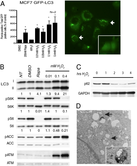
mTORC1 negatively regulates autophagy (22), a catabolic process in which cells deliver cytoplasmic components for degradation to the lysosome. Concomitant with mTORC1 repression by ROS, autophagy increased in cells treated with H2O2 (Fig. 2 and Fig. S2). LC3, the mammalian homolog of the yeast protein Atg8 (Aut7/Apg8p), is essential for autophagy in yeast (23), exists in two forms: a lower mobility LC3-I and lipidated higher mobility LC3-II. LC3-II associates with autophagosomes providing a necessary step in autophagy, thus levels of LC3-II correlate with the extent of autophagosome formation. MCF7 cells stably transfected with GFP-LC3 exhibited a significant increase in punctate cytoplasmic structures (autophagic vesicles) when treated with H2O2, comparable to that observed with rapamycin (Fig. 2A). H2O2 also induced an increase in LC3 mobility (LC3-II formation) (Fig. 2B), indicative of increased autophagy associated with mTORC1 repression and an increase in acridine orange fluorescence associated with acidic vesicles (AVO index) (Fig. S2). p62 protein binds to LC3 and is degraded by autophagy. The time-dependent decrease in p62 levels confirmed that ROS increased autophagic flux (Fig. 2C). Induction of autophagosomes was also directly visualized by electron microscopy in response to H2O2 (Fig. 2D).
Fig. 2.
mTORC1 repression induces autophagy (A) MCF7 cells stably transfected with GFP-LC3 were treated with H2O2 for 1 h and analyzed by microscopy for the presence of fluorescent puncta. Cells undergoing autophagy were quantitated as a percentage of total GFP positive cells. The graph shows the total number of puncta positive cells divided by total GFP positive cells, normalized to the vehicle control [n = 2, * P < 0.001 (χ2 test)]. (B and C) Western analysis of H2O2 treated SKOV-3 cells. (D) Electron microscopy images of H2O2 treated MEFs. Arrows indicate autophagosomes.
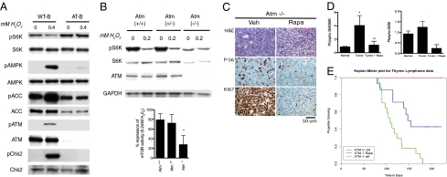
As expected, exposure of cells to DNA damaging agents (etoposide and IR) activated the ATM checkpoint pathway, inducing ATM autophosphorylation and p53 phosphorylation (Fig. S1 A and B). Importantly, we found ATM was also activated by ROS (Figs. 1, 2B, 3A, and 4B) with ATM, p53, and Chk2 phosphorylation increased in cells exposed to H2O2 (Figs. 1–3 and 4 and Figs. S1 and S3). Importantly, although early passage ATM-proficient lymphoblasts repressed mTORC1 in response to H2O2, in ATM-deficient lymphoblasts mTORC1 signaling was not repressed (Fig. 3A), and siRNA knockdown of ATM in MCF-7 cells also decreased mTORC1 repression by ROS (Fig. S3A). Furthermore, in multiple clonal isolates of Atm-proficient vs. Atm-deficient mouse embryonic fibroblasts (MEFs) isolated from Atm+/+, Atm+/-, and Atm-/- mice, Atm-/- MEFs (n = 3) exhibited a significantly diminished ability to repress mTORC1 following H2O2 treatment relative to Atm+/+ MEFs (n = 3) and Atm+/- MEFs (n = 3) (P < 0.05) (Fig. 3B).
Fig. 3.
Participation of ATM in mTORC1 repression by ROS (A) Western analysis of EBV-immortalized B-lymphocytes obtained from an AT-patient (AT-B), or nonaffected individual (WT-B) were treated with H2O2 for 1 h. (B) MEFs derived from Atm+/+, Atm+/-, or Atm-/- mice were treated with H2O2 for 1 h. Western analyses shown above and quantitation of the response (percent change in ratio of phosphorylated S6K/total S6K setting the NT control to 100%) of independent clonal isolates from Atm+/+ (n = 3), Atm+/- (n = 3), or Atm-/- (n = 3) mice shown in the graph below. *P < 0.05 (t test) (C) Immunohistochemistry of thymi from Atm-/- mice. The thymi of Atm-/- mice treated with rapamycin were markedly atrophic and hypocellular, with few if any residual tumor cells apparent (Right). (D) Quantitation of Western analysis to determine the ratio of phospho-S6K/total S6K (n = 5 normal, n = 9 tumors, n = 4 tumor+rapa), and phospho-S6/total S6 (n = 7 normal, n = 12 tumors, n = 6 tumor+rapa) in thymi in response to rapamycin. *P ≤ 0.05 (Mann-Whitney test, compared to normal) (E) Kaplan-Meier survival curves for Atm-/- mice treated with 15 mg/kg rapamycin, and control Atm-/- mice and Atm+/- mice. P < 0.001 (log-rank test).
Fig. 4.
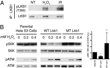
LKB1 mediates AMPK activation and mTORC1 repression by H2O2 (A) Immunoprecipitation showing that LKB1 is phosphorylated at Thr366 by ATM in response to H2O2 and 20Gy IR (positive control) in HEK293 cells. (B) Western analysis of LKB1-deficient HeLa S3 cells (parental) and stable clones expressing wild-type Lkb1 (WT) (n = 4) or T363A mutant Lkb1 (MT) (n = 2) treated with H2O2. *P < 0.03 versus parental. **P < 0.03 versus WT, 0.05 versus MT. (t test).
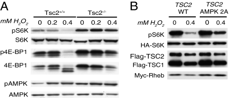
TSC2, an important repressor of mTORC1 activity (7), also participated in regulation of mTORC1 signaling by ROS, as repression of mTORC1 occurred in Tsc2+/+ but not Tsc2-/- MEFs in response to H2O2, (Fig. 5A) and siRNA knockdown of TSC2, attenuated mTORC1 repression by H2O2 by 70–90% (Fig. S3B). Importantly, both Tsc2-/- and Tsc2+/+ MEFs are p53-deficient (24), indicating that p53 was not required for repression of mTORC1 by ROS. This was also the case for p53 siRNA knockdown, with an equivalent mTORC1 repression induced by H2O2 in both p53-expressing and p53-deficient cells in response to H2O2 (Fig. S3C). Based on these data, we hypothesized that a p53-independent signaling pathway existed from ATM to TSC2, which regulated an mTORC1 stress response to ROS.
Fig. 5.
ROS induces AMPK activation of Tsc2 (A) Western analyses of Tsc2-proficient and deficient MEFs treated with H2O2. (B) Western analysis of HEK293 functional assay showing TSC2AMPK2A mutant is deficient in ROS-induced mTORC1 repression.
AMPK, one of many regulators of TSC2, activates TSC2 by phosphorylating it on several sites, including Thr1271 and Ser1387 (9). AMPK is activated by several AMPK kinases, including LKB1 (25–27). AMPK activation, as assessed by phosphorylation at Thr172 (the AMPK site phosphorylated by the LKB1 kinase) and phosphorylation of ACC at Ser79, a direct target of AMPK, occurred in response to H2O2 (Figs. 1 A–C, 2B, 3A, and 5A and Figs. S1D, S3B, S3D, S4A, and S5B) and preceded repression of mTORC1 (Fig. S1C). Treatment of cells with the AMPK inhibitor Compound C (28) attenuated repression of mTORC1 signaling in response to H2O2 (Fig. S4A).
In a HEK293 functional assay, using cells cotransfected with TSC2, the TSC2 activation partner TSC1, the GAP target Rheb, and the mTORC1 substrate S6K, ROS-induced repression of mTORC1 signaling to S6K was significantly attenuated when expressing a T1271/S1387A mutant (TSC2AMPK2A) that cannot be phosphorylated by AMPK compared with a wild-type TSC2 (TSC2WT) construct even in the presence of endogenous TSC2 (Fig. 5B). In addition, 2D phosphopeptide mapping revealed two peptides that were not present in TSC2AMPK2A mutant TSC2-expressing cells but were present in TSC2WT-expressing cells treated with H2O2 (Fig. S4B, green circle), and which increased in intensity and/or exhibited a mobility shift (Fig. S4B, red circle) relative to untreated TSC2WT-expressing cells.
However, activation of AMPK was not sufficient to repress mTORC1 in the absence of TSC2; Tsc2-null MEFs that did not repress mTORC1 have constitutively active AMPK (29) (Fig. 5A, compare lanes 1 and 4) and AMPK signaling was increased further in response to H2O2 in Tsc2-deficient cells (Fig. 5A), which were deficient in mTORC1 repression, indicating that TSC2 was an obligatory link between AMPK and mTORC1 repression.
LKB1 has been shown to be a target for ATM phosphorylation at Thr366 (human)/Thr363 (rodent) in response to DNA damage (30). In LKB1-proficient cells, ROS increased AMPK phosphorylation at Thr172, the site phosphorylated by LKB1 (Figs. 1 A–C, 3A, and 5A). Importantly, whereas ATM-proficient lymphoblasts exhibited increased phosphorylation of AMPK at Thr172 in response to ROS, phosphorylation of AMPK was barely detectable in AT-null lymphoblasts (which failed to repress mTORC1) (Fig. 3A). In LKB1-proficient HEK293 cells, LKB1 phosphorylation at the Thr366 ATM phosphorylation site (31) increased in response to ROS and IR (Fig. 4A), concordant with mTORC1 repression. In addition, siRNA knockdown of LKB1 attenuated mTORC1 repression by H2O2 (Fig S3D).
A direct role for LKB1 in repression of mTORC1 by ROS was further demonstrated in HeLa S3 cells that lack functional LKB1 but express AMPK (Fig. S5 A and B), whereas parental HeLa S3 cells could not repress mTORC1 in response to H2O2, reexpression of wild-type Lkb1 restored the ability to repress mTORC1 in response to ROS (Fig. 4B). In contrast, HeLa S3 cells reconstituted with a T363A mutant Lkb1 that cannot be phosphorylated by ATM failed to reconstitute the ability to repress mTORC1 when treated with H2O2, even though ATM was activated in clones expressing the T363A mutant Lkb1 (Fig. 4B).
However, in HeLa S3 cells, high concentrations of H2O2 (≥ 1 mM) can induce AMPK phosphorylation and mTORC1 repression, suggesting that LKB1-independent pathways for AMPK activation and mTORC1 repression can be induced under conditions of extreme oxidative stress (Fig S5C). Interestingly, although phosphorylation of AMPK correlated with LKB1 activity and mTORC1 repression, ACC phosphorylation did not: ACC was phosphorylated when AMPK was activated by ROS. However, in LKB1-deficient cells unable to phosphorylate AMPK and repress mTORC1, ACC phosphorylation still increased (Fig. S5B), and when AMPK activity was inhibited by Compound C, phosphoACC levels still increased with ROS (Fig. S5B). This suggests either Thr172 phosphorylation of AMPK by LKB1 contributes to substrate specificity (i.e., TSC2 vs. ACC) or that ACC phosphorylation at Ser79 is modulated by other kinase(s) or phosphatases that are redox-sensitive.
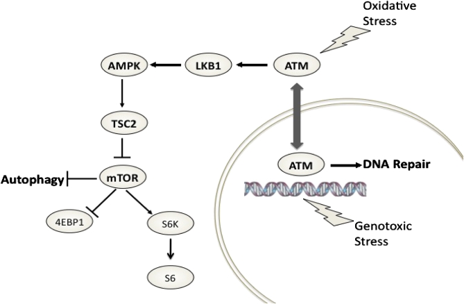
Although response to genotoxic stress is a nuclear function of ATM, TSC2 functions in the cytoplasm (32), raising the question as to the cellular compartment(s) for ATM signaling to TSC2 to repress mTORC1. The rapid repression of mTORC1 induced by H2O2 was not observed with the DNA damaging agent etoposide (Fig. S6A), although etoposide induced more robust DNA damage (phospho-H2AX accumulation) in the nucleus than H2O2 did (Fig. S6B). Furthermore, induction of DNA damage by etoposide did not activate AMPK nor did it result in repression of mTORC1 signaling (Fig. S6A). Subcellular fractionation revealed that although ATM was rapidly activated in the cytoplasm and the nucleus by H2O2 (consistent with accumulation of H2AX) (Fig. 1C and Fig. S6B), LKB1 remained localized to the cytoplasm in H2O2–treated cells. Moreover, AMPK phosphorylation by LKB1 and repression of mTORC1 occurred in the cytoplasmic fraction in response to ROS (Fig. 1C), indicating that this signaling node was localized to the cytoplasm. These data were confirmed with leptomycin B, a nuclear export inhibitor, which did not block mTORC1 repression by ROS (Fig. 1D) and equivalent ATM phosphorylation was observed in the cytoplasmic fraction of H2O2-treated MCF7 cells in the presence or absence of leptomycin B (Fig. S6C). These data identify a pathway for ROS-induced repression of mTORC1 in the cytoplasm, whereby a cytoplasmic pool of ATM activates TSC2 by engaging AMPK via the AMPK-K and ATM effector LKB1 (Fig. 6).
Fig. 6.
Schematic showing cytoplasmic signaling pathway from ATM to TSC2 via LKB1 and AMPK to repress mTORC1 and induce autophagy.
ROS is believed to contribute to lymphomagenesis in Atm-/- mice, which succumb to thymic lymphoma by 4–6 months of age (13, 33). mTORC1 hyperactivity has been shown to result in endoplasmic reticulum (ER) stress (34), which can lead to elevated ROS (35) suggesting a linkage between mTORC1 and production of ROS. As previously reported, we found that Tsc2-deficient MEFs and ATM-deficient human lymphoblasts had elevated levels of ROS relative to wild-type cells (Fig. S7A). Importantly, ROS levels in ATM-null lymphoblasts and Tsc2-null MEFs were reduced to wild-type levels by rapamycin (Fig. S7 A and B). In thymic lymphomas from Atm-deficient mice, we observed consistently elevated mTORC1 signaling when either fasted or fed ad libitum (Fig. 3 C and D). Short-term daily treatment of Atm-/- mice with rapamycin acutely reduced levels of mTORC1 activity in the thymi of these animals and induced thymic atrophy (Fig. 3 C and D). In addition, phospho-S6 levels dramatically decreased and proliferation (as measured by Ki67 staining) was also significantly decreased (Fig. 3C). Given these findings, rapamycin was administered daily to Atm-/- animals beginning at 2–3 months of age, and survival of treated animals compared to control Atm-/- and Atm+/- mice. Rapamycin treatment significantly increased survival of treated versus control Atm-/- mice (n = 17 control and n = 13 rapamycin-treated) with 100% of control Atm-/- animals dead by 200 days of age, whereas >50% of Atm-/- mice treated with rapamycin survived >200 days (P < 0.001) (Fig. 3E).
Discussion
Genotoxic stress has been reported to repress mTORC1 via activation of p53 (18). Recently, the p53-dependent pathway regulating mTORC1 in response to genotoxic stress was found to act via the p53 target genes, Sesn1 and Sesn2, in a redox-independent pathway that required TSC2 (21). We have now identified a p53-independent pathway that regulates mTORC1 in response to oxidative stress produced by reactive oxygen species (ROS). Identification of a cytoplasmic signaling node for LKB1/AMPK/TSC2 activation in response to oxidative stress mediated by ATM (Fig. 6) opens other avenues for understanding how defects in this signaling pathway may perturb redox homeostasis and contribute to disease pathogenesis.
Our finding that ROS potently and rapidly activates ATM in the cytoplasm is distinct from the classical pathway for ATM activation via DNA double-strand breaks in the nucleus, suggesting that additional mechanisms exist to activate ATM independently of DNA damage. The precise biochemical mechanism underlying activation of the ATM kinase by cytoplasmic ROS is at present unclear. ATM is a very large protein (approximately 350 kDa) containing many cysteine residues, and the possibility exists that ROS could directly induce conformational changes in ATM via oxidation of sulfhydryl groups and the formation of intra- or intermolecular disulfide bonds, an intriguing hypothesis that requires further exploration.
In addition to ATM and TSC2, we observed a dependence on LKB1 for mTOR repression in response to ROS. This contrasts with the earlier finding that in HeLa cells (which lack LKB1), AMPK could be activated by etoposide in an ATM-dependent manner to regulate mitochondrial biogenesis (36). Of interest mechanistically, however, this study required prolonged treatment with etoposide, again suggesting that multiple pathways exist to regulate AMPK and mTORC1 dependent on the type of cellular damage and perhaps in a cell-type dependent manner.
The function of the ATM phosphorylation site on LKB1 at Thr366 has been somewhat elusive beyond the observation that in G361 melanoma cells that lack LKB1, a mutant that lacks this site is threefold less efficient at regulating cell growth when overexpressed (37). Our data indicate that this site is functionally important and is required for ATM activation of the AMPK signaling node in response to ROS. Although LKB1 activation of AMPK is well-documented, its function as an obligate AMPK-kinase is controversial. In LKB1-deficient HeLa cells or Lkb1-/- MEFs, AMPK is not activated by agonists such as AICAR or metformin; however, AMPK possesses significant basal activation and phosphorylation at Thr172, suggesting that other AMPK-kinases can compensate to regulate the key metabolic processes controlled by AMPK (30). Our observation of discordance between LKB1-mediated activation of AMPK resulting in TSC2 activation and AMPK-mediated phosphorylation of ACC is particularly interesting, as it suggests that other determinants of AMPK activity beyond phosphorylation at Thr172 exist, perhaps involving pools of AMPK residing in different subcellular compartments. Clearly, additional work remains to be done to understand mechanisms underlying specific pathways for AMPK and TSC2 activation by different types of cellular stress.
An emerging picture suggests multiple pathways exist for regulation of mTORC1 and autophagy in response to cellular stress. The p53-regulated genes Sestrin 1 and Sestrin 2 were shown to activate AMPK and repress mTORC1 signaling in response to camptothecin-induced genotoxic stress in a redox-independent manner (21). This is in contrast to our observation of ATM signaling to AMPK and TSC2 to regulate mTORC1, which does not require p53 and is redox-regulated. The potential for ATM and/or TSC2-independent pathways to induce autophagy in response to ROS have not been ruled out especially at higher concentration of H2O2/ROS and/or at later (i.e., >1 hr) time points. In addition, activation of AMPK could directly suppress mTORC1 via phosphorylation of raptor (38), and, indeed, there is some suggestion of partial mTORC1 suppression even in Tsc2-/-; p53-/- cells in response to ROS.
Our finding that inhibition of mTORC1 in vitro reduces ROS levels and rapamycin rescues ATM-dependent lymphomagenesis suggests a mechanism by which dysregulation of mTORC1 may contribute to pathophysiology in the setting of ATM deficiency. However, it is not clear whether the therapeutic effect of rapamycin in Atm-/- mice results from rapamycin-induced decreases in ROS in thymic cells or rapamycin-induced death of premalignant, activated thymic cells by other known activities of rapamycin, including induction of autophagy, inhibition of protein synthesis, angiogenesis, and modulation of HIF-1α expression. It is clear, however, that elevated oxidative stress levels play a key role in the pathogenesis of lymphomas in Atm-deficient mice (13) because antioxidants suppress lymphomas, and treatment with N-acetyl cysteine acts as a chemoprevention agent via modulating levels of oxidative DNA lesions in these animals (39, 40). Regardless of mechanism, however, this is an observation that has potential therapeutic relevance for treatment of AT patients and warrants further definitive studies to determine whether oxidative damage is decreased in vivo in response to rapamycin.
As underscored by a recent large scale proteomic analysis of proteins involved in the DNA damage response (41), little is known about the physiological scope of damage responses mediated by ATM. The proteomic database generated in that study suggests that ATM has the potential to function in a much broader number of protein networks than previously appreciated. Interestingly, the insulin/insulin-like growth factor pathway was identified as a signaling network that interfaces with the ATM damage response (41), although how this pathway is linked to ATM or the biological significance of linkage to this key metabolic regulator was not explored. Analogous to the way cells activate AMPK in response to energy stress to repress mTORC1 and the energy-consuming process of protein synthesis when energy levels are low, activation of AMPK in cells by ATM would allow the cell to repress mTORC1 to decrease protein synthesis and induce autophagy in response to oxidative stress. Whether autophagy is activated as a survival mechanism in response to ROS or functions in an ATM-driven programmed cell death pathway remains to be explored. However, our findings establish a direct linkage to mTORC1 via ATM signaling to AMPK to limit mTORC1 signaling, a central controller of protein synthesis and cell growth, and point to a functional linkage between oxidative stress and a key metabolic pathway in the cell that can be activated by ATM to integrate damage response pathways with energy signaling, protein synthesis, and cell survival.
Methods
Cell Culture.
The origins and growth conditions of all of the cell lines used are described in SI Text.
GFP-LC3 Localization.
MCF7 cells stably transfected with GFP-LC3 construct were plated at 100 cells/mm2 on Labtek II chamber slides. Cells were exposed to vehicle, rapamycin, or H2O2 for 1 h and fixed for 10 min in 1:1 acetone/methanol. Coverslips were mounted using Prolong Gold antifade reagent (Invitrogen) and stored at −20 °C until use. Images were captured using the panoramic setting on a Zeiss confocal microscope, and analyzed manually for presence of more than five puncta per cell. Data are represented as puncta positive cells normalized to total number of GFP positive cells.
Plasmids and Mutagenesis.
Full-length human TSC1 and TSC2 cDNAs were subcloned into pCMV-Tag2 as reported in (32). Mutant constructs of TSC2 were generated by site-directed mutagenesis (Stratagene). The AMPK 2A mutant contains alanine substitutions at Thr1271 and Ser1387. Human Rheb was subcloned by PCR from GST-Rheb into pCMV-Tag3B to generate a Myc-tagged Rheb construct. Hemagglutinin (HA)-tagged S6K was a gift from J. Blenis (Harvard Medical School, Boston, MA). Histidine-tagged LKB1 was a gift from M. Zou (University of Oklahoma Health Sciences Center, Oklahoma City, OK) (42). All of the DNA constructs were verified by DNA sequencing.
Plasmid cotransfection was performed with Lipofectamine 2000 (Invitrogen) according to the manufacturer’s instructions. A total of 5 mg of DNA was used per well in six-well plates, in the following ratio: 3.3 mg TSC2, 0.9 mg TSC1, and 0.4 mg S6K and Rheb. Stable clones expressing wild-type LKB1 were generated by selection with 800 mg/mL genetecin (Invitrogen) for 3 weeks.
Immunoprecipitation.
HEK293 cell lysates prepared in lysis buffer as described in SI Text were precleared for 1 h with protein A agarose beads (Santa Cruz Biotechnology). The supernatant was incubated with LKB1 antibody at 1:100 dilution, using protein A beads, and Western analysis performed to detect phosphorylated LKB1.
Subcellular Fractionation.
Cytoplasmic and nuclear fractions were obtained as previously described (43).
Measurement of Intracellular ROS.
The cell-permeable dye 5,6-chloromethyl-2′,7′-dichlorodihydrofluorescein diacetate (CM-H2DCFDA; Molecular Probes) was used to detect H2O2. Detailed method available in SI Text.
2D Phosphopeptide Mapping.
Flag-TSC2 plasmids were transfected into HEK293 cells. The transfected cells were labeled with 32P-phosphate for 4 h and H2O2 was added 1 h before harvesting. Flag-TSC2 was immunoprecipitated and digested with trypsin, and 2D phosphopeptide mapping was performed as previously described (9).
Animals.
The care and handling of mice were in accordance with National Institutes of Health guidelines and Association for the Accreditation of Laboratory Animal Care-accredited facilities, and all protocols involving the use of these animals were approved by the M. D. Anderson Animal Care and Use Committee. Atm+/+, Atm+/-, and Atm-/- mice on a pure 129 background were maintained on-site, on a 12-h light/dark cycle (33). For experiments involving starvation, the animals were kept overnight without food, but allowed water ad libitum throughout. Rapamycin treatment was performed by daily i.p injection of 200 μL, equivalent to a dose of 15 mg/kg, with TPE (Tween-80, polyethylene glycol, and ethanol) used as vehicle.
Supplementary Material
Acknowledgments
We thank Drs. YiLing Lu and Qinghua Yu (University of Texas M. D. Anderson Cancer Center, Houston, TX) for MCF7 cell line stably expressing GFP-LC3; Drs. Celeste Simon, Dario Alessi, and David Johnson for critical review of the manuscript; Dr. Kevin Lin for biostatistical analyses; and Christine Wogan and Sarah Henninger for editorial assistance. We are also grateful for the assistance of Mr. Kenneth Dunner for assistance in electron microscopy image acquisition and analysis and Ms. Tia Berry and Mr. Sean Hensley for technical assistance. This work is supported in part by National Institutes of Health (NIH) Grant R01 CA63613 to C.L.W.; University of Texas M. D. Anderson Cancer Center Grants ES007784 and CA 16672; Children’s Hospital Boston Mental Retardation and Developmental Disabilities Research Center [NIH Grants P01 HD18655, R01 NS058956 (to M.S.), R01 CA71387, and R01 CA21765 (to M.B.K.)]; and the Sowell-Huggins Fellowship to A.A.
Footnotes
The authors declare no conflict of interest.
This article contains supporting information online at www.pnas.org/cgi/content/full/0913860107/DCSupplemental.
References
- 1.Hartwell LH, Kastan MB. Cell cycle control and cancer. Science. 1994;266:1821–1828. doi: 10.1126/science.7997877. [DOI] [PubMed] [Google Scholar]
- 2.Elledge SJ. Cell cycle checkpoints: Preventing an identity crisis. Science. 1996;274:1664–1672. doi: 10.1126/science.274.5293.1664. [DOI] [PubMed] [Google Scholar]
- 3.Kastan MB, Lim DS. The many substrates and functions of ATM. Nat Rev Mol Cell Biol. 2000;1:179–186. doi: 10.1038/35043058. [DOI] [PubMed] [Google Scholar]
- 4.Kastan MB, Bartek J. Cell-cycle checkpoints and cancer. Nature. 2004;432:316–323. doi: 10.1038/nature03097. [DOI] [PubMed] [Google Scholar]
- 5.Schneider JG, et al. ATM-dependent suppression of stress signaling reduces vascular disease in metabolic syndrome. Cell Metab. 2006;4:377–389. doi: 10.1016/j.cmet.2006.10.002. [DOI] [PubMed] [Google Scholar]
- 6.Crino PB, Nathanson KL, Henske EP. The tuberous sclerosis complex. N Engl J Med. 2006;355:1345–1356. doi: 10.1056/NEJMra055323. [DOI] [PubMed] [Google Scholar]
- 7.Wullschleger S, Loewith R, Hall MN. TOR signaling in growth and metabolism. Cell. 2006;124:471–484. doi: 10.1016/j.cell.2006.01.016. [DOI] [PubMed] [Google Scholar]
- 8.Inoki K, Corradetti MN, Guan KL. Dysregulation of the TSC-mTOR pathway in human disease. Nat Genet. 2005;37:19–24. doi: 10.1038/ng1494. [DOI] [PubMed] [Google Scholar]
- 9.Inoki K, Zhu T, Guan KL. TSC2 mediates cellular energy response to control cell growth and survival. Cell. 2003;115:577–590. doi: 10.1016/s0092-8674(03)00929-2. [DOI] [PubMed] [Google Scholar]
- 10.Hardie DG. New roles for the LKB1—>AMPK pathway. Curr Opin Cell Biol. 2005;17:167–173. doi: 10.1016/j.ceb.2005.01.006. [DOI] [PubMed] [Google Scholar]
- 11.Finlay GA, Thannickal VJ, Fanburg BL, Kwiatkowski DJ. Platelet-derived growth factor-induced p42/44 mitogen-activated protein kinase activation and cellular growth is mediated by reactive oxygen species in the absence of TSC2/tuberin. Cancer Res. 2005;65:10881–10890. doi: 10.1158/0008-5472.CAN-05-1394. [DOI] [PubMed] [Google Scholar]
- 12.Suzuki T, et al. Tuberous sclerosis complex 2 loss-of-function mutation regulates reactive oxygen species production through Rac1 activation. Biochem Biophys Res Commun. 2008;368:132–137. doi: 10.1016/j.bbrc.2008.01.077. [DOI] [PubMed] [Google Scholar]
- 13.Barzilai A, Rotman G, Shiloh Y. ATM deficiency and oxidative stress: A new dimension of defective response to DNA damage. DNA Repair (Amst) 2002;1:3–25. doi: 10.1016/s1568-7864(01)00007-6. [DOI] [PubMed] [Google Scholar]
- 14.Block ML, Zecca L, Hong JS. Microglia-mediated neurotoxicity: Uncovering the molecular mechanisms. Nat Rev Neurosci. 2007;8:57–69. doi: 10.1038/nrn2038. [DOI] [PubMed] [Google Scholar]
- 15.Benhar M, Engelberg D, Levitzki A. ROS, stress-activated kinases and stress signaling in cancer. EMBO Rep. 2002;3:420–425. doi: 10.1093/embo-reports/kvf094. [DOI] [PMC free article] [PubMed] [Google Scholar]
- 16.Veal EA, Day AM, Morgan BA. Hydrogen peroxide sensing and signaling. Mol Cell. 2007;26:1–14. doi: 10.1016/j.molcel.2007.03.016. [DOI] [PubMed] [Google Scholar]
- 17.Halliwell B, Gutteridge JMC. Reactive Species and Disease: Fact, Fiction or Filibuster? Free Radicals in Biology and Medicine. 4th Ed. Oxford: Oxford Univ Press; 2007. pp. 488–613. [Google Scholar]
- 18.Feng Z, Zhang H, Levine AJ, Jin S. The coordinate regulation of the p53 and mTOR pathways in cells. Proc Natl Acad Sci USA. 2005;102:8204–8209. doi: 10.1073/pnas.0502857102. [DOI] [PMC free article] [PubMed] [Google Scholar]
- 19.Tee AR, Proud CG. DNA-damaging agents cause inactivation of translational regulators linked to mTOR signalling. Oncogene. 2000;19:3021–3031. doi: 10.1038/sj.onc.1203622. [DOI] [PubMed] [Google Scholar]
- 20.Feng Z, et al. The regulation of AMPK beta1, TSC2, and PTEN expression by p53: Stress, cell and tissue specificity, and the role of these gene products in modulating the IGF-1-AKT-mTOR pathways. Cancer Res. 2007;67:3043–3053. doi: 10.1158/0008-5472.CAN-06-4149. [DOI] [PubMed] [Google Scholar]
- 21.Budanov AV, Karin M. p53 target genes sestrin1 and sestrin2 connect genotoxic stress and mTOR signaling. Cell. 2008;134:451–460. doi: 10.1016/j.cell.2008.06.028. [DOI] [PMC free article] [PubMed] [Google Scholar]
- 22.Shintani T, Klionsky DJ. Autophagy in health and disease: A double-edged sword. Science. 2004;306:990–995. doi: 10.1126/science.1099993. [DOI] [PMC free article] [PubMed] [Google Scholar]
- 23.Kabeya Y, et al. LC3, a mammalian homologue of yeast Apg8p, is localized in autophagosome membranes after processing. EMBO J. 2000;19:5720–5728. doi: 10.1093/emboj/19.21.5720. [DOI] [PMC free article] [PubMed] [Google Scholar]
- 24.Zhang H, et al. Loss of Tsc1/Tsc2 activates mTOR and disrupts PI3K-Akt signaling through downregulation of PDGFR. J Clin Invest. 2003;112:1223–1233. doi: 10.1172/JCI17222. [DOI] [PMC free article] [PubMed] [Google Scholar]
- 25.Witters LA, Kemp BE, Means AR. Chutes and Ladders: The search for protein kinases that act on AMPK. Trends Biochem Sci. 2006;31:13–16. doi: 10.1016/j.tibs.2005.11.009. [DOI] [PubMed] [Google Scholar]
- 26.Hardie DG. The AMP-activated protein kinase pathway—new players upstream and downstream. J Cell Sci. 2004;117:5479–5487. doi: 10.1242/jcs.01540. [DOI] [PubMed] [Google Scholar]
- 27.Forcet C, Billaud M. Dialogue between LKB1 and AMPK: A hot topic at the cellular pole. Sci STKE. 2007;2007(404):pe51. doi: 10.1126/stke.4042007pe51. [DOI] [PubMed] [Google Scholar]
- 28.Zhou G, et al. Role of AMP-activated protein kinase in mechanism of metformin action. J Clin Invest. 2001;108:1167–1174. doi: 10.1172/JCI13505. [DOI] [PMC free article] [PubMed] [Google Scholar]
- 29.Short JD, et al. AMP-activated protein kinase signaling results in cytoplasmic sequestration of p27. Cancer Res. 2008;68:6496–6506. doi: 10.1158/0008-5472.CAN-07-5756. [DOI] [PMC free article] [PubMed] [Google Scholar]
- 30.Alessi DR, Sakamoto K, Bayascas JR. LKB1-dependent signaling pathways. Annu Rev Biochem. 2006;75:137–163. doi: 10.1146/annurev.biochem.75.103004.142702. [DOI] [PubMed] [Google Scholar]
- 31.Sapkota GP, et al. Ionizing radiation induces ataxia telangiectasia mutated kinase (ATM)-mediated phosphorylation of LKB1/STK11 at Thr-366. Biochem J. 2002;368:507–516. doi: 10.1042/BJ20021284. [DOI] [PMC free article] [PubMed] [Google Scholar]
- 32.Cai SL, et al. Activity of TSC2 is inhibited by AKT-mediated phosphorylation and membrane partitioning. J Cell Biol. 2006;173:279–289. doi: 10.1083/jcb.200507119. [DOI] [PMC free article] [PubMed] [Google Scholar]
- 33.Barlow C, et al. Atm-deficient mice: A paradigm of ataxia telangiectasia. Cell. 1996;86:159–171. doi: 10.1016/s0092-8674(00)80086-0. [DOI] [PubMed] [Google Scholar]
- 34.Ozcan U, et al. Loss of the tuberous sclerosis complex tumor suppressors triggers the unfolded protein response to regulate insulin signaling and apoptosis. Mol Cell. 2008;29:541–551. doi: 10.1016/j.molcel.2007.12.023. [DOI] [PMC free article] [PubMed] [Google Scholar]
- 35.Haynes CM, Titus EA, Cooper AA. Degradation of misfolded proteins prevents ER-derived oxidative stress and cell death. Mol Cell. 2004;15:767–776. doi: 10.1016/j.molcel.2004.08.025. [DOI] [PubMed] [Google Scholar]
- 36.Fu X, Wan S, Lyu YL, Liu LF, Qi H. Etoposide induces ATM-dependent mitochondrial biogenesis through AMPK activation. PLoS ONE. 2008;3:e2009. doi: 10.1371/journal.pone.0002009. [DOI] [PMC free article] [PubMed] [Google Scholar]
- 37.Sapkota GP, et al. Identification and characterization of four novel phosphorylation sites (Ser31, Ser325, Thr336 and Thr366) on LKB1/STK11, the protein kinase mutated in Peutz-Jeghers cancer syndrome. Biochem J. 2002;362:481–490. doi: 10.1042/0264-6021:3620481. [DOI] [PMC free article] [PubMed] [Google Scholar]
- 38.Gwinn DM, et al. AMPK phosphorylation of raptor mediates a metabolic checkpoint. Mol Cell. 2008;30:214–226. doi: 10.1016/j.molcel.2008.03.003. [DOI] [PMC free article] [PubMed] [Google Scholar]
- 39.Reliene R, Fischer E, Schiestl RH. Effect of N-acetyl cysteine on oxidative DNA damage and the frequency of DNA deletions in atm-deficient mice. Cancer Res. 2004;64:5148–5153. doi: 10.1158/0008-5472.CAN-04-0442. [DOI] [PubMed] [Google Scholar]
- 40.Reliene R, Schiestl RH. Antioxidants suppress lymphoma and increase longevity in Atm-deficient mice. J Nutr. 2007;137(Suppl):229S–232S. doi: 10.1093/jn/137.1.229S. [DOI] [PubMed] [Google Scholar]
- 41.Matsuoka S, et al. ATM and ATR substrate analysis reveals extensive protein networks responsive to DNA damage. Science. 2007;316:1160–1166. doi: 10.1126/science.1140321. [DOI] [PubMed] [Google Scholar]
- 42.Xie Z, et al. Activation of protein kinase C zeta by peroxynitrite regulates LKB1-dependent AMP-activated protein kinase in cultured endothelial cells. J Biol Chem. 2006;281:6366–6375. doi: 10.1074/jbc.M511178200. [DOI] [PubMed] [Google Scholar]
- 43.Kim J, et al. Cytoplasmic sequestration of p27 via AKT phosphorylation in renal cell carcinoma. Clin Cancer Res. 2009;15:81–90. doi: 10.1158/1078-0432.CCR-08-0170. [DOI] [PMC free article] [PubMed] [Google Scholar]
Associated Data
This section collects any data citations, data availability statements, or supplementary materials included in this article.


