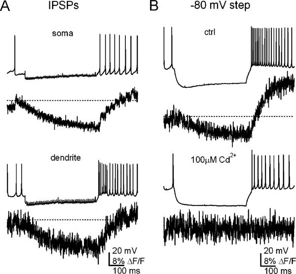
Fig. 4.
Ca signals during IPSPs and inhibition-like hyperpolarizations. a Spontaneous action potentials (no holding current) and IPSPs evoked at 100 Hz for 500 ms and somatic and dendritic Ca signals recorded with Fluo-4 (Kd=345 nM). ΔF/F values are calculated relative to the Ca signal when cells were held silent at –80 mV. Dotted lines indicate mean ΔF/F during spontaneous firing. Note that transients on the time scale of individual action potentials are evident. During IPSPs, Ca signals drop, indicating that firing neurons sustain a Ca load. After inhibition, Ca levels increase gradually at a rate proportional to the rate of firing, but do not rapidly exceed the value associated with spontaneous firing. b Experiments as in a, but with a current injection to –80 mV, a value about 5 mV negative to ECl, and about 10 mV hyperpolarized to the value reached during synaptic inhibition. With this hyperpolarization, the post-hyperpolarization dendritic Ca signal is large (top). The recording is repeated in the same cell (bottom) but with 100 μM Cd++ in the solution, which nearly fully blocks HVA channels but T-type channels by only 50%. The Ca changes are abolished, suggesting that they are mediated by HVA channels. Data from [45], reproduced with permission

