Abstract
An urgent requirement for 3D dosimetry has been recognized because of high failure rate (~25%) in RPC credentialing, which relies on point and 2D dose measurements. Comprehensive 3D dosimetry is likely to resolve more errors and improve IMRT quality assurance. This work presents an investigation of the feasibility of PRESAGE/optical-CT 3D dosimetry in the Radiologic Physics Center (RPC) IMRT H&N phantom. The RPC H&N phantom (with standard and PRESAGE dosimetry inserts alternately) was irradiated with the same IMRT plan. The TLD and EBT film measurement data from standard insert irradiation was provided by RPC. The 3D dose measurement data from PRESAGE insert irradiation was readout using the OCTOPUS™ 5X optical-CT scanner at Duke. TLD, EBT and PRESAGE dose measurements were inter-compared with Eclipse calculations to evaluate consistency of planning and delivery. Results showed that the TLD point dose measurements agreed with Eclipse calculations to within 5% dose-difference. Relative dose comparison between Eclipse dose, EBT dose and PRESAGE dose was conducted using profiles and gamma comparisons (4% dose-difference and 4 mm distance-to-agreement). Profiles showed good agreement between measurement and calculation except along steep dose gradient regions where Eclipse modelling might be inaccurate. Gamma comparisons showed that the measurement and calculation showed good agreement (>96%) if edge artefacts in measurements are ignored. In conclusion, the PRESAGE/optical-CT dosimetry system was found to be feasible as an independent dosimetry tool in the RPC IMRT H&N phantom.
1. Introduction
The Radiological Physics Center (RPC) Head and Neck (H&N) Intensity Modulated Radiation Therapy (IMRT) credentialing test is routinely taken by institutions that wish to participate in the H&N clinical trial protocols1. The dosimetry for credentialing comprises point and 2D dose measurements using TLDs and EBT Gafchromic film respectively. A lenient passing criterion of 7% dose-difference and 4 mm distance-to-agreement is used for comparison of measurement with planning calculations. It has been reported that 25% of the institutions fail the credentialing test at first attempt in spite of 2D measurements and generous verification criteria12. The standard of IMRT quality assurance could be improved if comprehensive 3D dosimetry is utilized.
The potential of the PRESAGE™/optical-CT system as a practical, 3D, dosimetric quality assurance tool in the clinic has been demonstrated3. This system has many favorable characteristics for optical-CT readout including stability in clinical settings, linear radiochromic (light absorbing not light-scattering) response to radiation and non-requirement of external containers3,4. In this study, the first implementation of the PRESAGE™/optical-CT 3D dosimetry system for 3D dosimetry in the RPC IMRT H&N phantom is demonstrated.
2. Materials and Methods
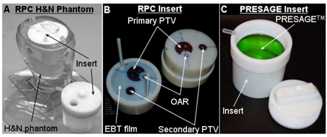
A picture of the RPC H&N phantom with the standard insert (henceforth called RPC insert) and a modified insert (for PRESAGE™ 3D dosimeter) is shown in fig 1. The components of the RPC insert are: an EBT film in the central axial plane, two EBT films in the sagittal plane, a primary planning target volume (PTV) structure, a secondary PTV structure, an organ at risk (OAR) structure and 8 TLDs dispersed in the three structures. The structures have different densities than the rest of the standard insert material such that they are clearly identified in an x-ray CT scan.
Fig 1. RPC H&N phantom and inserts.
A picture of the phantom with insert is shown in A. The central cross-section of the standard RPC insert is shown in B. The PRESAGE insert is shown in C.
Treatment planning and delivery for the H&N phantom (with standard insert as well as PRESAGE insert) followed the same procedure used for an actual patient treatment. After x-ray CT scans, an IMRT treatment plan was designed based on the constraints provided by the RPC. The only difference between the treatment plans for the RPC insert and PRESAGE insert was that the prescription dose was reduced to 4Gy for the PRESAGE insert from 6.6 Gy for the RPC insert. The reduction in prescription dose was required for optimal OD change in the PRESAGE dosimeter and did not change the relative dose distribution (see fig 2) and/or relative fluence. MapCheck IMRT QA was employed to check consistency of relative fluence between the RPC insert plan and PRESAGE insert plan. A gamma comparison with a 0.3% dose-difference and 0.3 mm distance-to-agreement criteria showed that the agreement was >98% for all beams. This facilitated direct comparison of relative dose distributions from PRESAGE™ measurement (PRESAGE dose), treatment planning system calculation (Eclipse dose) and EBT film measurement (EBT dose).
Fig 2. Reduction in prescription dose did not change relative dose distribution in RPC insert and PRESAGE insert.
After irradiation, the RPC standard insert was sent back to the RPC for dose readout from the TLDs and the axial EBT film whereas the PRESAGE dosimeter was scanned at Duke using the OCTOPUS™ 5X optical-CT scanner. The OCTOPUS™ 5X scanner is an upgraded version of the OCTOPUS scanner described in Guo et al3 with a 5x fast scanning capability. The 3D scan consisted of 20 axial slices (inter-slice spacing of 4 mm) where each slice comprised 600 linear projection scans (image matrix of 168 pixels with 1 mm pixel spacing) acquired at 0.60 angular increments. The 3D dose cube was reconstructed with a spatial resolution of 1 mm×1mm×4mm using an in house MATLAB code. After dose readout, the absolute point dose measurements form TLDs were compared with Eclipse calculations. The relative PRESAGE dose, Eclipse dose and EBT dose distributions were also compared for 2D and 3D analysis. Dose profiles and gamma maps were used for comparative analysis.
3. Results and Discussion
Successful RPC credentialing requires that the TLD measurements in the primary PTV and secondary PTV are within 7% of Eclipse calculations. Results showed that the TLD measurements comfortably passed the RPC threshold in primary PTV and secondary PTV where dose-difference was within 5% of Eclipse calculations. In the OAR, the pass criterion is that the distance-to-agreement (DTA) should be within 4 mm. TLD results showed that the DTA agreement was within 1mm thereby passing the credentialing test.
Fig 3 shows a central slice from PRESAGE dose measurement. Improvements in optics and acquisition technique resulted in lower noise (reduced to 2%) and reduced edge artefact (< 4 mm). The low noise and significantly reduced edge artefact facilitated highly accurate and precise dose measurement even at low dose levels and to within 4 mm of the edge.
Fig 3. PRESAGE dose measurement.
A) Central slice. B) Dose profiles along dotted lines in A show reduced noise and edge artefact.
Dose profile and gamma comparison between PRESAGE dose, EBT dose and Eclipse dose in the central axial plane is shown in fig 4. Profiles showed good agreement in the high dose region. In general, PRESAGE dose measurement showed better noise characteristics than EBT film dose measurement. Some differences were observed in dose gradient regions and near the edge. The differences in dose gradient regions were because of modeling errors of Eclipse while differences near the edge were because of edge artefacts in PRESAGE and EBT dose measurement. Gamma comparisons showed that all dose comparisons showed full agreement if a 4% dose-difference and 4 mm DTA criteria was used and if the outer 4 mm ring (edge artefact) is ignored. Similar agreement was observed for a full 3D gamma comparison between PRESAGE measurement and Eclipse calculation demonstrating the feasibility of accurate 3D dosimetry in the RPC IMRT H&N phantom.
Fig 4. Profile (A) and gamma comparison (B) between PRESAGE dose, EBT dose and Eclipse dose.
4. Conclusions
Significant failure rates for participating institutions have been reported in the 2D RPC credentialing tests even though generous verification criteria are used. Comprehensive 3D dosimetry (e.g. PRESAGE/optical-CT dosimetry system) has the potential to improve IMRT quality assurance and resolve more errors. This work is the first implementation of the PRESAGE/optical-CT dosimetry system for 3D dosimetry in the RPC IMRT H&N phantom. Noise level of PRESAGE measurement (2%) was significantly improved and was lesser than that of EBT film measurement used in RPC credentialing. Profile comparisons showed encouraging agreement of PRESAGE measurement with EBT film measurement and Eclipse calculations. Gamma map comparisons showed that if edge artefacts (significantly reduced to < 4mm) were ignored, PRESAGE measurement showed full agreement with EBT film measurement and Eclipse dose calculations to within 4% dose-difference and 4 mm distance-to-agreement constraints. In general, results showed that the PRESAGE/optical-CT system was a feasible tool for 3D dosimetry in the RPC H&N phantom and it has the potential to extend the RPC credentialing process to three dimensions.
Contributor Information
HS Sakhalkar, Department of Radiation Oncology Physics, Duke University Medical Center, Durham, NC.
D Sterling, Department of Radiation Oncology Physics, Duke University Medical Center, Durham, NC.
J Adamovics, Department of Chemistry and Biology, Rider University, Lawrenceville, NJ.
G Ibbott, Department of Radiation Physics, M. D. Anderson Cancer Center, Houston, Tx.
M Oldham, Email: mark.oldham@duke.edu, Department of Radiation Oncology Physics, Duke University Medical Center, Durham, NC.
References
- 1.Molineu A, et al. Design and implementation of an anthropomorphic quality assurance phantom for intensity-modulated radiation therapy for the Radiation Therapy Oncology Group. Int J Radiat Oncol Biol Phys. 2005;63:577–583. doi: 10.1016/j.ijrobp.2005.05.021. [DOI] [PubMed] [Google Scholar]
- 2.Radiological Physics Center Report #129. Report to the AAPM Therapy Physics Committee. 2008 [Google Scholar]
- 3.Guo P, Adamovics J, Oldham M. A practical three-dimensional dosimetry system for radiation therapy. Med Phys. 2006;33:3962–3972. doi: 10.1118/1.2349686. [DOI] [PMC free article] [PubMed] [Google Scholar]
- 4.Guo PY, Adamovics JA, Oldham M. Characterization of a new radiochromic threedimensional dosimeter. Med Phys. 2006;33:1338–1345. doi: 10.1118/1.2192888. [DOI] [PMC free article] [PubMed] [Google Scholar]


