

A composite digital photograph of the right retina of a 24-year-old Bangladeshi man with non-fatal cerebral malaria was taken on admission to hospital (Figure 1). He presented with 1 day of unconsciousness, pulmonary edema, and blackwater fever. Peripheral blood parasitemia was 79 Plasmodium falciparum per 1,000 red cells and hemoglobin was 10.9 g/dL. He recovered consciousness within 48 hours of starting intravenous artesunate and was discharged home after 6 days. Visual function and neurologic examination were normal on discharge.
Figure 1.
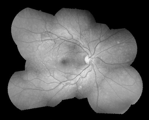
Composite digital photograph of the right retina of a 24-year-old man with cerebral malaria and severe malarial retinopathy. This figure appears in color at www.ajtmh.org.
The image is comprised of 14 separate photographs taken with a Genesis D (Kowa, Japan) handheld retinal camera. It shows severe malarial retinopathy with small patches of retinal whitening involving the entire macula and extensive areas of the peripheral retina, in particular temporal to the central fovea and nasal to the optic nerve head (some glare is present along the temporal vascular arcades). Malarial retinal whitening has a distribution that is unique to malaria1 and is present in 50% of children with cerebral malaria. 1 In adults, it has previously only been described in two Malawians with cerebral malaria, both of whom had mild changes. 2
The handheld retinal camera can be used at the bedside and, unlike conventional retinal photography with a table-top camera, does not require a cooperative, sitting patient.
Acknowledgments
The authors thank Dr. Arjen Dondorp for kind assistance with drafting the manuscript and the staff at Chittagong Medical College Hospital, Chittagong, Bangladesh, for assistance with this case.
Footnotes
Financial support: Mahidol-Oxford Tropical Medicine Research Unit is funded by the Wellcome Trust of Great Britain.
Disclosure: There are no conflicts of interest.
Ethical approval: Ethical permission for retinal photography was obtained from Chittagong Medical College Ethics Committee, Chittagong, Bangladesh, and Oxford Tropical Medicine Research Ethics Committee, Oxford, UK.
Authors' addresses: Richard J. Maude, Mahidol-Oxford Tropical Medicine Research Unit, Faculty of Tropical Medicine, Mahidol University, 3/F, 60th Anniversary Chalermprakiat Building, 420/6 Rajvithi Road, Rajthevee, Bangkok 10400, Thailand, E-mail: richardmaude@gmail.com or richard@tropmedres.ac. Mahtab Uddin Hassan, Department of Medicine, Chittagong Medical College Hospital, Chittagong, Bangladesh, E-mail: mahtabnipu@yahoo.com. Nicholas A. V. Beare, St. Paul's Eye Unit, Royal Liverpool University Hospital, Prescot Street, Liverpool, L7 8XP, UK, E-mail: nbeare@btinternet.com.
References
- 1.Beare NAV, Taylor TE, Harding SP, Lewallen S, Molyneux ME. Malarial retinopathy: a newly established diagnostic sign in severe malaria. Am J Trop Med Hyg. 2006;75:790–797. [PMC free article] [PubMed] [Google Scholar]
- 2.Beare NAV, Lewis DK, Kublin JG, Harding SP, Zijlstra EE. Retinal changes in adults with cerebral malaria. Ann Trop Med Parasitol. 2003;97:313–315. doi: 10.1179/000349803235001859. [DOI] [PubMed] [Google Scholar]