

Abstract
We report the synthesis of high density lipoprotein (HDL) bio-mimetic nanoparticles capable of binding cholesterol. These structures use a gold nanoparticle core to template the assembly of a mixed phospholipid layer and the adsorption of apolipoprotein A-I. These synthesized structures have the general size and surface composition of natural HDL, and importantly, bind free cholesterol (Kd = 4 nM). The determination of the Kd for these particles, with respect to cholesterol complexation, provides a key starting and comparison point for measuring and evaluating the properties of subsequently developed synthetic versions of HDL.
High density lipoprotein (HDL) is a dynamic serum nanostructure protective against the development of atherosclerosis and resultant illnesses such as heart disease and stroke.1 Increasing circulating serum HDL levels provides a promising therapeutic approach to preventing and, potentially, reversing atherosclerosis by augmenting reverse cholesterol transport.2-4 However, a facile route to synthetic HDL remains a challenge, as methods to control and mimic the size, surface chemistry, and activity of HDL have not yet been demonstrated. Due in part to their biocompatibility and tailorability, gold nanoparticle (Au NP) conjugates have shown promise as therapeutics,5,6 intracellular gene regulation agents,7,8 and in vitro diagnostic probes.9-11 Herein, we report a new Au NP core-shell structure, where the Au NP core serves as a size- and shape-controllable scaffold12 for constructing an HDL-like particle from phospholipids and apolipoprotein A-I (APOA1). Importantly, the proof-of-concept HDL-Au NP structure we report is designed to be within the size range of HDL and to mimic the general surface composition of HDL. Furthermore, we demonstrate that, like HDL, HDL-Au NPs are capable of binding cholesterol.
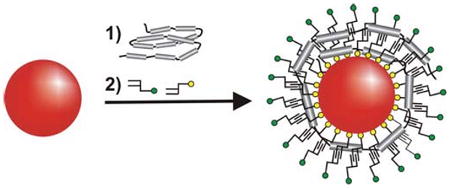
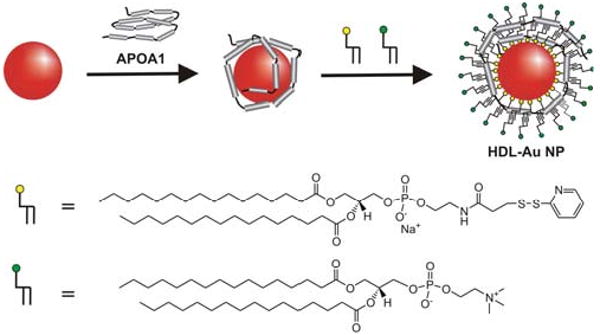
In a typical synthesis, an aqueous suspension of citrate-stabilized gold nanoparticles (5 ± 0.75 nm) is mixed with an aqueous solution of purified APOA1 and stirred overnight (Scheme 1).
Scheme 1.
Synthesis of Templated Spherical HDL Nanoparticles.
APOA1, comprised of 10 amphipathic alpha helices each with a hydrophobic domain and a negatively charged hydrophilic domain, is the main protein component of HDL and defines the structure and physiology of HDL in vivo.13-16 Next, a 1:1 solution of disulfide-functionalized lipid, 1,2-dipalmitoyl-sn-glycero-3-phosphoethanolamine-N-[3-(2-pyridyldithio)propionate], and amine functionalized lipid, 1-2-dipalmitoyl-sn-glycero-3-phosphocholine, was mixed in CHCl3 and added to the aqueous suspension of particles in 100-fold excess with respect to the Au NPs. While several ratios were tried, these ratios were found to give loadings of lipid and protein similar to that of natural HDL (vide infra). The disulfide lipid was selected since the disulfide functionality allows for chemisorption to the surface of the Au NP. The amine-modified lipid is a naturally occurring phospholipid known to electrostatically and hydrophobically associate with APOA1.13-16 This addition results in a two-phase mixture. Upon gradual heating, CHCl3 is evaporated and the lipids are transferred to the aqueous phase containing the dispersed Au NPs with APOA1. Note that if the disulfide lipid is added alone and without prior addition of APOA1, the particles precipitate since they become hydrophobic upon lipid adsorption to the Au NP surface.
Purification of the HDL-Au NPs is accomplished via repeated centrifugation and re-suspension in water or buffered saline solutions. UV-vis analysis of the purified HDL-Au NPs exhibits a band at 520 nm (Supporting Information), consistent with dispersed rather than aggregated HDL-Au NPs.11,12 Dynamic light scattering experiments were used to follow the Au NP surface modification process. The results demonstrate sequential growth of the HDL structures (Table 1). An unmodified gold colloid, (9.2 nm average hydrodynamic diameter) is first modified with APOA1 (11.0 nm), and subsequently the mixture of lipids (17.9 nm). The average size of the resulting HDL-Au NPs is similar to that for natural HDL.13,17,18
Table 1.
Hydrodynamic diameter of conjugates.
| Particles | Hydrodynamic Diameter (nm) |
|---|---|
| Au NP (5 nm diameter) | 9.2 ± 2.1 |
| Au NP + APOA1 | 11.0 ± 2.5 |
| Au NP + APOA1 + Phospholipids | 17.9 ± 3.1 |
Investigations have shown that there are between 2-5 copies of APOA1 and ~80-160 phospholipids per natural mature spherical HDL.15,16,19,20 To characterize the chemical composition of HDL-Au NPs, fluorophore labeled components (APOA1 and aminated phospholipids) were used to synthesize HDL-Au NPs as described. After dissolving the Au NPs using 0.04 M KCN, we quantitatively determined that the average number of proteins and aminated phospholipids per particle is 3 ± 1 and 83 ± 12, respectively (Supporting Information). Thus, these values correspond well to those reported for natural HDL.
We hypothesize that, in addition to modifying the exposed gold surface, the disulfide functionalized lipid interacts with the hydrophobic portions of APOA1 forming a complex that is water soluble. In our working model of this structure, the aminated lipid forms a tail-to-tail hydrophobic complex with the adsorbed disulfide functionalized lipid and an electrostatic complex with the adsorbed APOA1. The basis for this model is the known lipid organization capabilities of APOA1,13-16 the well precedented disulfide adsorption on gold chemistry,21 the water solubility of the resulting complex, and fluorescence measurements aimed at removing and quantifying the number of proteins and aminated phospholipids that make up the final structure.
Transport of cholesterol to the liver by HDL is a key mechanism by which HDL protects against the development of atherosclerosis.2 Thus, determining if HDL-Au NPs bind cholesterol is important for determining the potential of these structures as therapeutic agents. The binding of cholesterol to HDL-Au NPs was investigated with a fluorescent cholesterol analogue (25-{N-[(7-nitrobenz-2-oxa-1,3-diazol-4-yl)-methyl]amino}-27-norcholesterol, NBD-cholesterol). NBD-cholesterol fluorescence is weak in polar environments such as water; however, in non-polar matrices (such as a lipid membrane) NBD-cholesterol becomes fluorescent.22,23 Quenching by the Au NP24 causes the signal of the HDL-Au NP bound NBD-cholesterol to be partially dampened. However, titration of NBD-cholesterol into a solution of HDL-Au NPs provides a strong enough fluorescent signal to construct a binding isotherm (Figure 1). This isotherm was used to calculate a Kd of 3.8 ± 0.8 nM for NBD-cholesterol binding to HDL-Au NPs. Preliminary experiments indicate that NBD-cholesterol is irreversibly bound to HDL-Au NPs under the stated conditions; however, further experiments are underway to more thoroughly address this question. To the best of our knowledge, this the first example of a Kd measured for a synthetic HDL analogue. Interestingly, there is little information regarding the Kd of natural HDL for comparison purposes.
Figure 1.
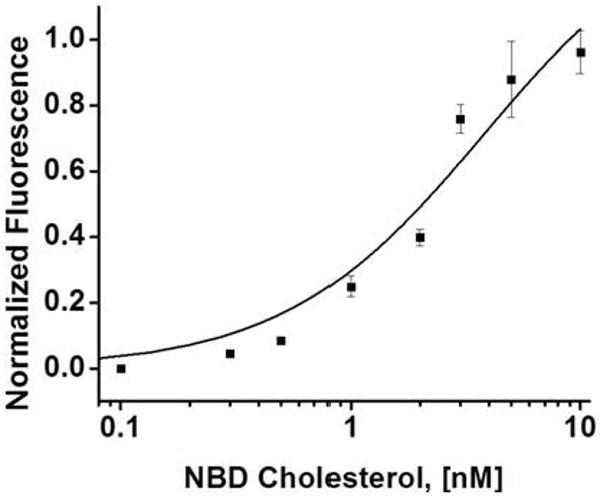
Binding isotherm of NBD-cholesterol to HDL-Au NPs. NBD-cholesterol was titrated into a 5 nM solution of HDL-Au NPs. The fluorescence versus NBD-cholesterol concentration was used to calculate the Kd of HDL-Au NPs.
In conclusion, we report the synthesis and characterization of a novel class of HDL-Au NPs, designed to mimic their biological HDL counterpart. Importantly, our data demonstrate that HDL-Au NPs can be used as biomimetic materials to bind cholesterol. Because it is being increasingly appreciated that the size, shape, and chemistry of HDL dictates in vivo physiology,25,26 this reported general approach may provide a novel method to fabricate and rationally tailor HDL structures. We anticipate that this synthetic method will provide a new and general approach for the development of templated nanomaterials designed to mimic HDL, and with future study and characterization in biologically relevant conditions, these compounds may prove interesting for the development of therapeutic agents. Finally, the determination of the Kd for these particles, with respect to cholesterol complexation, provides a key starting and comparison point for measuring and evaluating the cholesterol binding properties of HDL.
Supplementary Material
Acknowledgments
C.A.M. acknowledges a Cancer Center for Nanotechnology Excellence (NCI-CCNE) award for support of this research. C.A.M. is also grateful for a NIH Director's Pioneer Award. C.S.T would like to thank the Department of Urology at Northwestern University Feinberg School of Medicine for partial funding of this work.
Footnotes
Supporting Information Available: Experimental conditions and HDL-Au NP characterization. This material is available free of charge via the Internet at http://pubs.acs.org.
References
- 1.Kontush A, Chapman MJ. Nat Clin Pract Card. 2006;3:144–153. doi: 10.1038/ncpcardio0500. [DOI] [PubMed] [Google Scholar]
- 2.Joy T, Hegele RA. Nat Rev Drug Discov. 2008;7:143–155. doi: 10.1038/nrd2489. [DOI] [PubMed] [Google Scholar]
- 3.Nissen SE, Tsunoda T, Tuzcu EM, Schoenhagen P, Cooper CJ, Yasin M, Eaton GM, Lauer MA, Sheldon WS, Grines CL, Halpern S, Crowe T, Blankenship JC, Kerensky R. J Amer Med Assoc. 2003;290:2292–2300. doi: 10.1001/jama.290.17.2292. [DOI] [PubMed] [Google Scholar]
- 4.Tardif JC, Gregoire J, L'Allier PL, Ibrahim R, Lesperance J, Heinonen TM, Kouz S, Berry C, Basser R, Lavoie MA, Guertin MC, Rodes-Cabau J. J Amer Med Assoc. 2007;297:1675–1682. doi: 10.1001/jama.297.15.jpc70004. [DOI] [PubMed] [Google Scholar]
- 5.Huang XH, Jain PK, El-Sayed IH, El-Sayed MA. Nanomed. 2007;2:681–693. doi: 10.2217/17435889.2.5.681. [DOI] [PubMed] [Google Scholar]
- 6.Simberg D, Duza T, Park JH, Essler M, Pilch J, Zhang LL, Derfus AM, Yang M, Hoffman RM, Bhatia S, Sailor MJ, Ruoslahti E. P Natl Acad Sci USA. 2007;104:932–936. doi: 10.1073/pnas.0610298104. [DOI] [PMC free article] [PubMed] [Google Scholar]
- 7.Agbasi-Porter C, Ryman-Rasmussen J, Franzen S, Feldheim D. Bioconjugate Chem. 2006;17:1178–1183. doi: 10.1021/bc060100f. [DOI] [PubMed] [Google Scholar]
- 8.Rosi NL, Giljohann DA, Thaxton CS, Lytton-Jean L, Han MS, Mirkin CA. Science. 2006;312:1027–1030. doi: 10.1126/science.1125559. [DOI] [PubMed] [Google Scholar]
- 9.Cao CYW, Jin R, Mirkin CA. Science. 2002;297:1536–1540. doi: 10.1126/science.297.5586.1536. [DOI] [PubMed] [Google Scholar]
- 10.Wang Z, Heon Lee J, Lu Y. Adv Mater. 2008;20:3263–3267. [Google Scholar]
- 11.Elghanian R, Storhoff JJ, Mucic RC, Letsinger RL, Mirkin CA. Science. 1997;277:1078–1080. doi: 10.1126/science.277.5329.1078. [DOI] [PubMed] [Google Scholar]
- 12.Mirkin CA, Letsinger RL, Mucic RC, Storhoff JJ. Nature. 1996;382:607–609. doi: 10.1038/382607a0. [DOI] [PubMed] [Google Scholar]
- 13.Libby P, Bonow R, Mann D, Zipes D. Braunwald's Heart Disease: A Textbook of Cardiovascular Medicine. 8th. Saunders Elsevier; Philadelphia, PA: 2008. [Google Scholar]
- 14.Silva R, Huang R, Morris J, Fang J, Gracheva EO, Ren G, Kontush A, Jerome WG, Rye KA, Davidson WS. P Natl Acad Sci USA. 2008;105:12176–12181. doi: 10.1073/pnas.0803626105. [DOI] [PMC free article] [PubMed] [Google Scholar]
- 15.Ajees AA, Anantharamaiah GM, Mishra VK, Hussain MM, Murthy HMK. P Natl Acad Sci USA. 2006;103:2126–2131. doi: 10.1073/pnas.0506877103. [DOI] [PMC free article] [PubMed] [Google Scholar] [Retracted]
- 16.Thomas MJ, Bhat S, Sorci-Thomas MG. J Lipid Res. 2008;49:1875–1883. doi: 10.1194/jlr.R800010-JLR200. [DOI] [PMC free article] [PubMed] [Google Scholar]
- 17.Clay MA, Barter PJ. J Lipid Res. 1996;37:1722–1732. [PubMed] [Google Scholar]
- 18.Asztalos BF, Sloop CH, Wong L, Roheim PS. Biochim Biophys Acta. 1993;1169:291–300. doi: 10.1016/0005-2760(93)90253-6. [DOI] [PubMed] [Google Scholar]
- 19.Bhat S, Sorci-Thomas MG, Tuladhar R, Samuel MP, Thomas MJ. Biochemistry-US. 2007;46:7811–7821. doi: 10.1021/bi700384t. [DOI] [PMC free article] [PubMed] [Google Scholar]
- 20.Catte A, Patterson JC, Jones MK, Jerome WG, Bashtovyy D, Su ZC, Gu FF, Chen JG, Aliste MP, Harvey SC, Li L, Weinstein G, Segrest JP. Biophys J. 2006;90:4345–4360. doi: 10.1529/biophysj.105.071456. [DOI] [PMC free article] [PubMed] [Google Scholar]
- 21.Daniel MC, Astruc D. Chem Rev. 2004;104:293–346. doi: 10.1021/cr030698+. [DOI] [PubMed] [Google Scholar]
- 22.Mukherjee S, Chattopadhyay A. Biochemistry-US. 1996;35:1311–1322. doi: 10.1021/bi951953q. [DOI] [PubMed] [Google Scholar]
- 23.Pucadyil TJ, Mukherjee S, Chattopadhyay A. J Phys Chem B. 2007;111:1975–1983. doi: 10.1021/jp066092h. [DOI] [PubMed] [Google Scholar]
- 24.Dubertret B, Calame M, Libchaber AJ. Nat Biotechnol. 2001;19:365–370. doi: 10.1038/86762. [DOI] [PubMed] [Google Scholar]
- 25.Degoma EM, Degoma RL, Rader DJ. J Am Coll Cardiol. 2008;51:2199–2211. doi: 10.1016/j.jacc.2008.03.016. [DOI] [PMC free article] [PubMed] [Google Scholar]
- 26.Movva R, Rader DJ. Clin Chem. 2008;54:788–800. doi: 10.1373/clinchem.2007.101923. [DOI] [PubMed] [Google Scholar]
- 27.Cormode DP, Skajaa T, van Schooneveld MM, Koole R, Jarzyna P, Lobatto ME, Calcagno C, Barazza A, Gordon RE, Zanzonico P, Fisher EA, Fayed ZA, Mulder WJ. Nano Lett. 2008;8:3715–3723. doi: 10.1021/nl801958b. [DOI] [PMC free article] [PubMed] [Google Scholar]
Associated Data
This section collects any data citations, data availability statements, or supplementary materials included in this article.