Abstract
Background: Body mass index (BMI)–for-age has been recommended as a screening test for excess adiposity in children and adolescents.
Objective: We quantified the performance of standard categories of BMI-for-age relative to the population prevalence of high adiposity in children and adolescents overall and by race-ethnic group in a nationally representative US population sample by using definitions of high adiposity that are consistent with expert committee recommendations.
Design: Percentage body fat in 8821 children and adolescents aged 8–19 y was measured by using dual-energy X-ray absorptiometry in 1999–2004 as part of a health examination survey.
Results: With the use of several different cutoffs for percentage fat to define high adiposity, most children with high BMI-for-age (≥95th percentile of the growth charts) had high adiposity, and few children with normal BMI-for-age (<85th percentile) had high adiposity. The prevalence of high adiposity in intermediate BMI categories varied from 45% to 15% depending on the cutoff. The prevalence of a high BMI was significantly higher in non-Hispanic black girls than in non-Hispanic white girls, but the prevalence of high adiposity was not significantly different.
Conclusions: Current BMI cutoffs can identify a high prevalence of high adiposity in children with high BMI-for-age and a low prevalence of high adiposity in children with normal BMI-for-age. By these adiposity measures, less than one-half of children with intermediate BMIs-for-age (85th to <95th percentile) have high adiposity. Differences in high BMI ranges between race-ethnic groups do not necessarily indicate differences in high adiposity.
INTRODUCTION
Epidemiologic studies often use body mass index (BMI; in kg/m2) calculated from weight and height as an indicator of adiposity. For children and adolescents, definitions are based on BMI-for-age from a reference population. In the United States, the 2000 Centers for Disease Control and Prevention (CDC) growth charts are commonly used as the reference for sex-specific BMI-for-age percentiles (1–3). These CDC charts for ages 8–19 y were constructed by using data from nationally representative surveys covering the years 1963 through 1980. Expert committee recommendations (4–7) for terminology for BMI-for-age categories vary but consistently identify 2 levels of concern by using the 85th and the 95th percentiles of BMI-for-age from a reference population.
The underlying assumption for using BMI to assess adiposity is that, at a given height, higher weight is associated with increased fatness (8). However, BMI is an imperfect measure of body fatness (9, 10) because it cannot discriminate between lean mass and fat mass. The definition of obesity as “a condition that is characterized by excessive accumulation and storage of fat in the body” (11) has to do with fat, not weight or BMI. However, obesity may also be used as a label for a range of weight rather than of fat. To avoid confusion between different uses of the term obesity, we refer to high ranges of BMI-for-age as high BMI-for-age and to high values of percentage of body fat as high adiposity.
There are no widely accepted levels of body fatness that are considered to define high adiposity for children (12). Because expert committee reports on overweight children and obesity in children reflect expert judgment on the probability of excess fatness within BMI categories, we sought cutoffs of body fatness that would be consistent with those judgments.
We quantified the performance of standard categories of BMI-for-age relative to the population prevalence of high adiposity for children and adolescents, overall and by race-ethnic group, in a nationally representative US population sample by using definitions of high adiposity that are consistent with expert committee recommendations.
METHODS
In the 1999–2004 National Health and Nutrition Examination Survey (NHANES), a representative cross-sectional sample of the US civilian, noninstitutionalized population was selected by using a complex, multistage probability design. The survey included an interview in the household followed by an examination in a mobile examination center. NHANES 1999–2004 underwent institutional review board approval, and participants gave written informed consent to participate in the survey. This article uses data from NHANES 1999–2004 for 8821 nonpregnant participants, aged 8–19 y, who were measured for weight and height.
Age was calculated as the age in months at the time of the examination. Race and ethnicity were reported by the participants, and for the purposes of this study, race-ethnic groups were classified as non-Hispanic white, non-Hispanic black, Mexican American, and other. Weight and height (stature) were measured by using standardized techniques and equipment (13). Sex-specific BMI-for-age percentile values were calculated according to the 2000 CDC growth charts.
Dual-energy X-ray absorptiometry (DXA) is one of the most accurate and precise methods available to measure total body fat and lean soft tissue mass directly. Whole-body DXA scans were acquired with a Hologic QDR-4500A fan-beam densitometer (Hologic Inc, Bedford, MA) (13). Whole-body percentage body fat was calculated as the total body fat mass/total mass (from DXA) × 100. The scan for each survey participant was reviewed and analyzed by the Department of Radiology, University of California, San Francisco. Hologic Discovery software (version 12.1; Hologic Inc) was used to analyze the scans. The Discovery analysis algorithms automatically detect and measure very-low-density bone in children weighing ≤40 kg. The NHANES data were adjusted on the basis of on the results of an analysis of QDR-4500A DXA data from 7 research laboratories indicating that the QDR-4500A algorithm underestimated fat mass and overestimated lean mass (14). On the basis of the results of the analysis, the NHANES DXA lean mass was decreased by 5%, and an equivalent kilogram weight was added to the fat mass so that the total mass did not change. A detailed description of the procedures that were followed is provided as part of the documentation of the data files (15).
For our analyses, we used the NHANES 1999–2004 DXA multiple-imputation data files (15). The multiple imputations in which 5 DXA values were imputed for each missing DXA value reflect the uncertainty of the imputation procedure, and this uncertainty is incorporated into the standard errors and P values (16). The characteristics of the missing data and the imputation process for the NHANES 1999–2004 DXA multiple-imputation data files are both described in detail in the technical documentation (17). In 1999, women aged 8–17 y did not have institutional review board approval for DXA exams, and thus DXA data for 610 women with measured weight and height data in 1999 were imputed as part of the multiple-imputation project. In total, 1352 of the participants aged 8–19 y with measured weight and height data had some or all DXA data imputed.
BMI, obesity, and adiposity
According to the summary report of an American Medical Association (AMA) expert committee (5), “The use of 2 cutoff points, namely, BMI of 95th percentile and 85th percentile, captures varying risk levels and minimizes both overdiagnosis and underdiagnosis” (p S167). With the use of these outpoints, we divided BMI-for-age categories into 3 ranges: normal, intermediate, and high on the basis of percentiles of BMI-for-age from the CDC growth charts (Table 1). For descriptive purposes, these ranges were sometimes subdivided further (Table 1).
TABLE 1.
BMI categories and descriptors1
| Designation | Percentiles of BMI-for-age from the CDC growth charts | Percentile subgrouping | Nomenclature |
| High BMI | ≥95 | ≥97 | Very high |
| 95 to <97 | Moderately high | ||
| Intermediate BMI | 85 to <95 | 90 to <95 | High-intermediate |
| 85 to <90 | Low-intermediate | ||
| Normal BMI | <85 | 75 to <85 | High-normal |
| <75 | Low-normal |
CDC, Centers for Disease Control and Prevention.
To estimate sex-specific smoothed percentiles by age of percentage body fat from children measured in 1999–2004, a procedure consisting of a nonparametric double-kernel method and automatic bandwidth selection was used to estimate percentile curves for the DXA data (18). This approach extends a method of Yu and Jones (19) by incorporating sample weights into the curve estimation and bandwidth selection. We considered evenly spaced percentiles including the 65th, 70th, 75th, 80th, and 85th percentiles. The range of percentage of body fat between the 65th and 85th percentiles was 6 percentage points on average. For boys, the smoothed values of the 70th percentile of percentage body fat were ≈30% body fat at the youngest age (8 y) and decreased to 26% body fat at the oldest age (19.5 y); comparable values for girls were 35% and 40%, showing an increase rather than a decrease with age. Similar values for other percentiles of percentage body fat at the youngest and oldest ages ranged from 32% to 28% for boys and 36% to 41% for girls at the 75th percentile; 33–29% for boys and 37–42% for girls at the 80th percentile, and 36–31% for boys and 39–44% for girls at the 85th percentile. For both sexes at all ages, the overall difference between the 10th percentile and the 90th percentile was ≈20 percentage points.
With the use of several percentile cutoffs for body fatness, we calculated the prevalence of high adiposity overall and within BMI-for-age categories. We also calculated the mean percentage of body fat, fat mass, lean mass, weight, height, and BMI by race-ethnic group overall and within BMI categories.
Statistical methods
Analyses were conducted with PC-SAS (version 9.1; SAS Institute, Cary, NC) and SUDAAN (version 9.03; Research Triangle Institute, Research Triangle Park, NC). All analyses used sample weights and took into account the sample design and the multiple imputations in calculating statistical tests. Estimates were averaged over the 5 sets of imputations. Two-sample t tests were used to compare values. Statistical significance was determined on the basis of a 2-sided P value < 0.05 with Bonferroni adjustments. Results calculated without the imputed data were very similar to those calculated with the imputed data. All results presented in this report include the imputed data.
RESULTS
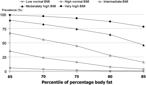
Basic descriptive information about the analytic sample is shown in Table 2. During the last several decades, expert committees on childhood-obesity assessment (4–7) consistently expressed the opinions that most children with high BMI-for-age are highly likely to have excess adiposity, children with intermediate BMI-for-age should be evaluated for excess adiposity, and relatively few children with normal BMI-for-age have excess adiposity. We calculated overall prevalence estimates of high adiposity within BMI categories for percentile cutoffs of 65, 70, 75, 80, and 85 for percentage body fat (Figure 1) to identify percentile cutoffs for high adiposity that would give prevalence estimates that approximately correspond to these statements.
TABLE 2.
Descriptive information (weighted estimates)
| Boys | Girls | |
| Unweighted sample size (n) | 4493 | 4322 |
| Weighted percentage | 51.0 | 49.0 |
| Age group (% total) | ||
| 8–11 y | 32.9 | 32.4 |
| 12–15 y | 34.1 | 36.6 |
| >16–19 y | 33.0 | 31.0 |
| Race-ethnic group (% total) | ||
| Non-Hispanic white | 61.0 | 60.4 |
| Mexican American | 14.8 | 14.9 |
| Non-Hispanic black | 11.1 | 11.1 |
| Other | 13.1 | 13.6 |
| BMI (kg/m2) | 21.8 ± 0.141 | 22.1 ± 0.15 |
| Body fat (%) | 25.4 ± 0.24 | 33.1 ± 0.21 |
| Prevalence of BMI-for-age categories (%) | ||
| ≥85th percentile | 33.9 ± 1.42 | 32.8 ± 1.20 |
| ≥95th percentile | 17.7 ± 0.98 | 16.6 ± 0.93 |
| ≥97th percentile | 12.7 ± 0.78 | 11.1 ± 0.91 |
Mean ± SE (all such values).
FIGURE 1.
Overall prevalence (%) of high adiposity within BMI categories by different percentile cutoffs for high adiposity.
The 70th percentile and particularly the 65th percentile of percentage body fat led to prevalence estimates for high adiposity in the high-normal BMI category that were potentially too high to be completely consistent with these expert committee statements. For these cutoffs, a fairly large proportion of children with high-normal BMI had high adiposity (35% of children with the 65th percentile cutoff and 24% of children with the 70th percentile cutoff.). The 65th percentile value would identify more children with high adiposity than all the children with intermediate and high BMI combined (33%).
We selected the 75th, 80th, and 85th percentiles of percentage body fat in 1999–2004 to provide possible estimates of high adiposity that would be reasonably consistent with expert committee recommendations. These percentiles covered a relatively small range of values of percentage body fat, with the average difference between the 75th percentile and the 85th percentile being 3.5 percentage points. Throughout this range of percentile cutoffs, the proportion of children with high adiposity at low-normal BMI was extremely low. For high-normal BMI, the prevalence of high adiposity ranged from 16% by using the 75th percentile of body fatness to 4% by using the 85th percentile of body fatness. For high BMI, the prevalence of high adiposity ranged from 87% by using the 75th percentile of body fatness to 69% by using the 85th percentile of body fatness. For very high BMI, the prevalence of high adiposity ranged from 93% by using the 75th percentile of body fatness to 79% by using the 85th percentile of body fatness. The choice of cutoff had the largest effects on the estimated prevalence of high adiposity in the intermediate BMI category; in that category, the difference in the prevalence of high adiposity between the 75th and 85th percentile cutoffs of percentage body fat was 29 percentage points (45% compared with 16%).
Race-ethnic comparisons of high BMI and high adiposity
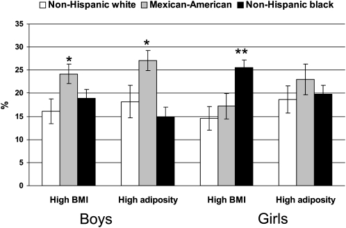
Comparisons of prevalences of high BMI and of high adiposity (on the basis of the 80th percentile of percentage body fat for this example) by race-ethnic group are shown separately for boys and girls in Figure 2. The patterns by race-ethnic group were different for high BMI and high adiposity.
FIGURE 2.
Prevalence (%) of high BMI-for-age and high adiposity (≥80th percentile) by race-ethnic group in boys and girls. *Mexican Americans were significantly different from non-Hispanic whites and non-Hispanic blacks by 2-sample t test: P < 0.0001. **Non-Hispanic blacks were significantly different from non-Hispanic whites and Mexican Americans by 2-sample t test: P < 0.0001.
For boys, the prevalence of high BMI-for-age did not differ significantly between non-Hispanic whites and non-Hispanic blacks (P = 0.10) but was significantly greater in Mexican Americans than in either of the other groups (P < 0.0001). Results for high adiposity at the 80th percentile range were similar to those for high BMI-for-age. For high adiposity, the differences between non-Hispanic whites and non-Hispanic blacks were not significant (P = 0.12), and the prevalence for Mexican Americans was significantly higher than for the other groups (P < 0.0001). For the 65th, 70th, and 75th percentile cutoffs for adiposity (data not shown), estimates were significantly higher for non-Hispanic whites than for non-Hispanic blacks (P < 0.001) and significantly higher for Mexican Americans than for non-Hispanic whites or blacks (P < 0.0001).
For girls, the prevalence of high BMI-for-age was significantly higher for non-Hispanic black girls than for non-Hispanic white girls (P < 0.0001) or Mexican-American girls (P < 0.0001) and did not differ between Mexican-American and non-Hispanic white girls (P = 0.22). The prevalence of high adiposity did not differ significantly between non-Hispanic white and non-Hispanic black girls at this cutoff (P = 0.50) or at lower percentile cutoffs. The prevalence of high adiposity did not differ significantly between Mexican-American girls and non-Hispanic white girls (P = 0.07) or non-Hispanic black girls (P = 0.09).
Race-ethnic comparisons of high adiposity by BMI category
The sex-specific prevalences of high adiposity within BMI-for-age categories by race-ethnic group by the 70th , 75th , 80th , and 85th percentile cutoffs of percentage body fat are shown in Table 3. These are positive predictive values showing the probability that children within a given BMI category have high adiposity. Within all BMI-for-age categories, for all percentile cutoffs, non-Hispanic black children, both boys and girls, had the lowest prevalences of high adiposity, although the differences were not always significant. Within BMI-for-age categories, there were no significant differences in the prevalence of high adiposity between non-Hispanic white and Mexican-American children.
TABLE 3.
High adiposity by sex, BMI category, and race-ethnic group: National Health and Nutrition Examination Survey (NHANES) 1999–20041
| Smoothed percentile of body fat, 1999–2004, used to define high adiposity | Normal BMI | Intermediate BMI | High BMI |
| Boys | |||
| 70th | |||
| Non-Hispanic white | 7.3 ± 1.2 | 57.7 ± 4.5 | 92.5 ± 1.9 |
| Mexican American | 6.9 ± 1.1 | 66.1 ± 3.7 | 95.5 ± 1.1 |
| Non-Hispanic black | 1.7 ± 0.42 | 30.0 ± 5.02 | 85.8 ± 2.43 |
| 75th | |||
| Non-Hispanic white | 4.5 ± 0.8 | 43.1 ± 3.4 | 87.2 ± 2.4 |
| Mexican American | 3.8 ± 0.7 | 46.9 ± 3.1 | 91.9 ± 1.1 |
| Non-Hispanic black | 1.1 ± 0.42 | 20.5 ± 3.92 | 77.4 ± 3.03 |
| 80th | |||
| Non-Hispanic white | 1.2 ± 0.4 | 27.2 ± 3.9 | 80.2 ± 2.7 |
| Mexican American | 1.9 ± 0.5 | 31.7 ± 2.2 | 83.5 ± 2.1 |
| Non-Hispanic black | 0.4 ± 0.23 | 14.3 ± 2.83 | 66.1 ± 3.73 |
| 85th | |||
| Non-Hispanic white | 0.4 ± 0.24 | 17.1 ± 2.8 | 69.1 ± 2.7 |
| Mexican American | 0.6 ± 0.24 | 17.3 ± 2.9 | 72.2 ± 3.1 |
| Non-Hispanic black | 0.2 ± 0.14 | 9.5 ± 2.5 | 57.0 ± 3.9 |
| Girls | |||
| 70th | |||
| Non-Hispanic white | 7.0 ± 1.3 | 57.6 ± 4.8 | 94.4 ± 2.5 |
| Mexican American | 12.5 ± 1.4 | 67.1 ± 3.3 | 94.6 ± 2.2 |
| Non-Hispanic black | 1.9 ± 0.62 | 30.2 ± 3.52 | 86.4 ± 2.4 |
| 75th | |||
| Non-Hispanic white | 3.6 ± 0.8 | 50.3 ± 4.3 | 91.6 ± 2.9 |
| Mexican American | 6.7 ± 0.9 | 57.4 ± 4.2 | 90.6 ± 2.7 |
| Non-Hispanic black | 0.9 ± 0.52 | 20.2 ± 2.92 | 78.6 ± 3.12 |
| 80th | |||
| Non-Hispanic white | 1.8 ± 0.6 | 30.8 ± 3.6 | 86.3 ± 3.6 |
| Mexican American | 3.4 ± 0.7 | 32.3 ± 4.5 | 85.7 ± 2.9 |
| Non-Hispanic black | 0.4 ± 0.33 | 11.8 ± 2.12 | 68.8 ± 3.82 |
| 85th | |||
| Non-Hispanic white | 1.0 ± 0.44 | 18.3 ± 3.5 | 74.1 ± 4.8 |
| Mexican American | 1.7 ± 0.64 | 17.0 ± 2.7 | 79.3 ± 2.7 |
| Non-Hispanic black | 0.3 ± 0.24 | 5.5 ± 1.82 | 56.6 ± 3.82 |
All values are prevalences (±SE) of high adiposity. Participants were aged 8–19 y and categorized according to different definitions of high adiposity on the basis of smoothed percentiles of percentage body fat.
Significantly different from non-Hispanic whites and Mexican Americans, P < 0.006 (2-sample t test).
Significantly different from Mexican Americans, P < 0.006 (2-sample t test).
Estimates had a relative SE >30% and did not meet standards of statistical reliability and precision.
Regardless of race-ethnic group, in the high BMI category most children had high adiposity, and in the normal BMI category relatively few children (ranging from 0.2% to 12.5%) had high adiposity. However, in the intermediate BMI category there were considerable differences by race-ethnic group; the prevalence of high adiposity among non-Hispanic black children was only about one-half of the prevalence among other children. These differences were significant in all but one case (Table 3).
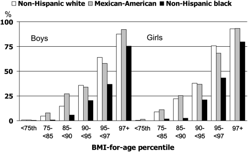
Examples of a more detailed breakdown by finer BMI categories for the 80th percentile cutoff of percentage body fat are shown separately for boys and girls in Figure 3. With this cutoff, fewer than half of non-Hispanic black boys or girls with high-intermediate BMI-for-age (90th to <95th percentile) had high adiposity, and for those with low-intermediate BMI-for-age (85th to <90th percentile), the proportion was well under 25%.
FIGURE 3.
Prevalence (%) of high adiposity (≥80th percentile) by race-ethnic group and BMI category in boys and girls.
Comparisons of body size and composition by race-ethnic group
Comparisons of percentage of body fat, total body fat mass, total body lean mass, height, weight, and BMI are shown by sex and race-ethnic group in Table 4. To control for possible differences in the age distribution by race-ethnic group, these values were standardized to the age distribution of the sample in 6-mo age groupings. However, the age standardization had little effect on the estimates. For boys, all 3 groups differed significantly in percentage body fat, with non-Hispanic blacks having the lowest percentage body fat and Mexican Americans having the highest percentage body fat. Non-Hispanic whites and non-Hispanic blacks did not differ significantly in mean fat mass, but non-Hispanic blacks had significantly higher average lean mass than non-Hispanic whites. Mexican Americans were significantly shorter on average and had significantly greater mean BMI and higher average percentage fat than the other 2 groups and significantly higher average fat mass and significantly lower average lean mass than non-Hispanic blacks.
TABLE 4.
Age-standardized values by race-ethnic group: National Health and Nutrition Examination Survey (NHANES) 1999–20041
| Fat | Fat mass | Lean mass | Height | Weight | BMI | |
| % | kg | kg | cm | kg | 2 | |
| Boys | ||||||
| All2 | 25.4 ± 0.2 | 15.4 ± 0.2 | 43.2 ± 0.2 | 160.3 ± 0.2 | 58.0 ± 0.4 | 21.8 ± 0.1 |
| Non-Hispanic white | 25.7 ± 0.334 | 15.5 ± 0.334 | 43.1 ± 0.3 | 160.8 ± 0.33 | 58.0 ± 0.6 | 21.6 ± 0.23 |
| Mexican American | 27.6 ± 0.24 | 16.9 ± 0.24 | 42.2 ± 0.24 | 158.2 ± 0.34 | 58.5 ± 0.4 | 22.6 ± 0.14 |
| Non-Hispanic black | 23.1 ± 0.3 | 14.7 ± 0.3 | 45.5 ± 0.3 | 161.4 ± 0.2 | 59.6 ± 0.5 | 22.1 ± 0.2 |
| Girls | ||||||
| All2 | 33.1 ± 0.2 | 18.9 ± 0.2 | 35.5 ± 0.1 | 154.5 ± 0.2 | 53.9 ± 0.4 | 22.1 ± 0.1 |
| Non-Hispanic white | 32.9 ± 0.33 | 18.5 ± 0.34 | 35.3 ± 0.234 | 155.0 ± 0.23 | 53.2 ± 0.54 | 21.7 ± 0.234 |
| Mexican American | 34.7 ± 0.24 | 19.5 ± 0.3 | 34.1 ± 0.24 | 151.8 ± 0.24 | 53.1 ± 0.44 | 22.5 ± 0.24 |
| Non-Hispanic black | 32.2 ± 0.2 | 20.3 ± 0.3 | 38.7 ± 0.2 | 155.6 ± 0.2 | 58.4 ± 0.4 | 23.6 ± 0.1 |
All values are means ± SEs standardized to the age distribution of the sample in 6-mo age groupings.
Includes race-ethnic groups not shown separately.
Significantly different from Mexican Americans, P < 0.017 (2-sample t test).
Significantly different from non-Hispanic blacks, P < 0.017 (2-sample t test).
Among girls, non-Hispanic black girls had significantly higher mean weights and BMIs than the other 2 groups. However, for percentage body fat, non-Hispanic black girls had the lowest mean percentage body fat of the 3 groups and did not differ significantly from non-Hispanic white girls. Non-Hispanic black girls had significantly greater mean fat mass and greater lean mass than non-Hispanic white girls. Mexican Americans were significantly shorter on average than the other 2 groups and had significantly lower average lean mass and a higher percentage of body fat.
DISCUSSION
We estimated the prevalence of high adiposity for children and adolescents in a large nationally representative sample. Because there are no standard values that are considered to represent high adiposity, we examined cutoffs including the 65th, 70th, 75th, 80th, and 85th percentiles of percentage body fat as possible definitions of high adiposity. On the basis of the reports of several expert committees (4–7), we used the 2 broad assumptions that children with high BMI (>95th percentile of BMI-for-age) were likely to have high adiposity and that children with normal BMI (<85th percentile of BMI-for-age) were relatively unlikely to have high adiposity.
Body fatness percentiles from the 75th to 85th percentiles, indicating a prevalence of high adiposity in the range of 15–25%, approximately corresponded to those assumptions. However, the 65th and 70th percentiles of percentage body fat led to relatively high prevalences of high adiposity among children with a high-normal BMI-for-age, suggesting that those percentile cutoffs may be too low, given that expert opinion identified children at this range as being of little concern for having high adiposity. None of the expert committees recommended any further BMI-based screening or interventions for children with a BMI-for-age <85th percentile. According to the summary report of an AMA expert committee (5), “When BMI is < 85th percentile, body fat levels are likely to pose little risk” (p S167).
A cutoff that gives a very high prevalence of high adiposity at high BMI ranges may lead to an overdiagnosis at intermediate or high-normal BMI ranges; conversely a cutoff that gives a low prevalence at high-normal BMI ranges may lead to an underdiagnosis at intermediate BMI ranges. The prevalence of high adiposity in the intermediate category of BMI-for-age varied considerably with the cutoff for high adiposity. According to expert committees (4, 6), this intermediate category of BMI-for-age was considered as being a group that should be evaluated for excess adiposity but with a lower level of suspicion than the category of high BMI. The difference in percentage body fat of 3.4 percentage points between the smoothed 75th and 85th percentiles translated into large differences (from 45% to 16%) in the prevalence of high adiposity for intermediate BMI ranges. According to a plausible range of possible cutoffs for high adiposity, fewer than half of children in the intermediate range of BMI-for-age have high adiposity.
Studies of diagnostic patterns suggest that pediatricians can clinically identify most children with high BMI as overweight/obese but a much smaller proportion of children with intermediate BMI (20–27). In one study, in which half the participants were African American, pediatricians identified 86% of children in the high BMI range as overweight or obese but only 26% of children in the intermediate BMI range as overweight or obese (20); the authors noted that “The appropriateness of the 26% identification rate between children with a BMI in the 85th to 94th percentile range is difficult to judge.” The current data may help explain this observation: between children with intermediate BMI, fewer than one-half appeared to have excessive adiposity, and for African American children at this BMI range, the prevalence of high adiposity was even lower.
Race-ethnic differences
Other studies (28–33) showed differences in body composition between black and white children in smaller nonrepresentative samples. In the current study, we showed that these differences in body composition translated into notable race-ethnic differences at the population level in the prevalence of high adiposity relative to BMI categories. In particular, although non-Hispanic black girls had a significantly higher prevalence of high BMI-for-age than non-Hispanic white girls, the prevalence of high adiposity did not differ significantly between the 2 groups. Thus, observations of higher BMI between non-Hispanic black girls should not necessarily be interpreted as reflecting higher adiposity.
Interpretation of the intermediate BMI category is particularly affected by these race-ethnic differences. For the range of percentage body fat cutoffs considered in the current study, the majority of non-Hispanic black boys or girls in the intermediate range did not have high adiposity. Even when using the 70th percentile definition of high adiposity, only ≈30% of non-Hispanic black boys and girls in the intermediate range of BMI had high adiposity compared with almost 60% of non-Hispanic whites.
For the purposes of this study, we defined high adiposity as a high percentage body fat. However, the variations in percentage body fat we observed for different race-ethnic groups are in part due to variations in lean mass rather than in fat mass. Differences in fat mass are less pronounced than differences in lean mass. For instance, non-Hispanic black girls have slightly higher fat mass, but considerably higher lean mass, than non-Hispanic white girls, resulting in a higher body weight but a lower percentage body fat on average. Comparisons between race-ethnic groups on the basis of BMI will give different results than comparisons on the basis of percentage of body fat.
Limitations
Our study has some limitations. Percentage body fat is based only on DXA measurements and not on more complex body-composition models such as a 4-compartment model (34). Multiple imputation methods were used to adjust for missing DXA data. As with any method that adjusts analyses for missing data, the validity of results depends on the validity of the assumptions about the missing data (35). The race-ethnic categories that we used, on the basis of participants’ own descriptions, should be interpreted with caution. We used percentage body fat as the main metric, but some other variable, such as absolute fat mass, may be a more important determinant of health risks. We used percentile values relative to age and sex rather than absolute values of percentage body fat as cutoffs. This is analogous to the use of BMI-for-age percentiles but, like BMI-for-age percentiles, will not capture variations in high adiposity across age and sex, although the variation in percentage body fat is not large across age. Our values for percentage body fat are not necessarily comparable with other published values. However, because our comparisons are internal to the ranking within the sample, our overall conclusions should not be affected by the absolute levels of percentage body fat. Body fat itself is not necessarily a precise measure of health risk. As stated by the recent AMA expert committee (5), “High levels of body fat are associated with increasing health risks. However, no single body fat value, whether measured as fat mass or as percentage of body weight, clearly distinguishes health from disease or risk of disease. Even if the body fat amount could be measured easily, other factors, such as fat distribution, genetics, and fitness, contribute to the health assessment.” Thus, these estimates should be interpreted cautiously in terms of an extrapolation to health risks.
Conclusions
A narrow range of cutoff percentiles of percentage body fat to define high adiposity identify a high prevalence of high adiposity in children with a high BMI and a low prevalence of high adiposity in children with a low-normal BMI. However, relatively small differences in the cutoff percentiles used to define high adiposity lead to large differences in the prevalence of high adiposity at intermediate BMI ranges. Comparisons by race-ethnic groups show that, at a given BMI range, non-Hispanic black children have a lower percentage body fat than non-Hispanic white or Mexican-American children and are less likely to have high adiposity. In particular, although non-Hispanic black girls have considerably higher prevalences of high BMI than non-Hispanic white girls, the prevalence of high adiposity does not differ between the 2 groups. Thus, particular caution should be exercised in interpreting comparisons of high BMI ranges between race-ethnic groups in terms of adiposity and in interpreting the prevalence of intermediate BMI ranges in terms of high adiposity.
Acknowledgments
The authors' responsibilities were as follows—KMF: design of the study and writing of the first draft of the manuscript; JAS and LGB: acquisition and interpretation of data from DXA scans; LGB and KMF: multiple imputation of DXA data; BIG: statistical analysis; and KMF, CLO, JAY, DSF, JAS, BIG, and LGB: analysis and interpretation of data and critical revision of the manuscript for important intellectual content. None of the authors had a personal or financial conflict of interest.
REFERENCES
- 1.Kuczmarski RJ, Ogden CL, Grummer-Strawn LM, et al. CDC growth charts: United States. Adv Data 2000;314:1–27 [PubMed] [Google Scholar]
- 2.Kuczmarski RJ, Ogden CL, Guo SS, et al. CDC growth charts for the United States: methods and development. Vital Health Stat 11 2000;246:1–190 [PubMed] [Google Scholar]
- 3.Ogden CL, Kuczmarski RJ, Flegal KM, et al. Centers for Disease Control and Prevention 2000 growth charts for the United States: improvements to the 1977 National Center for Health Statistics version. Pediatrics 2002;109:45–60 [DOI] [PubMed] [Google Scholar]
- 4.Barlow SE, Dietz WH. Obesity evaluation and treatment: Expert Committee recommendations. The Maternal and Child Health Bureau, Health Resources and Services Administration and the Department of Health and Human Services. Pediatrics 1998;102:E29. [DOI] [PubMed] [Google Scholar]
- 5.Barlow SE.Expert committee recommendations regarding the prevention, assessment, and treatment of child and adolescent overweight and obesity: summary report. Pediatrics 2007;120(suppl 4):S164–92 [DOI] [PubMed] [Google Scholar]
- 6.Himes JH, Dietz WH. Guidelines for overweight in adolescent preventive services: recommendations from an expert committee. The Expert Committee on Clinical Guidelines for Overweight in Adolescent Preventive Services. Am J Clin Nutr 1994;59:307–16 [DOI] [PubMed] [Google Scholar]
- 7.Krebs NF, Himes JH, Jacobson D, Nicklas TA, Guilday P, Styne D. Assessment of child and adolescent overweight and obesity. Pediatrics 2007;120(suppl 4):S193–228 [DOI] [PubMed] [Google Scholar]
- 8.Benn RT.Some mathematical properties of weight-for-height indices used as measures of adiposity. Br J Prev Soc Med 1971;25:42–50 [DOI] [PMC free article] [PubMed] [Google Scholar]
- 9.Roche AF, Sievogel RM, Chumlea WC, Webb P. Grading body fatness from limited anthropometric data. Am J Clin Nutr 1981;34:2831–8 [DOI] [PubMed] [Google Scholar]
- 10.Wellens RI, Roche AF, Khamis HJ, Jackson AS, Pollock ML, Siervogel RM. Relationships between the body mass index and body composition. Obes Res 1996;4:35–44 [DOI] [PubMed] [Google Scholar]
- 11.Merriam-Webster's Online Dictionary Obesity. Available from: http://www.merriam-webster.com/dictionary (cited 1 February 2010).
- 12.Physical status: the use and interpretation of anthropometry. World Health Organization Tech Rep Ser 1995;854:i–452 [PubMed] [Google Scholar]
- 13.National Center for Health Statistics National Health and Nutrition Examination Survey: Body composition procedures manual. 2008. Available from: http://www.cdc.gov/nchs/data/nhanes/bc.pdf (cited 1 February 2010).
- 14.Schoeller DA, Tylavsky FA, Baer DJ, et al. QDR 4500A dual-energy X-ray absorptiometer underestimates fat mass in comparison with criterion methods in adults. Am J Clin Nutr 2005;81:1018–25 [DOI] [PubMed] [Google Scholar]
- 15.National Center for Health Statistics The 1999-2004 dual energy X-ray absorptiometry (DXA) multiple imputation data files and technical documentation. 2008. Available from: http://www.cdc.gov/nchs/nhanes/dxx/dxa.htm (cited 1 February 2010).
- 16.Rubin DB.Multiple imputation for nonresponse in surveys. New York, NY: John Wiley, 1987 [Google Scholar]
- 17.National Center for Health Statistics National Health and Nutrition Examination Survey: technical documentation for the 1999-2004 dual energy X-ray absorptiometry (DXA) multiple imputation data files. 2008. Available from: http://www.cdc.gov/nchs/data/nhanes/dxa/dxa_techdoc.pdf (cited 1 February 2010).
- 18.Li Y, Graubard BI, Korn EL. Application of nonparametric quantile regression to body mass index percentile curves from survey data. Stat Med (Epub ahead of print 10 December 2009). [DOI] [PMC free article] [PubMed] [Google Scholar]
- 19.Yu K, Jones MC. Local linear quantile regression. J Am Stat Assoc 1998;93:228–37 [Google Scholar]
- 20.Barlow SE, Bobra SR, Elliott MB, Brownson RC, Haire-Joshu D. Recognition of childhood overweight during health supervision visits: Does BMI help pediatricians? Obesity (Silver Spring) 2007;15:225–32 [DOI] [PubMed] [Google Scholar]
- 21.Benson L, Baer HJ, Kaelber DC. Trends in the diagnosis of overweight and obesity in children and adolescents: 1999-2007. Pediatrics 2009;123:e153–8 [DOI] [PubMed] [Google Scholar]
- 22.Dilley KJ, Martin LA, Sullivan C, Seshadri R, Binns HJ. Identification of overweight status is associated with higher rates of screening for comorbidities of overweight in pediatric primary care practice. Pediatrics 2007;119:e148–55 [DOI] [PubMed] [Google Scholar]
- 23.Dorsey KB, Wells C, Krumholz HM, Concato J. Diagnosis, evaluation, and treatment of childhood obesity in pediatric practice. Arch Pediatr Adolesc Med 2005;159:632–8 [DOI] [PubMed] [Google Scholar]
- 24.Flower KB, Perrin EM, Viadro CI, Ammerman AS. Using body mass index to identify overweight children: barriers and facilitators in primary care. Ambul Pediatr 2007;7:38–44 [DOI] [PMC free article] [PubMed] [Google Scholar]
- 25.Huang JS, Donohue M, Golnari G, et al. Pediatricians' weight assessment and obesity management practices. BMC Pediatr 2009;9:19. [DOI] [PMC free article] [PubMed] [Google Scholar]
- 26.Miller JC, Grant AM, Drummond BF, Williams SM, Taylor RW, Goulding A. DXA measurements confirm that parental perceptions of elevated adiposity in young children are poor. Obesity (Silver Spring) 2007;15:165–71 [DOI] [PubMed] [Google Scholar]
- 27.Riley MR, Bass NM, Rosenthal P, Merriman RB. Underdiagnosis of pediatric obesity and underscreening for fatty liver disease and metabolic syndrome by pediatricians and pediatric subspecialists. J Pediatr 2005;147:839–42 [DOI] [PubMed] [Google Scholar]
- 28.Daniels SR, Khoury PR, Morrison JA. The utility of body mass index as a measure of body fatness in children and adolescents: differences by race and gender. Pediatrics 1997;99:804–7 [DOI] [PubMed] [Google Scholar]
- 29.Ellis KJ, Abrams SA, Wong WW. Monitoring childhood obesity: assessment of the weight/height index. Am J Epidemiol 1999;150:939–46 [DOI] [PubMed] [Google Scholar]
- 30.Ellis KJ, Abrams SA, Wong WW. Body composition of a young, multiethnic female population. Am J Clin Nutr 1997;65:724–31 [DOI] [PubMed] [Google Scholar]
- 31.Nelson DA, Barondess DA. Whole body bone, fat and lean mass in children: comparison of three ethnic groups. Am J Phys Anthropol 1997;103:157–62 [DOI] [PubMed] [Google Scholar]
- 32.Yanovski JA, Yanovski SZ, Filmer KM, et al. Differences in body composition of black and white girls. Am J Clin Nutr 1996;64:833–9 [DOI] [PubMed] [Google Scholar]
- 33.Freedman DS, Wang J, Thornton JC, et al. Racial/ethnic differences in body fatness among children and adolescents. Obesity (Silver Spring) 2008;16:1105–11 [DOI] [PubMed] [Google Scholar]
- 34.Wong WW, Hergenroeder AC, Stuff JE, Butte NF, Smith EO, Ellis KJ. Evaluating body fat in girls and female adolescents: advantages and disadvantages of dual-energy X-ray absorptiometry. Am J Clin Nutr 2002;76:384–9 [DOI] [PubMed] [Google Scholar]
- 35.Little RJA, Rubin DB. Statistical analysis with missing data. Hoboken, NJ: John Wiley & Sons, 2002 [Google Scholar]


