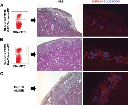
FIG. 4.
In vivo assessment of the autoreactive potential of GAD-autoreactive CD4 T-cells from patient 1. A peripheral blood sample (sample no. 10, Fig. 1C and D) from patient 1 yielded a very strong CD4 T-cell response to GAD; ∼7% of the CD4 T-cells were GAD autoreactive after in vitro stimulation and stained specifically with the DRB1*0405-GAD 555–567 tetramer (A). Approximately 15,000 tetramer-positive, GAD-autoreactive CD4 T-cells were purified by fluorescence-activated cell sorting and cotransplanted with human islets (1,300 islet equivalents), freshly isolated from an unrelated, deceased donor [HLA-A30, A33, B42, B70, DR8, DR17(3)], under the kidney capsule of a nondiabetic immunodeficient mouse. Control mice received islets with 15,000 CD4 T-cells from the same patient, which were stimulated with the HA control peptide and sorted after staining with a DRB1*0405-HA tetramer (B) or islets alone (C). H&E and insulin and glucagon stains representing the same areas of the graft reveal damaged islets and loss of insulin staining in the graft that received GAD-specific CD4 T-cells (A). Normal graft morphology and hormone staining patterns are seen in control mice receiving HA-specific CD4 T-cells (B) or islets alone (C). (A high-quality digital representation of this figure is available in the online issue.)

