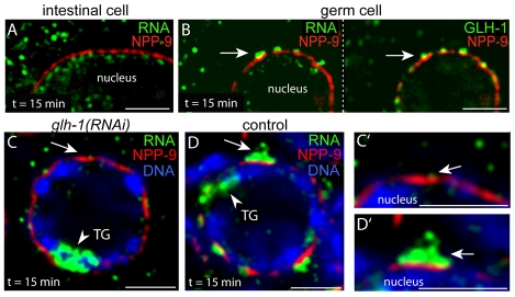
Fig. 7.
Perinuclear mRNA localization requires P granules. (A) Intestinal cell 15 minutes after heat shock, showing little or no mRNA (green) adjacent to the NPP-9 zone (red). (B) Germ cell in the same animals showing perinuclear foci of RNA (green, left panel) adjacent to the NPP-9 zone (red) and coincident with P granules (green, right panel). (C,C′) Single pachytene germ cell from a heat-shocked, glh-1(RNAi) animal. Staining as indicated for transgenic mRNA, NPP-9 and DNA. Note the very low level of perinuclear mRNA (arrow; shown at high magnification in C′). (D,D′) Germ cell from mock-treated control animal stained as indicated and showing a high level of perinuclear mRNA (arrow; shown at high magnification in D′). Scale bars: 25 μm in A,C,D; 2.5 μm in B,E-G.

