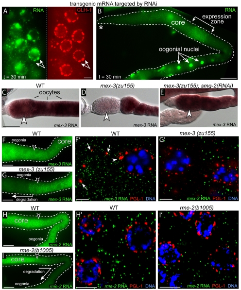
Fig. 8.
mRNAs targeted for degradation are not enriched in P granules. (A,B) FISH analysis of hsp16.2::gfp::pos-1 3′UTR expression on worms exposed to dsRNA for the pos-1 3′UTR. (A) Pachytene nuclei; double-headed arrow indicates an example of colocalized nascent mRNA (green) and P granules (red). (B) Low magnification view of the same gonad in A, showing that the induced mRNA has entered the gonad core and moved downstream to oogonia. (C-E) Panels show wild-type (WT) or mutant mex-3 mRNA detected by in situ hybridization in the strains indicated. mex-3(zu155) mRNA with a premature stop codon is degraded in the most mature oocytes, where it normally is translated (D, arrowhead), but persists when the NMD pathway is inhibited [smg-2(RNAi)] (E). (F-I′) FISH of the indicated mRNA in WT or mutant gonads (F-I); high magnification of the respective regions indicated by arrowheads are shown in F′-I′. Adjacent mRNA (green) and PGL-1 (red) signals are indicated in F′ for perinuclear P granules (arrowhead) and cytoplasmic P granules (arrows). Scale bars: 2.5 μm in A; 25 μm in B-I; 5 μm in F′-I′.

