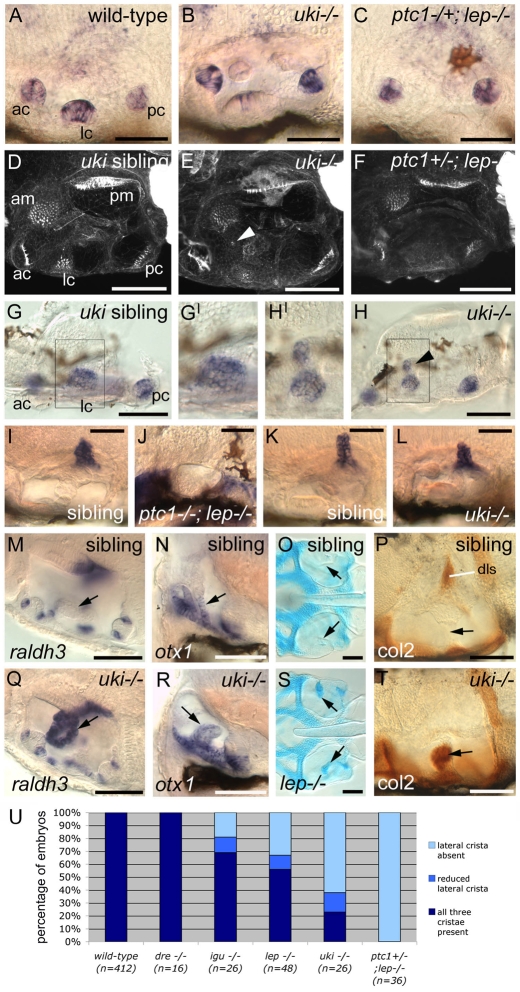
Fig. 2.
Dorsolateral patterning defects appear as the severity of otic phenotype increases in the Hh inhibitor mutants. (A-H′) Cristae are lost in the more severe Hh inhibitor mutants. (A-C,G-H) msxc in situ hybridisation at 3 dpf. (D-F) Confocal z-stacks of ears stained with FITC-phalloidin to reveal actin in the sensory hair bundles at 3 dpf. (A,D,G,G′) Three cristae (ac, lc, pc) are present in wild-type ears. (B,E,H,H′) uki mutants with reduced lateral cristae; the medial region of the lateral crista is sometimes displaced towards the anterior macula (arrowheads, E,H). (C,F) ptc1–/–; lep–/– mutants with no lateral cristae. (I-L) The endolymphatic duct is reduced in uki embryos and absent in ptc1–/–; lep–/– double mutants: in situ hybridisation to foxi1 at 68 hpf (I,J) and 72 hpf (K,L). (M-T) The ventral semicircular canal pillar (arrows) is abnormal in lep and uki homozygotes, with ectopic expression of raldh3 (M,Q) and Collagen type II protein (P,T) at 3 dpf, and ectopic Alcian Blue staining at 5 dpf (O,S). Expression of otx1 at 3 dpf in the ventral pillar is unaffected (N,R). igu embryos show similar defects in the ventral pillar (not shown). (A-C,I-L,P,T) Lateral views; anterior to left, dorsal to top. (D-H,M,Q) Dorsal views; anterior to left, medial to top. (O,S) Dorsal views; anterior to left. (N,R) Transverse hand-cut sections, ∼50 μm. Boxes in G,H show the region enlarged in G′,H′. Scale bars: 50 μm. (U) Chart showing the proportion of Hh inhibitor mutant embryos with lost or reduced lateral cristae. ac, anterior crista; am, anterior macula; dls, dorsolateral septum; lc, lateral crista; pc, posterior crista; pm, posterior macula.

