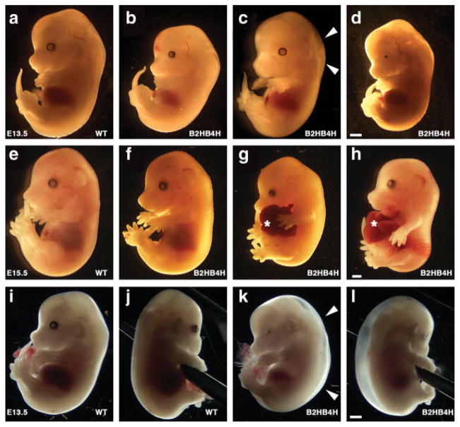
FIG. 2.
Abnormalities developed in the compound heterozygous embryos. Whole view of mouse embryos at E13.5 (a–d and i–l), E15.5 (e–h), wild type (a, e, i, and j), and compound heterozygous (b–d, f–h, and k, l) embryos. Compound heterozygous embryos showed no significant abnormality (b and f), edema (c and k, l, white arrowheads), small eye (d and k, l), and hernia (g and h, asterisk). i and j, and k and l are the same embryo, respectively. Scale bar, 1 mm. [Color figure can be viewed in the online issue, which is available at www.interscience.wiley.com.]

