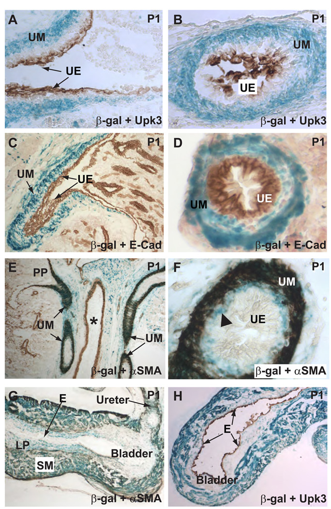
Figure 3. Tbx18-Cre expression is in the mesenchymal derivatives but not in the epithelium.
The urothelium (UE) was labeled by an anti-Uroplakin III (Upk3) antibody (A–B) and an E-Cadherin (E-Cad) antibody (C–D). LacZ expression (blue), indicative of loxP recombination, was in the mesenchymal derivatives but not in the epithelium as evidenced by the lack of colocalization of the blue signal and the epithelial markers (brown). Blue cells were largely absent in the kidney proper. UM: ureteric mesenchyme; UE: urothelium. E and F, most of the blue cells were also α-SMA-positive (brown) urinary tract SMCs. Vascular SMCs in the adjacent blood vessels did not have significant loxP recombination. PP: Papilla of the kidney. * indicates the lumen of a blood vessel. In addition to urinary tract SMCs, the ureteric stromal cells were also positive for β-galactosidase (triangle in F), indicating Cre transgene expression in this cell population or their progenitors. G–H, within the bladder, SMCs and the α-SMA-negative cells within the lamina propria (LP) had extensive lacZ expression while the transitional epithelium (E) is completely void of any staining.

