Abstract
A 21-year-old man presented in a state of delirium with compulsive, pathologic laughter and no focal neurologic findings. Brain imaging revealed a lenticulostriate artery infarct of the caudate nucleus. The absence of neurological signs made the diagnosis difficult, but the presence of delirium was a clue to the existence of structural brain disease.
Keywords: brain imaging, MRI scan, compulsive laughter, caudate nucleus infarct, lenticulostriate artery infarct, delirium, encephalopathy, impaired cognition, focal neurological examination
Introduction
The caudate nucleus is important in modulation of motor functions and may influence associative and cognitive processes.1 Pathologic laughter is a compulsive, exaggerated type of laughter unrelated to emotions. It is occasionally documented following multiple, bilateral, cerebral lesions as a manifestation of pseudobulbar palsy.2–4 Pathologic laughter is not commonly associated with unilateral infarcts in the region of the caudate.
A lenticulostriate artery infarct usually causes significant motor and sensorimotor deficits,5 and neuroimaging evidences that decreased activation of the caudate can be related to obsessive-compulsive behaviors.6 The clinical vignette presents a case of pathologic laughter in the absence of focal neurologic deficits. This unusual finding with confusing clinical features could be mistaken for a psychiatric disorder and result in delays at diagnosis and intervention.
Clinical Vignette
A 21-year-old, right-handed, male patient with abnormal behavior of 10 days duration was hospitalized on the psychiatry service. Originally, this patient went to a clinic with complaints of headache and nausea. His past medical and family history revealed no neurologic or psychiatric disorders and no diabetes or hypertension. The physician noted pathological laughter, which was felt to be of psychogenic origin or malingering. The physician prescribed mefenamic acid and metoclopramide, and the headache improved, but vomiting and pathological laughter worsened. Odd, hyperactive behavior and dysphasia with repetitive word usage developed and led to hospitalization on the psychiatry ward with a neurosurgical consultation.
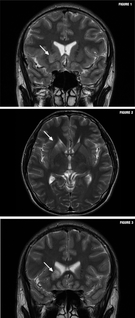
On mental status examination, the patient was uncooperative and displayed agitation and inappropriate behavior. Poor eye contact with decreased attention and disorientation to time was observed. Short-term memory impairment was prominent. Speech was fluent otherwise, but thought processes were circumstantial. Response to verbal and written commands was slow and dysfunctional. The patient remained tense; affect was blunted, but interrupted by bursts of sudden, inappropriate laughter. The physical and neurologic examination was unremarkable. Routine laboratory reports were all within normal ranges. A head scan by magnetic resonance imaging (MRI) revealed an acute unilateral infarction in the right medial lenticulostriate artery territory.
Discussion
Psychiatric presentations can be a result of cerebrovascular accidents.7 Lateralization of cerebral lesions with organic mood disorders are reported with an association between psychiatric symptoms and right sided strokes.8–10
FIGURES 1–3.
Axial and coronal fast-spin echo T2-weighted images (1.5 Tesla, TR 4000ms, TE 125ms, slice thickness 5mm, 320 x 224 matrix, NEX 2) revealed a high signal intensity lesion in the lower portion of the head of the right caudate nucleus, inferior portion of the anterior limb of the right sided internal capsule, and the anterior third of the right putamen. The shape and location of this lesion matched the medial lenticulostriate artery territory, which is the cone shaped region extending from the anterior perforated substance to lower portion of the caudate nucleus with lateralized convex curvature.
This case presented with acute delirium, ultimately found to be due to a right caudate nucleus infarction etiology. His mood was euthymic, which differentiates pathologic laughter from mania that more characteristically follows bilateral cerebral lesions; yet, pathological laughter is also documented in patients with a unilateral stroke.11 Out of 13 subjects with pathological laughter and unilateral brain infarcts, six had right-sided cerebral lesions and seven had a left sided stroke.12 One case of a left caudate nucleus infarct with depressive features was reported.13 That suggests that lateralization may be more important than anatomical region in the synthesis of pathologic laughter.
Unilateral lesions of the caudate nucleus or neighboring structures characteristically induce restlessness or behavioral changes with agitation and hyperactivity.14 Strokes can precipitate delirium and may be associated with brain-specific lesions in the caudate nucleus or thalamic structures.15 Hyperactive delirium is characterized by confusion with increased motor activity or even agitation.16 Quickly identifying and correcting the underlying cause of a delirium is very important to improve clinical outcome. This is true in cases of post-stroke delirium since that diagnosis is associated with significant mortality and morbidity.17
Our clinical vignette presented with confusion and the “psychiatric symptoms”; of inappropriate laughter and hyperactivity. Absence of abnormal neurological signs made the diagnosis more difficult to identify. Presence of disorientation, abnormal speech, and confusion characterized a delirium. This sign of encephalopathy was the clue that precipitated an investigation for brain pathology and discovery of a caudate nucleus infarct diagnosis. The patient never met criteria for psychiatric illnesses.
The caudate nucleus participates as a gatekeeper in control of goal-directed motor activities. Failure of the caudate nucleus to function normally can result in aberrant behaviors, which might simulate a psychiatric disturbance.1 Pathologic laughter should be differentiated from mania by its euthymic nature. Mental illness symptoms may also mask neurologic conditions. Although the incidence of brain infarct is low in young adults, psychiatrists should always consider brain pathology in evaluating patients with new-onset psychiatric issues, particularly and always when signs of encephalopathy are present.
Specific guidelines to help physicians in deciding when to order brain imaging studies on psychiatric patients include focal neurological findings, abnormal electroencephalographic readings, movement disorders, unexplained cases of confusion, prolonged catatonia, first episode of psychosis, first affective episode or change in personality after age 50, history of significant head trauma, onset of seizures, and impaired cognition on mental status examinations.18 When head scans are done without such abnormal findings, images are usually unremarkable, but with objective abnormalities discovered, results most often reveal overt brain pathology.19
Conclusion
Acute delirium and pathologic laughter without focal neurologic findings was induced by a unilateral caudate nucleus infarction. This pathology indicates a possible role of specific structures in the right caudate nucleus in the expression of pathologic laughter. To enhance diagnostic precision and to provide prompt treatment, a brain imaging study should be immediately performed to rule out disease when structural brain pathology is in the differential diagnosis.
Contributor Information
Sangsoo Lee, Dr. Lee is from the Department of Psychiatry, Armed Forces Gangneung Hospital, South Korea.
Dae Yoon Kim, Dr. DY Kim is from the Department of Radiology, Armed Forces Gangneung Hospital, South Korea.
Jung Soo Kim, Dr. JS Kim is from the Department of Neurosurgery, Armed Forces Gangneung Hospital, South Korea.
Sainath Manda, Drs. Manda are from the Department of Psychiatry, University of Louisville School of Medicine, Louisville, Kentucky..
Lilia Danilov, Danilov are from the Department of Psychiatry, University of Louisville School of Medicine, Louisville, Kentucky..
Mostafa El-Refai, El-Refai are from the Department of Psychiatry, University of Louisville School of Medicine, Louisville, Kentucky..
Steven Lippmann, Lippmann are from the Department of Psychiatry, University of Louisville School of Medicine, Louisville, Kentucky..
References
- 1.Sadock BJ, Sadock VA. Synopsis of Psychiatry. 10. Baltimore, MD: Lippincott Williams & Wilkins; 2007. pp. 77–78. [Google Scholar]
- 2.Davison C, Kelman H. Pathologic laughing and crying. Arch Neurol Psychiatry. 1939;42:595–604. [Google Scholar]
- 3.Wilson SAK. Some problems in neurology II. Pathological laughing and crying. J Neurol Neurosurg Psychiatry. 1924;16:299–333. [Google Scholar]
- 4.Stern K, Dancey TE. Glioma of the diencephalon in manic patients. Am J Psychiatry. 1942;98:716–719. [Google Scholar]
- 5.Akkus DE. Pure mutism due to simultaneous bilateral lenticulostriate artery territory infarction. CNS Spectr. 2006;11(4):257–259. doi: 10.1017/s1092852900020745. [DOI] [PubMed] [Google Scholar]
- 6.Guehl D, Benazzouz A, Aouizerate B, et al. Neuronal correlates of obsessions in the caudate nucleus. Biologic Psychiatry. 2008;63(6):557–562. doi: 10.1016/j.biopsych.2007.06.023. [DOI] [PubMed] [Google Scholar]
- 7.Goyal R, Sameer M, Chandrasekaran R. Mania secondary to right-sided stroke-responsive to olanzapine. Gen Hospital Psychiatry. 2006;28:262–263. doi: 10.1016/j.genhosppsych.2006.01.003. [DOI] [PubMed] [Google Scholar]
- 8.Robinson RG, Boston JD, Starkstein SE, Price TR. Comparison of mania and depression after brain injury: causal factors. Am J Psychiatry. 1988;145:172–178. doi: 10.1176/ajp.145.2.172. [DOI] [PubMed] [Google Scholar]
- 9.Fawcett RG. Cerebral infarct presenting as mania. J Clin Psychiatry. 1991;52(8):352–353. [PubMed] [Google Scholar]
- 10.Benke T, Kurzthaler I, Schmidauer C, et al. Mania caused by a diencephalic lesion. Neuropsychologia. 2002;40:245–252. doi: 10.1016/s0028-3932(01)00108-7. [DOI] [PubMed] [Google Scholar]
- 11.Black DW. Pathological laughter: a review of the literature. J Nerv Ment Dis. 1982;170:67–71. [PubMed] [Google Scholar]
- 12.Kim JS. Pathologic laughter after unilateral stroke. J Neurologic Sci. 1997;148:121–125. doi: 10.1016/s0022-510x(96)05323-3. [DOI] [PubMed] [Google Scholar]
- 13.Nagaratnam N, Nagaratnam K, Ng K, Diu P. Akinetic mutism following stroke. J Clin Neurosci. 2004;11(1):25–30. doi: 10.1016/j.jocn.2003.04.002. [DOI] [PubMed] [Google Scholar]
- 14.Chung CS, Lee HS, Caplan LR. Caudate infarcts and hemorrhages. In: Bougausslausky J, Caplan L, editors. Stroke Syndromes. Second. Cambridge, MA: Cambridge University Press; 2001. pp. 469–478. [Google Scholar]
- 15.McManus J, Pathansali R, Stewart R, et al. Delirium post-stroke. Age Ageing. 2007;36:613–618. doi: 10.1093/ageing/afm140. [DOI] [PubMed] [Google Scholar]
- 16.Lee YM, Lee BD, Park JM. Clinical implication of delirium subtype. J Korean Neuropsychiatr Assoc. 2009;48:123–129. [Google Scholar]
- 17.Henon H, Lebert F, Durieu I, et al. Confusional state in stroke, relation to preexisting dementia, patient characteristics and outcome. Stroke. 1999;30:773–779. doi: 10.1161/01.str.30.4.773. [DOI] [PubMed] [Google Scholar]
- 18.Rale RE, Yudofsky SC. Textbook of Clinical Psychiatry. Fourth. Washington DC: American Psychiatric Publishing, Inc; 2003. p. 277. [Google Scholar]
- 19.Pary R, Lippmann S. Clinical review of head CT scans in psychiatric patients. VA Practitioner. 1986;3:48–53. [Google Scholar]


