Abstract
Stable infection of Bacillus anthracis laboratory strains with environmental bacteriophages confers survival phenotypes in soil and earthworm intestinal niches (R. Schuch and V. A. Fischetti, PLoS One 4:e6532, 2009). Here, the natural occurrence of two such B. anthracis-infective bacteriophages, Wip1 and Wip4, was examined in the intestines of Eisenia fetida earthworms as part of a 6-year longitudinal study at a Pennsylvania forest site. The Wip1 tectivirus was initially dominant before being supplanted by the Wip4 siphovirus, which was then dominant for the next 3 years. In a host range analysis of a wide-ranging group of Bacillus species and related organisms, Wip1 and Wip4 were both infective only toward B. anthracis and certain B. cereus strains. The natural host of Wip4 remained constant for 3 years and was a B. cereus strain that expressed a B. anthracis-like surface polysaccharide at septal positions on the cell surface. Next, a novel metagenomic approach was used to determine the extent to which such B. cereus- and B. anthracis-like strains are found in worms from two geographical locations. Three different enrichment strategies were used for metagenomic DNA isolation, based either on the ability of B. cereus sensu lato to form heat-resistant spores, the sensitivity of B. anthracis to the PlyG lysin, or the selective amplification of environmental phages cocultured with B. anthracis. Findings from this work indicate that B. cereus sensu lato and its phages are common inhabitants of earthworm intestines.
The notorious animal pathogen Bacillus anthracis is considered to be a genetically monomorphic variant of the large, well-studied B. cereus sensu lato (s.l.) lineage of often ubiquitous soil organisms like B. cereus sensu stricto (referred to here as simply B. cereus), B. thuringiensis, B. pseudomycoides, B. weihenstephanensis, and B. mycoides (20, 21, 25, 38). Despite a high degree of chromosomal DNA sequence similarity and synteny within B. cereus s.l., it is nonetheless a highly pleomorphic group of saprophytes, intestinal symbionts, and both opportunistic and frank pathogens of many animals (19, 39). The niche-specific adaptive behaviors associated with B. cereus s.l. are often attributed to highly variable sets of potentially transmissible megaplasmids (20, 25). For example, the pXO1 and pXO2 plasmids of B. anthracis encode both the virulence genes directly responsible for anthrax and the transcriptional regulatory proteins involved in plasmid and chromosomal gene expression (23, 40). The presence of large plasmids alone, however, does not sufficiently explain the ability of B. cereus s.l. organisms to elaborate different behaviors. Rather, species-specific chromosomal loci, accounting for ca. 15% of the genes in each sequenced B. cereus s.l. genome (18), may also have significant phenotypic impacts. Included among the highly variable loci of B. cereus s.l. are a diverse range of mobile genetic elements, including small plasmids, insertion sequences, transposons, introns and, importantly, bacteriophages (21).
Bacteriophages have a very well-described impact on bacterial genetic diversity and behavior through the introduction of virus-encoded factors that alter bacterial phenotypes in a process called lysogenic conversion (7). Despite the presence of species-specific bacteriophages throughout the B. cereus s.l. lineage, either in chromosomes as prophages or as independently replicating episomes, the possibility of lysogenic conversion has, until recently (30), received little attention. Toward this end, we recently described a set of bacteriophages with the ability to enhance environmental survival phenotypes of B. cereus s.l. (30). Lysogeny with any of a series of soil-, fern rhizosphere-, and earthworm gut-derived phages was found to reduce or block sporulation, favor biofilm formation, and promote long-term survival in both soil microcosms and the intestinal tract of Eisenia fetida worms (30). This effect was observed with both laboratory B. anthracis strains and environmental B. cereus isolates. Furthermore, the earthworm colonization phenotype was driven solely by expression of phage-encoded bacterial sigma factors which can activate the expression of at least one (but likely more) B. anthracis chromosomal loci (30). Here, bacteriophages promote environmental functioning in a process by which phage-encoded transcription factors activate host-encoded phenotypes.
The initial goal of the present study was to describe two B. anthracis-infective phages originally found in the intestines of E. fetida worms from a forest leaf litter site in Pennsylvania. We followed the occurrence of these phages over 6 years and characterized one natural bacterial host. Based on the identification of B. cereus s.l. organisms and phages in the worm niche, metagenomic methods were developed and used to specifically examine the extent to which such bacteria and phages are found in worms at a separate location, in New York City.
MATERIALS AND METHODS
Bacterial strains and growth conditions.
The majority of bacterial strains in the present study were previously described (29, 30, 32). Additional strains include Staphylococcus aureus Newman (3), B. cereus E33L (12), and B. thuringiensis Al Hakam (8). All organisms, except Escherichia coli, were grown on brain heart infusion (BHI; Difco) broth or agar plates at 30°C according to standard protocols. E. coli strain TOP10 (Invitrogen, Inc.) was grown on Luria-Bertani (LB; Sigma-Aldrich, Inc.) broth or agar plates at 37°C according to standard protocols.
Virus manipulations.
Wγ was obtained from the Felix d'Herelle Reference Center for Bacterial Viruses at Laval University in Quebec, Canada. Phage propagation, storage, and host range analyses were performed exactly as described previously (29). Phage plaque assays were always performed with a “no phage” control, to account for a potential background of induced prophage.
Worm and soil collection and analysis.
E. fetida worms and surrounding soils were taken from two sites: a forest floor in Stroudsburg, Pennsylvania (PA), and an outdoor garden in New York City, New York (NYC). In PA, on the first Saturday of October over a 6-year period from 2003 until 2008, ∼5 g of soil and two worms were removed from each of six positions at roughly three meter increments extending nine meters north and east of a decaying tree stump. In NYC, a total of 12 worms were collected once in June 2009. In all cases, worms and/or soil were taken from within ∼12 cm of the surface. Soil from both the NYC garden and PA forest was always humuslike (i.e., dark brown, porous, and spongy), with a pH between 6 and 8. All samples were immediately placed in separate 50-ml Falcon tubes and transported to the laboratory for processing within 24 h. Recovery and identification of bacteriophages and bacterial organisms from worms and soil was performed as described previously (30) in the exact same manner every year. For PCR analyses of worm and soil samples, 25 μl of each environmental slurry was mixed with 25 μl of 0.5 M NaOH for 15 min at room temperature and diluted with 25 μl of 1 M Tris (pH 8.0) and 1.0 ml of distilled H2O to yield a PCR template. Then, 1.0 μl of each template was used in a 100-μl PCR containing the Taq PCR master mix (Qiagen, Inc.). Phage-specific DNA primers were identical to those previously used (30). DNA samples were stored at −20°C between analyses.
Search for natural phage lysogens.
A bacterial culture-based method was used to search for the natural hosts of both Wip1 (from all worms sampled from 2003 to 2005) and Wip4 (from all worms sampled from 2006 to 2008). Extracted worm gut samples from each year were plated undiluted (and three 100-fold serial dilutions thereof) on 150-mm BHI agar plates. After overnight incubation, individual colonies exhibiting characteristic B. anthracis- or B. cereus-like morphologies (37) were recovered, subcultured on BHI agar plates, and examined by PCR in the manner described with Wip1- or Wip4-specific PCR primers (lysogens are positive for these markers). PCR-positive organisms were subcultured twice and reexamined by PCR to confirm the presence of phage.
Sequence analyses of RS1045.
The Wip4-positive strain RS1045 was used as DNA template source for 16S rRNA sequencing with primers 16S-up (5′-AGAGTTTGATCCTGGCTCAG-3′) and 16S-down (5′-ACGGCTACCTTGTTACGACTT-3′) in a manner previously described (27). The plcR-papR locus of RS1045 was first amplified with the flanking primers PlcRup (5′-GTCTAATTCTTGACATGAATAGCTTC-3′) and PapRdown (5′-GTAAAGACGTTTGGATGTTACTCC-3′), and the product was sequenced with PlcRup, PapRdown, and the forward and reverse versions of PlcRmid1 (5′-GCTCAATCAACAATTGGCAGGAATAG-3′), PlcRmid2 (5′-TGACATTGATTGGAGTTATATTATCG-3′), and PlcRmid3 (5′-GGAAGAATATCATCCCGAGCTG-3′). Sequence analysis was performed by Genewiz, Inc. The resulting data were assembled by using SeqMan Pro (DNASTAR, Inc.) and analyzed by using BlastN.
Genomic fingerprinting.
M13 fingerprint analysis was performed as described previously (2). Briefly, an M13 primer was used (5′-GAGGGTGGCGGCTCT-3′) to generate size pattern variations that are diagnostic for bacterial species and phages. PCRs were denatured for 10 min at 95°C, followed by 40 cycles of 15 s at 96°C, 15 s at 36°C, and 2 min at 72°C. Resulting PCR products were separated on 1.0% agarose gels and visualized with ethidium bromide.
Sequence alignments.
Multiple sequence alignments were obtained by using the CLUSTAL W server (http://www.ch.embnet.org/software/CLUSTALW.html) and visualized by using BOXSHADE 3.21 (http://www.ch.embnet.org/software/BOX_form.html).
Phase-contrast and fluorescence microscopy.
The construction and purification of GFP-PlyGBD was described previously (29, 30). For labeling, strains were grown at 30°C for 16 h in 5 ml of BHI (in 50-ml Falcon tubes) with aeration. Overnight cultures were diluted 100-fold in 5 ml of BHI and grown with aeration at 30°C for 3 h. Cells were then washed in 1× phosphate-buffered saline (PBS) and concentrated 10-fold in PBS. Aliquots containing 0.25 ml of cells were mixed with an equal volume of purified GFP-PlyGBD material (∼0.25 μg), incubated for 5 min at 24°C, washed once in PBS, and mounted on glass slides. Samples were visualized at 2,000× magnification with or without UV irradiation (the excitation and emission wavelengths were 480 and 535, respectively) by using an Eclipse E400 microscope (Nikon, Inc.) and QCapture Pro version 5.1 imaging software. Images were assembled by using Adobe Photoshop version 10.0.1.
PlyG sensitivity assay.
The assay was performed as described previously (32). Briefly, 5-ml log-phase RS1045 cultures, growing at 30°C in BHI, were washed with PBS and resuspended in 2.5 ml of PBS. Cells were added as 0.1-ml aliquots to 96-well Corning Costar plates and mixed with an equivalent volume of either a 6.4-mg ml−1 stock of purified PlyG and seven 2-fold serial dilutions thereof (or PBS alone as a control). Absorbance at 600 nm (A600) was followed for 15 min at 24°C by using a Spectramax Plus 384 spectrophotometer (Molecular Devices).
Metagenomic analysis.
Four random shotgun libraries were constructed from pooled NYC worm gut extracts using a variation of a previously described method (28) for producing expressible linker-amplified shotgun libraries. For the “total bacterial” library, gut samples were first extracted with successive phenol-chloroform and chloroform treatments and then precipitated with 3 volumes of 100% ethanol and washed with 1 volume of 70% ethanol. Resulting DNA was gel purified to remove impurities and partially digested for 5 min at 65°C with Tsp509I (New England Biolabs, Inc.). After organic solvent extraction and ethanol precipitation, digested DNA was ligated to a linker with a complementary 5′ overhang (5′-AATTCGGCTCGAG-3′, where the overhang is underlined). Ligated DNA was then used as a template in a PCR using a linker-specific primer Lsp2 (5′-CCATGACTCGAGCCGAATT-3′) and conditions of 95°C for 1 min; 95°C for 30 s, 55°C for 30 s, and 72°C for 5 min (40 cycles); and 72°C for 10 min. Resulting products were then ligated into an arabinose-inducible pBAD plasmid-expression vector (using the pBAD TOPO TA expression kit of Invitrogen, Inc.) and transformed into Escherichia coli TOP10 cells. Colonies appearing on LB agar supplemented with 100 μg of ampicillin ml−1 were randomly chosen for plasmid purification and DNA sequence analysis.
For the production of a “spore-enhanced” metagenomic DNA library, a variation was introduced into the above protocol prior to DNA isolation. Here, the worm gut material was first washed twice with PBS, resuspended in 0.5 ml of PBS, and heated at 95°C for 30 min to destroy all nonspore forms. After heating, samples were placed on ice for 5 min, diluted into 2 ml of BHI and incubated with aeration at 37°C for 3 h. The 3-h incubation is sufficient to promote both spore germination and outgrowth (4), as well as the subsequent vegetative expansion of surviving forms (with clear samples becoming turbid). After this treatment, samples were washed twice with PBS (to remove remaining exogenous DNA) and resuspended in 0.5 ml of PBS prior to processing as described above for the analysis of metagenomic DNA.
For the “PlyG-released” metagenomic DNA library, worm gut material was first washed five times with PBS, resuspended in 0.5 ml of PBS, and mixed with 0.5 ml of a purified PlyG preparation (6.4-mg ml−1 stock concentration) for 2 h at 37°C. Samples were then pelleted by centrifugation (4,000 rpm for 5 min in an Eppendorf Model 5415D Microcentrifuge), and the resulting supernatants were passed through a 0.22-μm-pore-size filter to remove contaminating material. The DNA-bearing lysate was extracted with phenol-chloroform and precipitated with ethanol prior to metagenomic library construction as described above.
For the production of “B. anthracis phage-enhanced” metagenomic DNA, another variation was used whereby phage-bearing supernatants of pelleted crude intestinal extracts were added to a culture of B. anthracis ΔSterne growing in 5 ml of BHI at 30°C with agitation. After an overnight incubation, potentially phage-bearing culture supernatants were collected, filtered to remove bacteria, and used to infect another culture of B. anthracis ΔSterne growing in 5 ml of BHI at 30°C with agitation. After overnight incubation (i.e., a second round of phage amplification), culture supernatants were recovered, filtered, extracted with phenol-chloroform, and precipitated with ethanol prior to metagenomic library construction as described above.
Fifty clones were sequenced for each library. The number of resulting high-quality sequences varied with each library and included 47 for the total bacteria, 25 for the spore enhanced, 48 for the PlyG released, and 24 for the B. anthracis phage enhanced. Duplicate sequences were not observed. All missing sequences were clones with no library inserts.
Nucleotide sequence accession numbers.
Sequences of both the entire plcR-papR locus and the partial 16S rRNA locus of RS1045 were submitted to GenBank (under accession numbers GU142941 and GU142940, respectively). Sequences of the Wip1 and Wip4 lysins (GU142942 and GU142943, respectively), as well those identified in the metagenomic analyses (GU142944, GU142945, GU142946, GU142947, and GU142948), were submitted as well.
RESULTS
Longitudinal survey: sample recovery.
Every first weekend of October over a 6-year period, two worms were recovered from each of six positions at 3-m increments extending both 9 m north and east of a central, decaying tree stump. The 12 animals were returned to the laboratory, washed separately in distilled H2O to remove adherent dirt particles, and processed for the recovery of gut contents. The contents were centrifuged, and the resulting supernatants were filtered to yield bacterium-free extracts containing worm-gut phages.
Longitudinal survey: bacteriophage detection.
Bacteriophages in both worm and soil extracts were sought as PFU in two ways. First, extract aliquots were incubated for 16 h on BHI soft-agar overlays containing freshly plated lawns of organisms, including B. anthracis, B. thuringiensis, B. cereus, Staphylococcus aureus, Streptococcus pyogenes, and Pseudomonas aeruginosa. From each worm isolated throughout this experiment (except for in 2006 when no PFU were found), 1 × 10³ to 5 × 10³ PFU were recovered using the B. anthracis ΔSterne reporter. All other reporter organisms (including B. thuringiensis HD1, B. cereus ATCC 14579 and ATCC 10987, S. aureus Newman, and P. aeruginosa PAO1) yielded no PFU. In addition, all soil extracts were free of phages infecting the reporter organisms, including B. anthracis (see Table S1 in the supplemental material).
For the second method to detect PFU, each reporter organism was subjected to two 24-h infections with worm gut extracts. After this amplification step, infected culture supernatants were collected, filtered to remove bacteria, and titered on agar plates freshly overlaid with the corresponding reporter strain. This “phage-enrichment” method was previously used to detect otherwise undetectable phages in environmental samples (30). Here, 106 to 108 PFU per worm were found every year (including 2006) using the B. anthracis ΔSterne reporter. No plaques were observed on other reporter strains, including B. cereus and B. thuringiensis.
Surprisingly, no PFU were detected in the extracts of PA forest soil from which phage-bearing worms were isolated. Free intestinal phages should be released in worm excreta referred to as castings. Although released phages could have adsorbed to the humuslike soil or casting material, the preparation methods used here should have released some detectable phages into solution. Previously, we successfully isolated B. anthracis-infective phages (Slp1 and Frp1) from the soil using the same technique (30). To confirm here that the amplification step does indeed work, soil from three additional environments was examined (see Table S1 in the supplemental material). Although the primary extracts from these soils yielded no PFU on indicator strains (with two exceptions), the phage enrichment method did yield PFU for most Bacillus strains tested. Thus, while multiple soil samples do contain Bacillus phages, none are detectable in the soil containing PA worms in the present study.
Longitudinal survey: bacteriophage identification.
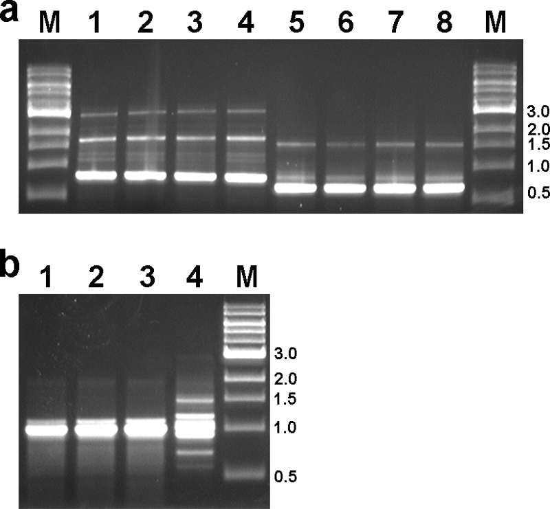
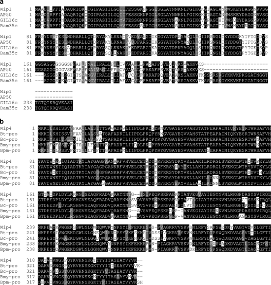
To study B. anthracis-infective, worm intestinal phages, a single plaque was first obtained from the primary extract of a worm isolated in 2003 in PA. This phage, called Wip1 (for worm intestinal phage 1), was expanded and used for the identification of a unique phage DNA sequence based on a genetic screen for lysin activity (31). Thus, Wip1 genomic DNA was used to generate an inducible expression library for a lytic activity screen against B. anthracis. DNA sequences of 15 lytic clones revealed a common 657-bp open reading frame encoding a putative 218-residue protein similar to the glucosaminidases of Tectiviridae phages of Gram-positive bacteria (Fig. 1a). Two distinct Wip1 lysin gene-specific primer pairs were then used to screen both the primary gut extracts and the 125 resulting plaques derived from every worm between 2003 and 2008. Ultimately, Wip1-specific PCR products were observed with every gut extract and individual plaque isolated between 2003 and 2005. After 2005, Wip1 was never detected again. The genetic similarity of the Wip1 phages from 2003 to 2005 (25 phages per year) was confirmed by M13 genetic fingerprinting. DNA fingerprints of phage isolates from both 2004 and 2005 were identical to each other and to Wip1 from 2003 (Fig. 2a). Wip1 was, therefore, a common B. anthracis phage in the worm gut environment for 3 years (2003 to 2005).
FIG. 1.
(a) Sequence alignment of lysins encoded by the Tectiviridae phages of B. cereus s.l. The Wip1 lysin is compared to that of B. anthracis phage AP50 (GenBank accession number EU408779) and B. thuringiensis phages GIL16c (GenBank accession number AY701338) and Bam35c (GenBank accession number AY257527). (b) Sequence alignment of the Wip4 lysin with the lysins of prophages from B. thuringiensis BGSC 4CC1 (Bt-pro), B. cereus G9842 (Bc-pro), B. mycoides Rock3-17 (Bmy-pro), and B. pseudomycoides DSM 12442 (Bpm-pro) bearing the GenBank accession numbers ZP_04078846, YP_002446312, ZP_04160784, and ZP_04152425, respectively.
FIG. 2.

(a) M13 fingerprint patterns obtained with purified genomic DNA isolated from the dominant worm gut phage isolated at each yearly time point in northeastern PA. The samples from 2003 to 2005 (lanes 1 to 3, respectively) ultimately corresponded to different Wip1 isolates. Lane 4 is a second distinct Wip1 isolate from 2005. The samples from 2006 to 2008 (lanes 5 to 7, respectively) corresponded to different Wip4 isolates. Lane 8 is a second distinct Wip4 isolate from 2008. (b) M13 fingerprint patterns obtained with genomic DNA isolated from a distinct Wip4 lysogen isolated every year from 2006 to 2008 (lanes 1 to 3, respectively). The sample in lane 4 is purified DNA from an unrelated B. cereus lab strain ATCC 4342, included as a control. All PCR products were separated by electrophoresis in a 1.0% agarose gel and visualized by staining with ethidium bromide. Lanes M contain a molecular weight marker (1-kb Plus DNA Ladder; Invitrogen, Inc.), and the relevant band sizes are indicated in kilobases to the right of each gel.
The presence of Wip1-negative PFU after 2005 suggested the presence of a new dominant phage(s). To identify new phages, a high-titer lysate was first generated from a single plaque obtained from one worm in 2006. Genomic DNA was ultimately purified from the resulting phage (referred to as Wip4) and used to generate an expression library for a lysin activity screen as described above. Sequences of 10 lytic clones revealed a common 1,056-bp locus encoding a 351-residue protein most similar to N-acetylmuramoyl-l-alanine amidases in prophages of B. thuringiensis, B. cereus, B. mycoides, and B. pseudomycoides (Fig. 1b). Two distinct primer pairs specific for the Wip4 lysin gene were used in a PCR screen of all gut extract samples (from 2003 to 2008) and 150 plaques derived from each worm extract recovered after 2005. Although each pre-2006 sample was PCR negative for Wip4 (yet positive for Wip1), all of the 2006 to 2008 samples were Wip4-positive and Wip1-negative. M13 genetic fingerprints of the 2006 to 2008 phages were also identical (and distinct from the year 2003 to 2005 phages) (Fig. 2a), thus confirming a close genetic relationship. As a control, a PCR screen was also performed with primers specific to four previously described B. anthracis phages (Wβ, Frp2, Bcp1, and Wip2) known to be genetically distinct from Wip1 and Wip4 (30); these primers yielded no products using DNA templates obtained from worms between 2003 and 2008.
Host range analysis.
Previously, phages corresponding to Wip1 and Wip4 were found to infect either of two lab strains of B. anthracis, but not the B. cereus strain ATCC 14579 (30). Since many B. anthracis phages (including Wγ and Wβ) can also infect certain B. cereus strains (29, 32), we decided to greatly expand our host range study to include more closely and distantly related organisms. For comparison, an analysis of the standard B. anthracis diagnostic phage used in clinical laboratories (1), Wγ, was included. High-titer phage preparations were generated on B. anthracis ΔSterne and tested for PFU formation on the strains in Table 1. Each phage was largely infective for only B. anthracis and the B. cereus strains ATCC 4342 and CDC32805. ATCC 4342 has previously been shown to support replication of phage Wγ (32). Wip1 was notably the most specific phage, infecting all three B. anthracis strains. Surprisingly, the Wγ diagnostic phage did not infect the environmental B. anthracis strain RS1615 and yielded plaques on ΔSterne only after 5 days. Thus, while Wip1 and Wip4 appear to be at least as selective as Wγ, Wip1 may be the superior diagnostic tool.
TABLE 1.
Host range analysisa
| Strain | Infectivity (PFU/ml of phage stock)b |
||
|---|---|---|---|
| Wip1 | Wip4 | Wγ | |
| Bacillus anthracis | |||
| Sterne | 8 × 10⁹ (c) | 4 × 10⁹ (c) | 4 × 10⁹ (c) |
| ΔSterne | 6 × 10⁹ (c) | <10 | <10 (3 × 10⁹ [t]) |
| RS1615 | 1 × 10⁴ (c) | <10 | <10 |
| Bacillus cereus | |||
| ATCC 4342 | <10 | 4 × 10⁹ (c) | 1 × 10⁵ (t) |
| CDC32805 | 3 × 10⁷ (c) | 5 × 10⁷ (c) | 4 × 10⁷ (c) |
| ATCC 10987 | <10 | <10 | <10 |
| E33L | <10 | <10 | <10 |
| ATCC 14579 | <10 | <10 | <10 |
| ATCC 11950 | <10 | <10 | <10 |
| RS1045 | <10 | <10 | <10 |
| Bacillus thuringiensis | |||
| HD1 | <10 | <10 | <10 |
| HD73 | <10 | <10 | <10 |
| Al Hakam | <10 | <10 | <10 |
| 97-27 | <10 | <10 | <10 |
| Bacillus megaterium | |||
| ATCC 8245 | <10 | <10 | <10 |
| RS77 | <10 | <10 | <10 |
| Bacillus mycoides BGSC 6A11 | <10 | <10 | <10 |
| Bacillus pumilis SL4680 | <10 | <10 | <10 |
| Bacillus subtilis SL4 | <10 | <10 | <10 |
| Bacillus brevis | <10 | <10 | <10 |
| Sporosarcina ureae | <10 | <10 | <10 |
| Brevibacillus laterosporus | <10 | <10 | <10 |
High-titer phage stocks were obtained using B. anthracis Sterne and titered on the the indicated strains. PFU were generated by drop dilution and evaluated at 1 day.
Mean averages are shown for three independent experiments. “<10” indicates PFU were not detected. “(c)” indicates clear plaques were observed. “(t)” indicates turbid plaques were observed. “No phage” controls were included for each strain to confirm that PFU were not the result of native prophage induction. The values in parentheses indicates PFU observed after a 5-day incubation.
Natural hosts of Wip1 and Wip4.
After the detection of Wip1 PFU and Wip1 loci by PCR in the worm gut, a natural host of Wip1 was sought. Gut contents of multiple distinct worms (from 2003 to 2005) were first plated on BHI agar. Of the different B. cereus s.l.-like colonies recovered, ∼100 were chosen each year and screened by PCR with Wip1-specific primers (data not shown). Despite the presence of “free” Wip1 in worm guts, no Wip1 lysogen was subsequently identified. A host may either have been obscured by background organisms, not expressed a B. cereus s.l.-like colony morphology, or existed in a nonculturable state.
In contrast to Wip1, natural Wip4 lysogens were repeatedly identified in the guts of all worms from 2006 to 2008. Each of the worms from 2006 to 2008 was enriched for a B. cereus-like strain that was PCR positive for all Wip4-specific primers and present at an estimated concentration of ∼6 × 10³ CFU per worm. This is similar to previously reported values for steady-state colonization of the worm gut with vegetative forms of different B. cereus s.l. strains (30). The M13 genetic fingerprints of Wip4 lysogens isolated from 2006 to 2008 were all identical (Fig. 2b), suggesting the same bacterial host was present for all 3 years.
In a previous study, a Wip4 lysogen was originally found to have some B. cereus-like phenotypes, including colony morphology, beta-hemolysis, and resistance to the antibiotic fosfomycin (30). In the present study, DNA sequence analysis of the Wip4 lysogen strain (now referred to as RS1045) revealed the 5′ half of the 16S rRNA locus to be 99% identical to sequences of only B. anthracis, B. cereus and B. thuringiensis. The plcR-papR locus of RS1045, encoding a quorum-sensing system of B. cereus s.l. (33), was also sequenced and found to be most similar (between 92 and 93% identical) to B. cereus s.l. animal pathogenic strains, including B. anthracis, B. thuringiensis 97-27, B. cereus 03BB102, and others (see Fig. S1 in the supplemental material). In addition, the protein sequences of PlcR and PapR were most similar (with 98 and 100% identity, respectively) to the products of B. cereus strain AH1273, a human blood infection isolate from Iceland (13). The last five residues of PapR encoded by RS1045 were VPFEY, indicating a pherotype present in all B. anthracis strains (33).
RS1045 was observed to be a rod-shaped bacillus (Fig. 3a) that binds GFP-PlyGBD at an exclusively septal position (Fig. 3b). GFP-PlyGBD consists of the C-terminal cell wall binding domain of the γ phage lysin, PlyG, fused to a fluorescent protein; the resulting molecule specifically directs fluorescence to the surface polysaccharides of B. anthracis and some closely related B. cereus strains such as ATCC 4342 (29, 30). The septal binding of GFP-PlyGBD to RS1045, stands in distinct contrast to the whole-cell binding of B. anthracis (Fig. 4d) and the polar-septal binding of ATCC 4342 (Fig. 3c). Such binding patterns clearly reflect differences in the surface localization of B. anthracis-like cell wall polysaccharides. Despite the unique binding of GFP-PlyGBD to RS1045, the strain was nonetheless susceptible to the lytic action of PlyG in a liquid killing assay (Fig. 3e). Sensitivity to PlyG is a phenotype specific to B. anthracis and B. cereus strains such as ATCC 4342 (32) and, apparently, RS1045. Our findings here confirmed the presence of a B. anthracis-like B. cereus strain, RS1045, which also served as the source of one B. anthracis-infective, worm gut bacteriophage (Wip4) identified in the present study.
FIG. 3.
Susceptibility of RS1045 to the binding and lytic activities of the PlyG lysin. All images were obtained at 2,000× magnification. A phase-contrast micrograph of exponential-phase RS1045 is shown (a), with a corresponding fluorescence image (b), obtained with a 1-s exposure time, showing surface labeling with GFP-PlyGBD. Surface labeling of both B. cereus ATCC 4342 and B. anthracis ΔSterne with GFP-PlyGBD is shown in fluorescence micrographs obtained with 1-s (c) and 0.1-s (d) exposures, respectively. (e) Sensitivity of RS1045 to the lytic activity of purified PlyG lysin. Aliquots of RS1045 were mixed with 100 μg of PlyG (▪) or PBS alone as a control (▴), and the optical density was monitored for 15 min at room temperature in a SpectraMax Plus (Molecular Devices, Inc.) plate reader.
FIG. 4.
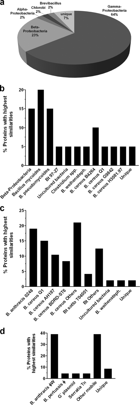
Relative abundances of bacterial and viral components in the intestinal tract of Eisenia fetida worms. The distribution of BlastN or BlastX homolog sequences obtained from the total bacterial (a), spore-enriched (b), PlyG-released (c), and B. anthracis phage-enhanced (d) metagenomic DNA libraries are shown. The data are organized according to prokaryotic phyla, class, genus, species, or strain. Sequences that are similar to nothing in the database are classified as “unique.” Abbreviations: Bt, B. thuringiensis; weihensteph., weihenstephanensis; G−, Gram-negative; Tn, transposon.
Bacterial flora of the worm gut environment.
Having identified B. anthracis phages in PA earthworms, our next step was to assess the extent to which B. anthracis-like organisms (like RS1045) were found in worms from another location—an outdoor garden bed in NYC. For this, we developed a novel three-tiered analytical method, distinct from the phage amplification- and bacterial culture-based methods used with PA worms. This method incorporated a metagenomic approach (28), and ultimately enabled specific access to B. cereus s.l. organisms and phages in the worm gut microbiome.
Initially, the intestinal contents of 12 NYC worms were combined in a 3-ml PBS suspension and total DNA was extracted for construction of a plasmid vector-based library with insert sizes of 0.5 to 2 kb. Forty-seven DNA sequences were ultimately identified and assigned to a phylum, class, or genus (Fig. 4a) based on the most statistically significant similarity from a BlastN analysis. The total DNA pool from NYC worms was found to consist predominantly (62%) of Gammaproteobacteria sequences, including Shewanella, Citrobacter, Aeromonas, Pseudomonas, Salmonella, Acinetobacter, Thiomicrospira, Klebsiella, Enterobacter, Marinomonas, and others (Fig. 4a). Aeromonas spp. accounted for 20% of the sequences and was the most common genus identified (data not shown). While one sequence was most similar to the Gram-positive firmicute Brevibacillus brevis, no B. cereus s.l. homologs were observed. These findings differ slightly from a report for E. fetida from Long Island (28), in which 13% of the sequences were Firmicutes (although no B. cereus s.l. was reported). B. thuringiensis and B. mycoides are the only previously described B. cereus s.l. organisms recovered from earthworms (in Denmark and Germany) (10, 14, 17).
Since the absence of B. cereus s.l. sequences from NYC worms could reflect a minority status in the worm gut, an initial enrichment for heat-resistant endospores was next performed prior to DNA isolation. As such, “spore-enriched” pools were generated by heating gut extracts at 95°C for 30 min to kill everything except spores; heated samples were then resuspended in BHI and grown at 37°C to promote the spore germination and outgrowth necessary for bacterial lysis, DNA recovery, and metagenomic library construction. Of 25 analyzed sequences, ∼75% were now B. cereus s.l. group members, including B. mycoides, B. pseudomycoides, B. weihenstephensis, B. cereus, and B. thuringiensis (Fig. 4b). Surprisingly, 15% of the sequences were of non-spore-forming Betaproteobacteria that survived heat treatment.
The possibility of B. anthracis-like organisms being present as vegetative forms in the worm gut was addressed as part of the next enrichment strategy. Here, the sensitivity of vegetative B. anthracis and certain B. cereus strains to the lytic action of PlyG (32) was exploited prior to DNA isolation. PlyG treatment of gut extracts should specifically release only DNA from sensitive organisms into solution for subsequent metagenomic analysis. As expected, more than 90% of the inserts from the “PlyG-released” library were now from B. cereus s.l. organisms (Fig. 4c). Importantly, 19% of the inserts were 100% identical to a variety of B. anthracis loci. Thus, an organism similar to B. anthracis or B. cereus is a member of the NYC worm gut flora. These findings also show the specificity of phage lysins can be harnessed to focus metagenomic analyses on specific and/or rare organisms of interest.
Bacteriophage flora of the worm gut environment.
Considering the presence of B. anthracis-like sequences, B. anthracis-infective phages should have been present in the NYC worms as well. To investigate this, worm intestinal extracts were first filtered to remove bacteria and retain free phages. When plated directly to a B. anthracis reporter, however, the filtrate yielded no PFU. Similarly, metagenomic DNA isolated from the filtrate also yielded no B. anthracis phage sequences (data not shown). While such findings could suggest the absence of B. anthracis-infective phages, they may simply be in an extreme minority or otherwise difficult to detect. A strategy to enrich for B. anthracis-infective phages was therefore used, whereby exponential phase ΔSterne cultures were inoculated with worm gut extracts and incubated for 24 h. This infection cycle could amplify rare phages to detectable levels. Indeed, when NYC worm gut extracts were used to infect ΔSterne overnight, the resulting supernatants became enriched for B. anthracis-infective phages (∼106 PFU per ml). Sequence analysis of a metagenomic DNA library generated from phage-bearing supernatant revealed 39% of the inserts to be most similar to B. anthracis phages of φW siphovirus family, including Wβ and three distinct Wγ isolates (Fig. 4d) (11, 29). Interestingly, Wip4 (from PA) is genetically similar to the φW family as well. In all, these findings show phages infecting B. anthracis are indeed present in the NYC worms. Interestingly, the amplification step here also yielded sequences (51.9% of the total) similar to mobile genetic elements of Gram-negative organisms.
DISCUSSION
In the present study, the occurrence of B. anthracis-infective bacteriophages in earthworms recovered directly from the forest leaf litter in Pennsylvania was examined. Starting in 2003, phages infecting both B. anthracis and certain B. cereus strains were found in each of 12 different worms collected annually for 6 years over a 9-m radius around a central decaying tree stump. Interestingly, one phage species (Wip1) was replaced by another (Wip4) over time. RS1045 was found to be the host of Wip4 from 2006 to 2008 and could not be detected by PCR in the whole worm gut extracts recovered before 2006. The loss of one phage and/or lysogen over another could reflect changing environmental conditions (i.e., soil humidity, temperature, and the nature of decaying organic matter in the soil) and an altered equilibrium in the worm gut microbiota. Nevertheless, a total of 72 E. fetida worms obtained over a 6-year period provided an environmental niche for at least two B. anthracis-infective phages.
The extreme host specificities of Wip1 and Wip4, suggested the presence of B. anthracis-like isolates (perhaps similar to B. cereus strains such as ATCC 4342 and CDC32805) in the intestines of worms from what is essentially an area where anthrax is not endemic. Indeed, RS1045, the Wip4 host, was found to be a B. cereus s.l. strain phylogenetically clustered with mammalian pathogens, including B. anthracis (based on its plcR-papR sequence). As with B. anthracis ΔSterne and B. cereus ATCC 4342, RS1045 expressed a B. anthracis-like surface polysaccharide structure and was sensitive to lysis after exposure to the PlyG lysin. RS1045 was unique, however, since the GFP-PlyGBD-binding location (and thus the location of a B. anthracis-like surface polysaccharide structure) was restricted to a septal position. Non-B. anthracis strains of B. cereus s.l. expressing a range of B. anthracis phenotypes and genotypes are present throughout the environment (12, 15, 21, 25, 36) and are likely the source of all B. anthracis-infective phages in areas where anthrax is not endemic, such as Pennsylvania and New York.
A second entirely distinct, metagenomic-based method was used in this work to supplement analysis of worm-associated B. cereus s.l. strains and phages. Here, the focus was on worms from a second nonendemic site: garden soil in NYC. Sequence analysis of 47 random clones from a metagenomic DNA library prepared from NYC worm intestines, revealed organisms of the Gammaproteobacteria as dominant intestinal forms. Similar results were found with worms from Long Island (28), Japan (34), and Germany (17). Although the percentage of Firmicutes in NYC worms was low compared to reports for Long Island worms, the organic matter on which worms feed can transiently influence the composition of intestinal flora (6). Initial decomposition processes favor Proteobacteria which can exploit soluble organic compounds (carbohydrates, amino acids, etc.), while a second successional phase favors Bacillus species which can degrade cellulose and hemicelluloses (6). The apparent ratio of Proteobacteria to Firmicutes can thus vary over time.
The initial absence of B. cereus s.l. sequences from NYC worm intestines was the impetus for developing three new methods to generate input DNA for metagenomic library construction. Here, the goal was to select for bacterial or viral subsets of interest. First, the ability of Bacillus and Clostridium species to form dormant endospores that resist temperatures which destroy other organisms was exploited. Although the resulting heat treatment method was only partially successful, the spore-enhanced metagenomic library was heavily biased toward B. cereus s.l. (based on analysis of 25 sequences). B. cereus s.l. was therefore the dominant spore-former in NYC worms. This method should provide an excellent means to access the diversity of spore-formers in any particular environment.
The absence of B. anthracis-like sequences in the spore fraction of NYC worms led us to develop a second method for generating input DNA for metagenomic library construction. Here, the focus was only on vegetative B. anthracis-like organisms sensitive to lysis by the PlyG lysin (32). PlyG is a cell wall hydrolase that selectively lyses B. anthracis and some B. cereus strains, even in complex samples containing a vast excess of lysin-resistant organisms. Treatment of washed worm gut extracts with PlyG was, therefore, expected to release only B. anthracis DNA and the DNA of other sensitive B. cereus s.l. organisms present in a vegetative state in worm guts. Of the 48 sequences examined from the “PlyG-released” metagenomic DNA library, >90% corresponded to B. cereus s.l. organisms, and 20% of the inserts were 100% identical to the B. anthracis chromosome. These findings indicate the presence of organisms very similar to B. anthracis (like some B. cereus strains) in the NYC worm intestinal tract niche. These findings also suggest lysins are useful for the construction of species-specific metagenomic libraries.
Given the presence of B. anthracis-like sequences in NYC worms, B. anthracis-infective phages should have been detected as well. To identify such phages, a B. anthracis phage-enhanced metagenomic library was created from the total viral pool of extracted worm guts. This “enhancement” was essentially attained after a 24-h cycle of phage amplification in the presence of a B. anthracis host. Based on sequences from 24 library members, the resulting “phage” DNA pool yielded a variety of mobile DNA elements, including phages, plasmids, and transposons. Significantly, ca. 40% of the sequences corresponded to a variety of inserts from the W family of B. anthracis-infective phages, including Fah, β, d'Herelle, Cherry, LSU, Porton, and USAMRIID (5, 11, 24, 29). The W family of phages are diagnostic for B. anthracis and have evolved from a common ancestor over decades of use in clinical labs (22). The identification of W sequences in a B. anthracis-phage enhanced metagenomic library is consistent with the presence W-sensitive B. cereus organisms, such as ATCC 4342 and CDC32805, in NYC worms.
Our findings suggest that B. cereus strains with limited B. anthracis-like phenotypes are present as spore and vegetative forms in earthworm intestines. To continue with our investigation of the complex ecology of B. cereus s.l, the next step will be to screen for such organisms and phages in worms from areas where anthrax is endemic and from anthrax carcass burial sites. In this manner, the question of whether worm intestines provide a reservoir for bona fide B. anthracis may be answered. Several observations from environmental surveys in areas of anthrax endemicity already infer a role for earthworms in the ecology of B. anthracis (9, 16, 26, 35). Most notably, increases in earthworm abundance and diversity are associated with soil and climatic conditions considered to be predictive of enzootic risks for anthrax. As such, slightly alkaline soils with elevated calcium levels and rich in organic matter favor both anthrax persistence (16, 35) and high earthworm densities (9, 26). In addition, anthrax outbreaks are also known to occur after rainy seasons or periods of flooding, when earthworms are driven to the soil surface. To establish a link between B. anthracis (including its vegetative and spore forms), earthworms and the occurrence of anthrax outbreaks will again require direct sampling at enzootic areas.
Supplementary Material
Acknowledgments
We thank Natalie Wolkow for help with phage sequencing and organic solvent susceptibility assays. We thank Ryann Russell and Shiwei Zhu for technical assistance, Eleana Sphicas for electron microscopy, and current and past members of the Fischetti lab for helpful discussions.
This study was carried out with the financial support of USPHS grant AI057472.
Footnotes
Published ahead of print on 29 January 2010.
Supplemental material for this article may be found at http://aem.asm.org/.
REFERENCES
- 1.Abshire, T. G., J. E. Brown, and J. W. Ezzell. 2005. Production and validation of the use of gamma phage for identification of Bacillus anthracis. J. Clin. Microbiol. 43:4780-4788. [DOI] [PMC free article] [PubMed] [Google Scholar]
- 2.Andersen, G. L., J. M. Simchock, and K. H. Wilson. 1996. Identification of a region of genetic variability among Bacillus anthracis strains and related species. J. Bacteriol. 178:377-384. [DOI] [PMC free article] [PubMed] [Google Scholar]
- 3.Baba, T., T. Bae, O. Schneewind, F. Takeuchi, and K. Hiramatsu. 2008. Genome sequence of Staphylococcus aureus strain Newman and comparative analysis of staphylococcal genomes: polymorphism and evolution of two major pathogenicity islands. J. Bacteriol. 190:300-310. [DOI] [PMC free article] [PubMed] [Google Scholar]
- 4.Boydston, J. A., L. Yue, J. F. Kearney, and C. L. Turnbough, Jr. 2006. The ExsY protein is required for complete formation of the exosporium of Bacillus anthracis. J. Bacteriol. 188:7440-7448. [DOI] [PMC free article] [PubMed] [Google Scholar]
- 5.Brown, E. R., and W. B. Cherry. 1955. Specific identification of Bacillus anthracis by means of a variant bacteriophage. J. Infect. Dis. 96:34-39. [DOI] [PubMed] [Google Scholar]
- 6.Brown, G. G., and B. M. Doube. 2004. Functional interactions between earthworms, microorganisms, organic matter, and plants, p. 213-239. In C. A. Edwards (ed.), Earthworm ecology, 2nd ed. CRC Press, Inc., Boca Raton, FL.
- 7.Brussow, H., C. Canchaya, and W. D. Hardt. 2004. Phages and the evolution of bacterial pathogens: from genomic rearrangements to lysogenic conversion. Microbiol. Mol. Biol. Rev. 68:560-602. [DOI] [PMC free article] [PubMed] [Google Scholar]
- 8.Challacombe, J. F., M. R. Altherr, G. Xie, S. S. Bhotika, N. Brown, D. Bruce, C. S. Campbell, M. L. Campbell, J. Chen, O. Chertkov, C. Cleland, M. Dimitrijevic, N. A. Doggett, J. J. Fawcett, T. Glavina, L. A. Goodwin, L. D. Green, C. S. Han, K. K. Hill, P. Hitchcock, P. J. Jackson, P. Keim, A. R. Kewalramani, J. Longmire, S. Lucas, S. Malfatti, D. Martinez, K. McMurry, L. J. Meincke, M. Misra, B. L. Moseman, M. Mundt, A. C. Munk, R. T. Okinaka, B. Parson-Quintana, L. P. Reilly, P. Richardson, D. L. Robinson, E. Saunders, R. Tapia, J. G. Tesmer, N. Thayer, L. S. Thompson, H. Tice, L. O. Ticknor, P. L. Wills, P. Gilna, and T. S. Brettin. 2007. The complete genome sequence of Bacillus thuringiensis Al Hakam. J. Bacteriol. 189:3680-3681. [DOI] [PMC free article] [PubMed] [Google Scholar]
- 9.Curry, J. P. 2004. Factors affecting the abundance of earthworms in the soil, p. 91-113. In C. A. Edwards (ed.), Earthworm ecology, 2nd ed. CRC Press, Inc., Boca Raton, FL.
- 10.Drake, H. L., and M. A. Horn. 2007. As the worm turns: the earthworm gut as a transient habitat for soil microbial biomes. Annu. Rev. Microbiol. 61:169-189. [DOI] [PubMed] [Google Scholar]
- 11.Fouts, D. E., D. A. Rasko, R. Z. Cer, L. Jiang, N. B. Fedorova, A. Shvartsbeyn, J. J. Vamathevan, L. Tallon, R. Althoff, T. S. Arbogast, D. W. Fadrosh, T. D. Read, and S. R. Gill. 2006. Sequencing Bacillus anthracis typing phages gamma and cherry reveals a common ancestry. J. Bacteriol. 188:3402-3408. [DOI] [PMC free article] [PubMed] [Google Scholar]
- 12.Han, C. S., G. Xie, J. F. Challacombe, M. R. Altherr, S. S. Bhotika, N. Brown, D. Bruce, C. S. Campbell, M. L. Campbell, J. Chen, O. Chertkov, C. Cleland, M. Dimitrijevic, N. A. Doggett, J. J. Fawcett, T. Glavina, L. A. Goodwin, L. D. Green, K. K. Hill, P. Hitchcock, P. J. Jackson, P. Keim, A. R. Kewalramani, J. Longmire, S. Lucas, S. Malfatti, K. McMurry, L. J. Meincke, M. Misra, B. L. Moseman, M. Mundt, A. C. Munk, R. T. Okinaka, B. Parson-Quintana, L. P. Reilly, P. Richardson, D. L. Robinson, E. Rubin, E. Saunders, R. Tapia, J. G. Tesmer, N. Thayer, L. S. Thompson, H. Tice, L. O. Ticknor, P. L. Wills, T. S. Brettin, and P. Gilna. 2006. Pathogenomic sequence analysis of Bacillus cereus and Bacillus thuringiensis isolates closely related to Bacillus anthracis. J. Bacteriol. 188:3382-3390. [DOI] [PMC free article] [PubMed] [Google Scholar]
- 13.Helgason, E., N. J. Tourasse, R. Meisal, D. A. Caugant, and A. B. Kolsto. 2004. Multilocus sequence typing scheme for bacteria of the Bacillus cereus group. Appl. Environ. Microbiol. 70:191-201. [DOI] [PMC free article] [PubMed] [Google Scholar]
- 14.Hendriksen, N. B., and B. M. Hansen. 2002. Long-term survival and germination of Bacillus thuringiensis var. kurstaki in a field trial. Can. J. Microbiol. 48:256-261. [DOI] [PubMed] [Google Scholar]
- 15.Hoffmaster, A. R., K. K. Hill, J. E. Gee, C. K. Marston, B. K. De, T. Popovic, D. Sue, P. P. Wilkins, S. B. Avashia, R. Drumgoole, C. H. Helma, L. O. Ticknor, R. T. Okinaka, and P. J. Jackson. 2006. Characterization of Bacillus cereus isolates associated with fatal pneumonias: strains are closely related to Bacillus anthracis and harbor B. anthracis virulence genes. J. Clin. Microbiol. 44:3352-3360. [DOI] [PMC free article] [PubMed] [Google Scholar]
- 16.Hugh-Jones, M., and J. Blackburn. 2009. The ecology of Bacillus anthracis. Mol. Aspects Med. 30:356-367. [DOI] [PubMed] [Google Scholar]
- 17.Ihssen, J., M. A. Horn, C. Matthies, A. Gossner, A. Schramm, and H. L. Drake. 2003. N2O-producing microorganisms in the gut of the earthworm Aporrectodea caliginosa are indicative of ingested soil bacteria. Appl. Environ. Microbiol. 69:1655-1661. [DOI] [PMC free article] [PubMed] [Google Scholar]
- 18.Ivanova, N., A. Sorokin, I. Anderson, N. Galleron, B. Candelon, V. Kapatral, A. Bhattacharyya, G. Reznik, N. Mikhailova, A. Lapidus, L. Chu, M. Mazur, E. Goltsman, N. Larsen, M. D'Souza, T. Walunas, Y. Grechkin, G. Pusch, R. Haselkorn, M. Fonstein, S. D. Ehrlich, R. Overbeek, and N. Kyrpides. 2003. Genome sequence of Bacillus cereus and comparative analysis with Bacillus anthracis. Nature 423:87-91. [DOI] [PubMed] [Google Scholar]
- 19.Jensen, G. B., B. M. Hansen, J. Eilenberg, and J. Mahillon. 2003. The hidden lifestyles of Bacillus cereus and relatives. Environ. Microbiol. 5:631-640. [DOI] [PubMed] [Google Scholar]
- 20.Koehler, T. M. 2009. Bacillus anthracis physiology and genetics. Mol. Aspects Med. 30:386-396. [DOI] [PMC free article] [PubMed]
- 21.Kolsto, A. B., N. J. Tourasse, and O. A. Okstad. 2009. What sets Bacillus anthracis apart from other Bacillus species? Annu. Rev. Microbiol. 63:451-476. [DOI] [PubMed] [Google Scholar]
- 22.McCloy, E. 1951. Unusual behaviour of a lysogenic Bacillus strain. J. Gen. Microbiol. 5:xiv-xv. [PubMed]
- 23.Mignot, T., M. Mock, D. Robichon, A. Landier, D. Lereclus, and A. Fouet. 2001. The incompatibility between the PlcR- and AtxA-controlled regulons may have selected a nonsense mutation in Bacillus anthracis. Mol. Microbiol. 42:1189-1198. [DOI] [PubMed] [Google Scholar]
- 24.Minakhin, L., E. Semenova, J. Liu, A. Vasilov, E. Severinova, T. Gabisonia, R. Inman, A. Mushegian, and K. Severinov. 2005. Genome sequence and gene expression of Bacillus anthracis bacteriophage Fah. J. Mol. Biol. 354:1-15. [DOI] [PubMed] [Google Scholar]
- 25.Rasko, D. A., M. R. Altherr, C. S. Han, and J. Ravel. 2005. Genomics of the Bacillus cereus group of organisms. FEMS Microbiol. Rev. 29:303-329. [DOI] [PubMed] [Google Scholar]
- 26.Reich, P. B., J. Oleksyn, J. Modrzynski, P. Mrozinski, S. E. Hobbie, D. M. Eissenstat, J. Chorover, O. A. Chadwick, C. M. Hale, and M. G. Tjoelker. 2005. Linking letter calcium, earthworms and soil properties: a common garden test with 14 tree species. Ecol. Lett. 8:811-818. [Google Scholar]
- 27.Sacchi, C. T., A. M. Whitney, L. W. Mayer, R. Morey, A. Steigerwalt, A. Boras, R. S. Weyant, and T. Popovic. 2002. Sequencing of 16S rRNA gene: a rapid tool for identification of Bacillus anthracis. Emerg. Infect. Dis. 8:1117-1123. [DOI] [PMC free article] [PubMed] [Google Scholar]
- 28.Schmitz, J. E., A. Daniel, M. Collin, R. Schuch, and V. A. Fischetti. 2008. Rapid DNA library construction for functional genomic and metagenomic screening. Appl. Environ. Microbiol. 74:1649-1652. [DOI] [PMC free article] [PubMed] [Google Scholar]
- 29.Schuch, R., and V. A. Fischetti. 2006. Detailed genomic analysis of the Wbeta and gamma phages infecting Bacillus anthracis: implications for evolution of environmental fitness and antibiotic resistance. J. Bacteriol. 188:3037-3051. [DOI] [PMC free article] [PubMed] [Google Scholar]
- 30.Schuch, R., and V. A. Fischetti. 2009. The secret life of Bacillus anthracis: bacteriophage-mediated ecological adaptations. PLoS One 4:e6532. [DOI] [PMC free article] [PubMed] [Google Scholar]
- 31.Schuch, R., V. A. Fischetti, and D. C. Nelson. 2009. A genetic screen to identify bacteriophage lysins. Methods Mol. Biol. 502:307-319. [DOI] [PMC free article] [PubMed] [Google Scholar]
- 32.Schuch, R., D. Nelson, and V. A. Fischetti. 2002. A bacteriolytic agent that detects and kills Bacillus anthracis. Nature 418:884-889. [DOI] [PubMed] [Google Scholar]
- 33.Slamti, L., and D. Lereclus. 2005. Specificity and polymorphism of the PlcR-PapR quorum-sensing system in the Bacillus cereus group. J. Bacteriol. 187:1182-1187. [DOI] [PMC free article] [PubMed] [Google Scholar]
- 34.Toyota, K., and M. Kimura. 2000. Microbial community indigenous to the earthworm Eisenia fetida. Biol. Fertil. Soils 31:187-190. [Google Scholar]
- 35.Turnbull, P. C. 2002. Introduction: anthrax history, disease, and ecology. Curr. Top. Microbiol. Immunol. 271:1-19. [DOI] [PubMed] [Google Scholar]
- 36.Turnbull, P. C., R. A. Hutson, M. J. Ward, M. N. Jones, C. P. Quinn, N. J. Finnie, C. J. Duggleby, J. M. Kramer, and J. Melling. 1992. Bacillus anthracis but not always anthrax. J. Appl. Bacteriol. 72:21-28. [DOI] [PubMed] [Google Scholar]
- 37.Turnbull, P. C. B. 1999. Definitive identification of Bacillus anthracis: a review. J. Appl. Microbiol. 87:237-240. [DOI] [PubMed] [Google Scholar]
- 38.Van Ert, M. N., W. R. Easterday, L. Y. Huynh, R. T. Okinaka, M. E. Hugh-Jones, J. Ravel, S. R. Zanecki, T. Pearson, T. S. Simonson, J. M. U'Ren, S. M. Kachur, R. R. Leadem-Dougherty, S. D. Rhoton, G. Zinser, J. Farlow, P. R. Coker, K. L. Smith, B. Wang, L. J. Kenefic, C. M. Fraser-Liggett, D. M. Wagner, and P. Keim. 2007. Global genetic population structure of Bacillus anthracis. PLoS One 2:e461. [DOI] [PMC free article] [PubMed] [Google Scholar]
- 39.Vilas-Boas, G. T., A. P. Peruca, and O. M. Arantes. 2007. Biology and taxonomy of Bacillus cereus, Bacillus anthracis, and Bacillus thuringiensis. Can. J. Microbiol. 53:673-687. [DOI] [PubMed] [Google Scholar]
- 40.White, A. K., J. A. Hoch, M. Grynberg, A. Godzik, and M. Perego. 2006. Sensor domains encoded in Bacillus anthracis virulence plasmids prevent sporulation by hijacking a sporulation sensor histidine kinase. J. Bacteriol. 188:6354-6360. [DOI] [PMC free article] [PubMed] [Google Scholar]
Associated Data
This section collects any data citations, data availability statements, or supplementary materials included in this article.


