Abstract
Autophagy is a catabolic membrane-trafficking process whereby cells recycle cytosolic proteins and organelles under stress conditions or during development. This degradative process is mediated by autophagy-related (ATG) proteins that have been described in yeast, animals, and more recently in plants. In this study, we report the molecular characterization of autophagy in the unicellular green alga Chlamydomonas reinhardtii. We demonstrate that the ATG8 protein from Chlamydomonas (CrATG8) is functionally conserved and may be used as a molecular autophagy marker. Like yeast ATG8, CrATG8 is cleaved at the carboxyl-terminal conserved glycine and is associated with membranes in Chlamydomonas. Cell aging or different stresses such as nutrient limitation, oxidative stress, or the accumulation of misfolded proteins in the endoplasmic reticulum caused an increase in CrATG8 abundance as well as the detection of modified forms of this protein, both landmarks of autophagy activation. Furthermore, rapamycin-mediated inhibition of the Target of Rapamycin signaling pathway, a major regulator of autophagy in eukaryotes, results in identical effects on CrATG8 and a relocalization of this protein in Chlamydomonas cells similar to the one observed upon nutrient limitation. Thus, our findings indicate that Chlamydomonas cells may respond to stress conditions by inducing autophagy via Target of Rapamycin signaling modulation.
Protein turnover is essential for the adaptation of cells to variable environmental conditions. Similar to other eukaryotes, plants have developed two distinct mechanisms to regulate protein degradation, a selective ubiquitin/26S proteasome pathway (Vierstra, 2009) and macroautophagy (hereafter referred to as autophagy), a nonselective membrane-trafficking process (Bassham, 2009). During autophagy, a large number of cytosolic components, including entire organelles, organelle fragments, and protein complexes, are enclosed in bulk within a double-membrane structure known as the autophagosome and delivered to the vacuole/lysosome for degradation to recycle needed nutrients or degrade toxic components (Xie and Klionsky, 2007; Nakatogawa et al., 2009). The autophagosomes appear to arise from isolation membranes usually observed in close proximity to the vacuole called the preautophagosomal structure (PAS). These membranes expand and fuse to encircle portions of the cytoplasm, generating an autophagosome that is targeted to the vacuole. The outer membrane of the autophagosome then fuses with the vacuole membrane, and the remaining vesicle, known as the autophagic body, is finally released to the vacuole for its degradation (Xie and Klionsky, 2007).
The evolutionary conservation of autophagy among eukaryotes indicates that structural and regulatory components of this cellular process must be also conserved. Accordingly, a significant number of autophagy-related (ATG) genes that participate in autophagy regulation and autophagosome formation have been identified, initially through genetic approaches in yeast and subsequently in higher eukaryotes, including mammals, insects, protozoa, and plants (Bassham et al., 2006; Bassham, 2007; Meijer et al., 2007). In yeast, two protein conjugation systems composed of the ubiquitin-like proteins ATG8 and ATG12 and the three enzymes ATG3, ATG7, and ATG10 play an essential role in autophagosome formation and seem to be conserved through evolution (Geng and Klionsky, 2008). ATG8 becomes modified with the lipid molecule phosphatidylethanolamine (PE) by the consecutive action of the ATG7 and ATG3 enzymes in a process mechanistically similar to ubiquitination (Ichimura et al., 2000). Prior to this modification, ATG8 must be cleaved by the Cys protease ATG4 to expose a C-terminal Gly residue that is conjugated to PE (Kirisako et al., 2000; Kim et al., 2001). ATG12 becomes covalently attached to the ATG5 protein in a conjugation reaction that is catalyzed by ATG7 and ATG10 (Mizushima et al., 1998). ATG8-PE and ATG12-ATG5 conjugates localize to autophagy-related membranes and are required for the initiation and expansion of autophagosomal membrane and hemifusion of this membrane with the vacuolar membrane (Hanada et al., 2007; Nakatogawa et al., 2007, 2009; Fujita et al., 2008; Geng and Klionsky, 2008; Xie et al., 2008).
Our understanding of the autophagy process has substantially increased with the development of specific markers for autophagy. In plants, two markers for autophagosomes have been described, the monodansylcadaverine dye and GFP-ATG8 fusion protein (Yoshimoto et al., 2004; Contento et al., 2005; Thompson et al., 2005). As in other species, binding of ATG8 to autophagosomes has been used to monitor autophagy in plants. In contrast to yeast, where a single ATG8 gene is present, plants appear to contain a small gene family with several ATG8 isoforms, suggesting that autophagy is more complex in these photosynthetic organisms. For example, Arabidopsis (Arabidopsis thaliana) and maize (Zea mays) encode nine and five ATG8 genes, respectively (Doelling et al., 2002; Hanaoka et al., 2002; Ketelaar et al., 2004; Chung et al., 2009). However, despite the high complexity of the ATG8-conjugating system in plants, important findings have been recently reported on the molecular characterization of autophagy using ATG8 as an autophagy marker in these organisms. The use of specific markers for autophagy in plants has revealed that this process is active at a basal level under normal growth and is induced upon nitrogen- or carbon-limiting conditions as well as in response to oxidative stress (Yoshimoto et al., 2004; Thompson et al., 2005; Xiong et al., 2005, 2007; Chung et al., 2009). Reverse genetic approaches have also been applied to a number of Arabidopsis ATG genes using T-DNA insertional mutants or RNA interference in order to investigate the physiological roles of autophagy in plants. The initial characterization of autophagy-deficient plants demonstrated that the ATG system is not essential under nutrient-rich conditions. However, a detailed analysis of these mutants indicated that autophagy is required for the proper response of the plant to nutrient limitation or pathogen infection. Plants lacking the ATG4, ATG5, ATG7, ATG9, or ATG10 gene display premature leaf senescence and are hypersensitive to nitrogen or carbon limitation (Doelling et al., 2002; Hanaoka et al., 2002; Yoshimoto et al., 2004; Thompson et al., 2005; Phillips et al., 2008). Arabidopsis plants with reduced levels of ATG18, which is required for autophagosome formation, are more sensitive to methyl viologen treatment and accumulate high levels of oxidized proteins, demonstrating that autophagic processes participate in the response of the plant to oxidative stress (Xiong et al., 2005, 2007). Plants deficient in the autophagy genes ATG6/Beclin1, ATG3, ATG7, and ATG9 exhibit unrestricted hypersensitive response lesions in response to pathogen infection (Liu et al., 2005; Hofius et al., 2009). These findings implicate autophagy as a prosurvival mechanism to restrict programmed cell death associated with the pathogen-induced hypersensitive response in plants. Arabidopsis ATG6 has also been shown to mediate pollen germination in a manner independent of autophagy (Fujiki et al., 2007).
As mentioned above, autophagy is triggered among other factors by a reduction in the availability of nutrients. This starvation signal is transmitted to the autophagic machinery by important regulatory factors, including the Ser/Thr kinases Target of Rapamycin (TOR), ATG1, and SNF1 and the phosphatidylinositol 3-kinase ATG6/Beclin1 (Diaz-Troya et al., 2008b; Bassham, 2009; Cebollero and Reggiori, 2009). TOR has been identified as a negative regulator of autophagy in yeast, mammals, and fruit flies (Diaz-Troya et al., 2008b). The pharmacological inhibition of TOR by rapamycin leads to autophagy activation through a mechanism that requires the activation of the ATG1 kinase (Kamada et al., 2000). It has been recently demonstrated in mammals and fruit flies that a rapamycin-sensitive TOR signaling complex termed TORC1 directly phosphorylates and inhibits the ATG1 kinase and its regulatory protein ATG13 (Chang and Neufeld, 2009; Hosokawa et al., 2009; Jung et al., 2009). These regulatory proteins are conserved in plants, although except for ATG6 (Liu et al., 2005), there is no direct evidence for regulation of autophagy by these signaling pathways.
The unicellular green alga Chlamydomonas reinhardtii has been used as a model for the study of important cellular and metabolic processes in photosynthetic organisms (Harris, 2001). More recently, Chlamydomonas has also been proposed as a useful system for the characterization of the TOR signaling pathway in photosynthetic eukaryotes based on the finding that, unlike plants, Chlamydomonas cell growth is sensitive to rapamycin (Crespo et al., 2005; Diaz-Troya et al., 2008a). Treatment of Chlamydomonas cells with rapamycin results in a pronounced increase of vacuole size that resembles autophagy-like processes (Crespo et al., 2005). However, a role of TOR in autophagy regulation could not be demonstrated due to the absence of an autophagy marker in Chlamydomonas. Actually, no studies have been reported on any autophagy-related protein in green algae, despite the high conservation of ATG genes in Chlamydomonas (Diaz-Troya et al., 2008b).
This study reports the molecular and cellular characterization of autophagy in the green alga Chlamydomonas. We demonstrate that the ATG8 protein from Chlamydomonas (CrATG8) may be used as a specific autophagy marker. Nutrient limitation and cell aging trigger an autophagic response that can be traced as an increase at the level of CrATG8, the detection of modified forms of CrATG8, and a change in the cellular localization of this protein. Furthermore, we demonstrate that autophagy is inhibited by a rapamycin-sensitive TOR cascade in Chlamydomonas.
RESULTS
Identification of Chlamydomonas ATG8 cDNA
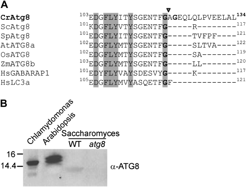
A recent survey of the Chlamydomonas genome has revealed the conservation of a single ATG8 gene in this alga (Diaz-Troya et al., 2008b). The putative CrATG8 gene spans about 2 kb of genomic DNA and consists of five exons and four introns. The full-length ATG8 cDNA was cloned by PCR from a Chlamydomonas cDNA library (see “Materials and Methods”). The CrATG8 gene encodes 134 amino acids with a predicted molecular mass of 15 kD. An evolutionary study of this protein evidenced a high sequence identity to plant ATG8s that ranges from 47% to 74% (data not shown). The critical Gly-116 residue of yeast ATG8 that is processed by the ATG4 protease is conserved in CrATG8 and corresponds to Gly-120 (Fig. 1A). However, a C-terminal extension of 14 residues that is not usually conserved among ATG8 proteins from other organisms was found after Gly-120 (Fig. 1A). To characterize CrATG8, we generated a polyclonal antibody against recombinant CrATG8 (see “Materials and Methods”) that reveals a protein of about 15 kD from Chlamydomonas soluble extracts (Fig. 1B). The anti-CrATG8 antibody cross-reacted with other putative ATG8 proteins from Arabidopsis and weakly recognized yeast ATG8 in total extracts (Fig. 1B).
Figure 1.
A, Amino acid sequence alignment of the C-terminal segments of the ATG8 protein from Chlamydomonas and other organisms. Identical residues are shaded. The arrowhead indicates the processing site recognized by the ATG4 protease that exposes the C-terminal Gly for lipidation. The conserved Gly is shaded and in boldface, and Gly-122, present only in Chlamydomonas, is shown in boldface. The number of amino acids for each protein is indicated at the end of the sequence. Species abbreviations are as follows: Cr, Chlamydomonas reinhardtii; Sc, Saccharomyces cerevisiae; Sp, Schizosaccharomyces pombe; At, Arabidopsis thaliana; Zm, Zea mays; Hs, Homo sapiens. The sequence from CrATG8 was taken from the Chlamydomonas database (http://www.chlamy.org/chlamydb.html). The rest of the sequences were obtained from the National Center for Biotechnology Information (http://ncbi.nlm.nih.gov). B, Cross-reactivity of anti-CrATG8 antibody with ATG8 proteins from Arabidopsis and yeast. Total extracts from Chlamydomonas, Arabidopsis, or S. cerevisiae (wild-type [WT] and atg8 strains) were prepared and resolved by 15% SDS-PAGE. ATG8 proteins were detected by immunoblot analysis with anti-CrATG8. Molecular mass markers (kD) are indicated on the left.
CrATG8 Functionally Replaces Yeast ATG8
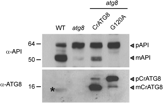
To investigate the possible role of CrATG8 in autophagy, we studied the functionality of this protein expressed in yeast mutant cells lacking endogenous ATG8. Yeast ATG8 is essential in both cytoplasm-to-vacuole targeting and autophagy pathways; therefore, atg8 mutant cells fail to deliver proaminopeptidase I (pAPI) from the cytosol to the vacuole, where pAPI is processed to its mature form (mAPI; Kirisako et al., 1999; Huang et al., 2000). Therefore, processing of pAPI to mAPI in atg8 mutant cells expressing CrATG8 was examined in order to study the functionality of CrATG8. Our results indicate that CrATG8 is able to functionally replace yeast ATG8, since mAPI was detected in atg8 cells expressing CrATG8 (Fig. 2). To further demonstrate that the mAPI band detected in atg8 cells expressing CrATG8 is due to the activity of this protein, processing of pAPI was also examined in cells transformed with a mutant form of CrATG8 where Gly-120 has been replaced by an Ala residue (CrG120A). A similar mutation in yeast ATG8 results in the complete loss of protein activity due to its inability for membrane attachment (Kirisako et al., 2000). As expected, no mAPI protein was detected in atg8 cells expressing CrG120A (Fig. 2). To demonstrate that wild-type and mutant forms of CrATG8 were correctly expressed, we analyzed the transformant yeast extracts by western blot using the anti-CrATG8 antibody. Both CrATG8 and CrG120A were efficiently expressed, and due to the presence of the C-terminal extension in CrATG8, it was possible to distinguish the precursor (pCrATG8) and the mature (mCrATG8) forms of the protein (Fig. 2). Similar to what has been reported in yeast ATG8 (Kirisako et al., 2000), CrATG8 is predominantly processed when expressed in yeast cells, whereas the CrG120A mutant cannot be maturated. Overall, these results demonstrate that CrATG8 is cleaved at Gly-120 and presumably binds to membranes to functionally replace yeast ATG8 in delivering pAPI from the cytosol to the vacuole.
Figure 2.
Complementation of the yeast atg8 mutant with CrATG8 from Chlamydomonas. Western-blot analysis of transformants using anti-pAPI (top panel) or anti-CrATG8 (bottom panel) antibodies. Top, Wild-type (WT) and atg8 strains were transformed with p426TEF empty plasmid or the same plasmid containing CrATG8 (ATG8 wild-type protein) or CrG120A (Gly-to-Ala mutant protein). Wild-type and atg8 yeast strains were used as positive and negative controls, respectively. The pAPI (precursor protein) and mAPI (mature protein) bands are marked with arrowheads. Bottom, Western-blot analysis of the same transformants with anti-CrATG8 antibody. The pCrATG8 (precursor protein) and mCrATG8 (mature protein) bands are marked with arrowheads. Molecular mass markers (kD) are indicated on the left.
In Vitro Processing of CrATG8 by Yeast ATG4 at Gly-120
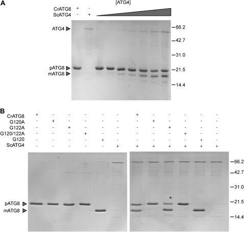
Yeast complementation assays of atg8 mutant cells suggested that CrATG8 must be processed by yeast ATG4 at the conserved Gly-120 residue. To demonstrate that CrATG8 is recognized by yeast ATG4 (ScATG4), in vitro cleavage assays were performed using purified CrATG8 and ScATG4 proteins. The His6 tag was fused to the N terminus of these proteins, and recombinant CrATG8 and ScATG4 were purified from Escherichia coli cells by nickel affinity chromatography (see “Materials and Methods”). Processing of CrATG8 was monitored by the different mobilities in SDS-PAGE of the precursor and mature forms of the protein. Our results indicate that ScATG4 cleaves CrATG8, presumably at its C terminus (Fig. 3A). To investigate the participation of Gly-120 in CrATG8 processing, we studied the in vitro cleavage of the G120A mutant. In agreement with the in vivo analysis of this mutant form of CrATG8 performed in yeast (Fig. 2), substitution of Gly-120 by Ala (G120A) completely abrogated processing of this protein by ScATG4 (Fig. 3B). Moreover, we investigated whether another Gly residue localized at the C terminus of CrATG8 at position 122, which is not conserved in other ATG8 proteins (Fig. 1A), may be recognized by ScATG4. Cleavage assays revealed that the G122A mutant was processed by ScATG4 whereas the double mutant G120/122A was not, indicating that substitution of Gly-122 by Ala has no effect on CrATG8 cleavage by ScATG4 and confirming that Gly-120 is the ScATG4 target (Fig. 3B).
Figure 3.
CrATG8 of Chlamydomonas is processed in vitro by yeast ATG4 at Gly-120. A, SDS-PAGE analysis of CrATG8 processing by different amounts of ScATG4 (0.25, 0.50, 1, 2, 5, 10, or 20 μg) for 3 h. B, Recombinant wild-type (CrATG8) and mutant forms (G120A, G122A, G120122A, and G120, where G120 is an ATG8 truncated protein at Gly-120 used as a processed-like form control) of Chlamydomonas ATG8 were incubated in an assay mixture that was protease free (left panel) or in the presence (right panel) of the protease ScATG4 (5 μg) for 3 h. The typical reaction mixture contained 5 μg of ScATG4, 5 μg of ATG8, 1 mm 1,4-dithiothreitol, and 1 mm EDTA in 100 μL of TBS buffer, and reactions were performed at 30°C for 3 h. The precursor (p) and mature (m) forms of CrATG8 are marked with arrowheads. Molecular mass markers (kD) are indicated on the right.
CrATG8 Is Processed at the C Terminus by an ATG4 Protease Activity in Chlamydomonas
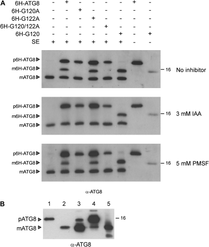
Our results demonstrated that CrATG8 is functional in a yeast system (Fig. 2). To investigate the functionality of CrATG8 in Chlamydomonas, we designed an ATG4 proteolytic assay in Chlamydomonas total extracts by taking advantage of the different sizes of the endogenous CrATG8 protein and recombinant His6-tagged CrATG8. Total extracts were obtained from Chlamydomonas cells grown in acetate medium and then mixed with a fixed amount of different forms of recombinant CrATG8. Incubation of wild-type CrATG8 with Chlamydomonas extract resulted in the fast processing of the recombinant protein. A band with the same size as a truncated form of CrATG8 lacking the last 14 residues (6H-G120) was readily visible, indicating that CrATG8 was processed at Gly-120 (Fig. 4A). Mutation of Gly-120 to Ala abolished CrATG8 cleavage, while the substitution of Gly-122 to Ala had no effect in CrATG8 processing (Fig. 4A). As expected, the G120/122A double mutant could not be processed (Fig. 4A). These results indicated that Chlamydomonas total extracts have a proteolytic ATG4 activity that recognizes and processes the C terminus of CrATG8 at Gly-120. ATG4 proteases contain a catalytic Cys involved in substrate hydrolysis and therefore are sensitive to iodoacetamide (Kirisako et al., 2000; Kim et al., 2001). To investigate if the CrATG8 processing detected in Chlamydomonas extracts is mediated by a Cys protease, proteolytic assays were performed in the presence of iodoacetamide. CrATG8 cleavage was fully abolished by iodoacetamide, while the Ser protease inhibitor phenylmethanesulfonyl fluoride (PMSF) had no effect (Fig. 4A), indicating that the CrATG8 proteolytic activity in Chlamydomonas extracts is mediated by a Cys protease.
Figure 4.
ATG4 protease activity in cell-free extracts of Chlamydomonas. A, Ten nanograms of the different recombinant ATG8 proteins (CrATG8, G120A, G122A, G120122A, or G120) was incubated with 30 μg of cell-free extracts (SE) of Chlamydomonas during 45 min on ice. When required, cell-free extracts were previously incubated with the corresponding protease inhibitor for 45 min on ice. The reaction was stopped by the addition of loading buffer and boiling at 100°C. Aliquots of the reaction mixtures were analyzed by western blotting with the anti-CrATG8 antibody. Exogenous, recombinant proteins contained the His6 tag and were labeled as 6H to distinguish them from endogenous CrATG8. The precursor (p) and mature (m) forms of CrATG8 are marked with arrowheads. The top panel corresponds to control (without protease inhibitor) assays, while the middle and bottom panels correspond to experiments carried out in the presence of the protease inhibitor 3 mm iodoacetamide or 5 mm PMSF, respectively. B, CrATG8 is constitutively processed in Chlamydomonas. Western-blot analysis of the CrATG8 protein. Lane 1, His tag-free ATG8 purified protein; lane 2, His tag-free G120 purified protein; lane 3, total extract from atg8 yeast cells expressing CrATG8; lane 4, total extract from atg8 yeast cells expressing G120A; lane 5, cell-free extract from Chlamydomonas. The precursor (p) and mature (m) forms of CrATG8 are marked with arrowheads. A molecular mass marker (kD) is indicated on the right.
Having shown that Chlamydomonas cells have ATG4 activity, we next investigated if the CrATG8 band of 15 kD detected in total extracts corresponds to the mature form of the protein. To this aim, purified unprocessed CrATG8 and a C-terminal truncated form of the protein (G120), together with Chlamydomonas total extracts and extracts of yeast cells expressing wild-type or the G120A mutant of CrATG8, were resolved by SDS-PAGE and analyzed by western blot. Our results indicate that the band detected in Chlamydomonas extracts with the anti-CrATG8 antibody corresponds to the C-terminal mature form of CrATG8 (Fig. 4B). Moreover, unprocessed CrATG8 was not detected under these and other experimental conditions (Fig. 4B; data not shown), suggesting that newly synthesized CrATG8 is immediately cleaved by an ATG4 protease to expose the Gly residue, as described previously in yeast and plant cells (Kirisako et al., 2000; Kim et al., 2001; Yoshimoto et al., 2004).
CrATG8 Is Reversibly Modified during Stationary Growth Phase
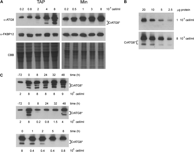
Previous studies in yeast, mammals, and plants demonstrated that the modified form of ATG8 generated upon autophagy activation migrates faster than unmodified ATG8 (Kabeya et al., 2000; Kirisako et al., 2000; Yoshimoto et al., 2004). We observed that besides the main 15-kD CrATG8 band detected by western-blot analysis in Chlamydomonas cell extracts, two less abundant bands of lower apparent molecular mass were visible (Figs. 1B and 4). To investigate whether these two bands may be generated as a result of CrATG8 modification, we performed western-blot analysis of Chlamydomonas cells under different growth conditions. First, we analyzed CrATG8 modification in exponential and stationary phase cells in acetate-rich or minimal medium. In addition to mature CrATG8, two bands became visible as cells approached the stationary phase in acetate-rich medium, whereas these bands were almost undetectable in exponential cells (Fig. 5A). Similar to what has been described in other systems, these forms likely represent lipidated CrATG8, although we cannot exclude the possibility that one of these bands may be the result of a different modification. A significant and progressive increase in CrATG8 abundance was also found in stationary phase cells compared with exponential cells, suggesting that the function of this protein is required when cells age. Western-blot analysis of the cytosolic protein FKBP12 and Coomassie Brilliant Blue staining of the gel were used as loading controls (Fig. 5A). Modified CrATG8 was also observed in minimal medium, although the faster migrating band was faintly detected (Fig. 5A). As in acetate-grown cells, the level of CrATG8 increased during the growth phase in minimal medium (Fig. 5A). Since the lower molecular mass variant of CrATG8 was concomitantly detected with an increase in the abundance of mature CrATG8, dilution series of total extracts obtained from log or stationary cells were analyzed by western blot in order to have similar amounts of the mature form from both conditions. Our results indicated that under uniform levels of mature CrATG8, modified forms are specifically detected in stationary cells (Fig. 5B).
Figure 5.
Analysis of CrATG8 during the growth phase in Chlamydomonas. A, Induction and modification of CrATG8 during the growth phase in Chlamydomonas. Thirty micrograms of total extracts of Chlamydomonas cells was resolved by 15% SDS-PAGE followed by western blotting with the anti-CrATG8 antibody. Cells were grown in acetate medium (Tris-acetate phosphate [TAP]) or minimal medium (Min) from 0.2 × 10⁶ cells mL−1 to 8 × 10⁶ cells mL−1, and the CrATG8 protein profile was analyzed by western blotting after SDS-PAGE. Western-blot analyses with anti-FKBP12 and Coomassie Brilliant Blue (CBB) staining were used as loading controls. B, Dilution series of total soluble extracts obtained from Chlamydomonas cells growing at exponential (1 × 10⁶ cells mL−1) or stationary (8 × 10⁶ cells mL−1) phase. Different amounts (2.5, 5, 10, and 20 μg) of soluble extracts were resolved by SDS-PAGE and subjected to western-blot analysis. C, Autophagy induction is a reversible process in Chlamydomonas. Stationary cells (8 × 10⁶ cells mL−1) grown in acetate-rich medium were transferred to a fresh medium to a cell density of 2 × 10⁵ cells mL−1 (middle panel), 4 × 10⁵ cells mL−1 (bottom panel), or maintained under similar growth conditions (top panel). Soluble extracts were prepared from samples obtained at different times, and CrATG8 modification was analyzed by western blotting. CrATG8* denotes modified CrATG8.
To further characterize the process of CrATG8 modification associated with cell growth, CrATG8 was analyzed by western blot in Chlamydomonas cultures that have reached the stationary growth phase and then transferred to fresh medium to initiate a new cell growth cycle or maintained in the same medium. While modified CrATG8 was clearly detectable in cells that remained in the stationary growth phase, CrATG8 was delipidated within 6 h when stationary cells were transferred to new medium (Fig. 5C, middle and lower panels). Modified CrATG8 was again detected after 48 h of reinoculation (Fig. 5C, middle panel). None of the three bands detected correspond to unprocessed CrATG8, since this protein is immediately cleaved (Fig. 4). Taken together, these results indicate that CrATG8 modification can be efficiently reversed under optimal growth conditions.
Modification of CrATG8 under Different Stress Conditions
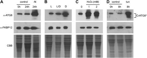
The participation of CrATG8 in the cellular response to different stresses such as nitrogen or carbon limitation and oxidative stress was examined by monitoring the modification state of this protein. To investigate whether nitrogen limitation may induce CrATG8 modification and autophagy activation in Chlamydomonas, exponentially growing cells were subjected to nitrogen depletion and CrATG8 modification was tested by western-blot analysis. Compared with untreated control cells, nitrogen limitation resulted in a substantial increase in CrATG8 levels as well as the detection of modified CrATG8 forms (Fig. 6A).
Figure 6.
Analysis of CrATG8 under different autophagy-activating conditions. A, CrATG8 modification upon nitrogen starvation. Chlamydomonas cells growing exponentially (1 × 10⁶ cells mL−1) in Tris-acetate phosphate medium were washed with a nitrogen-free medium and grown under these conditions for 24 h. Cells grown in nitrogen-containing medium were used as controls. B, CrATG8 modification under light/dark transitions. Exponential cells grown under continuous light in minimal medium were maintained under similar conditions (L) or subjected to 8-h-light/16-h-dark cycles (L/D) or continuous darkness (D). Next, cultures were diluted to 4 × 10⁵ cells mL−1 and maintained under the same conditions until a cell density of 1 × 10⁶ cells mL−1. C, Analysis of CrATG8 modification under oxidative stress conditions. Chlamydomonas cells growing exponentially (1 × 10⁶ cells mL−1) were treated with 1 or 2 mm H2O2 for 8 h. Untreated cells were used as controls. D, CrATG8 modification in Chlamydomonas cells subjected to ER stress. Chlamydomonas cells growing at exponential phase (1 × 10⁶ cells mL−1) were treated with 5 μg mL−1 tunicamycin (tun) for 8 h. Untreated cells at different times were used as controls. In all experiments, 30 μg of total extracts was resolved by 15% SDS-PAGE followed by western blotting with the anti-CrATG8 antibody. Western-blot analyses with anti-FKBP12 and Coomassie Brilliant Blue (CBB) staining were used as loading controls. CrATG8* corresponds to CrATG8 modifications.
Carbon starvation induced by darkness has been shown to induce autophagy in plants (Thompson et al., 2005; Phillips et al., 2008; Chung et al., 2009). To test whether carbon-limiting conditions also trigger autophagy in Chlamydomonas, cells grown under continuous light in minimal medium were shifted to darkness or maintained under a short-day photoperiod (8 h of light/16 of h dark) and the presence of modified CrATG8 was examined by western blot. Fixed-carbon limitation induced by darkness resulted in an increase in CrATG8 abundance and in the detection of modified CrATG8 forms (Fig. 6B). However, no effect was observed in cells grown under a short-day photoperiod compared with cells maintained under continuous light (Fig. 6B).
Autophagy is involved in degrading oxidized proteins during oxidative stress in plants (Xiong et al., 2007). The possible participation of CrATG8 in oxidative stress was analyzed by hydrogen peroxide (H2O2) treatment, an established oxidative stress inducer, in Chlamydomonas cells. Modified CrATG8 was readily detected upon treatment with H2O2 (Fig. 6C). Moreover, as in nutrient-starved cells, the level of CrATG8 significantly increased under oxidative stress conditions (Fig. 6C). Therefore, autophagy might contribute to the degradation of oxidized proteins during oxidative stress in Chlamydomonas.
In addition to nutrient or oxidative stress, we investigated the activation of CrATG8 in response to a specific stress in the endoplasmic reticulum (ER) by treating Chlamydomonas cells with tunicamycin, which raises the level of unfolded polypeptides at this compartment by inhibiting protein glycosylation. Compared with control, untreated cells, tunicamycin strongly increased CrATG8 lipidation and abundance (Fig. 6D), indicating that autophagy is involved in the degradation of unfolded proteins in the ER.
TOR Inhibits Autophagy in Chlamydomonas
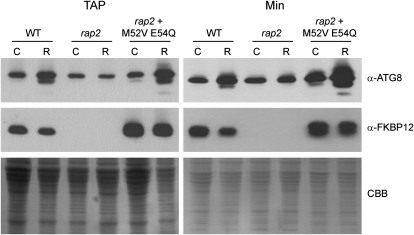
In a previous study, we showed that rapamycin-mediated inhibition of TOR signaling caused increased vacuolization (Crespo et al., 2005), a typical morphological autophagy effect. To confirm that the TOR pathway controls autophagy in Chlamydomonas, we took advantage of the autophagy monitoring assay using the anti-CrATG8 antibody and examined the presence of modified CrATG8 forms in rapamycin-treated cells. As shown in Figure 7, rapamycin treatment resulted in CrATG8 modification and an increase in the level of this protein. The rapamycin-induced effects were more pronounced in a Chlamydomonas mutant strain (Crespo et al., 2005) that exhibits high sensitivity to rapamycin (Fig. 7). As expected, no effect on CrATG8 was observed in the rapamycin-resistant mutant rap2 strain (Fig. 7), which lacks the rapamycin primary target FKBP12 (Crespo et al., 2005). Thus, our results demonstrate that CrATG8 is under the control of a rapamycin-sensitive TOR signaling pathway in Chlamydomonas.
Figure 7.
TOR signaling inhibition induces autophagy in Chlamydomonas cells. Chlamydomonas cells from different strains (wild type [WT], rap2, and rap2 + M52V E54Q) growing exponentially in acetate-rich (Tris-acetate phosphate [TAP]) or minimal (Min) medium were treated with 500 nm rapamycin for 16 h. The CrATG8 protein profile was analyzed by western blot with the anti-CrATG8 antibody. Western blot analyses with anti-FKBP12 and Coomassie Brilliant Blue (CBB) staining were used as loading controls. C, Control cells; R, rapamycin-treated cells.
ATG8 Associates with Membranes in Chlamydomonas
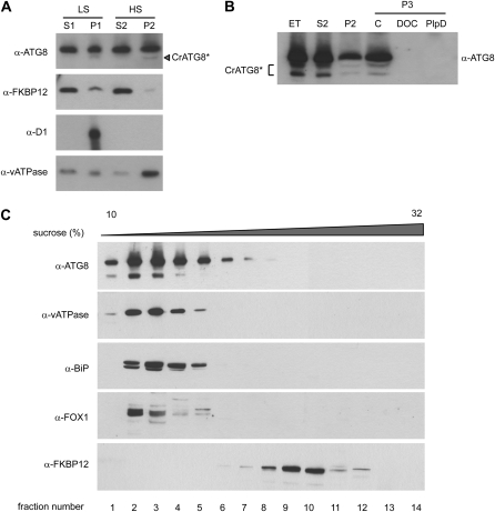
ATG8 proteins from yeast, mammals, and plants have been shown to be membrane associated (Kirisako et al., 1999; Kabeya et al., 2000; Yoshimoto et al., 2004; Chung et al., 2009). To investigate whether CrATG8 is associated with membranes, we performed subcellular fractionation by ultracentrifugation. Total soluble extracts were obtained from exponentially growing Chlamydomonas cells, and the presence of CrATG8 in the different soluble and membrane-enriched fractions was analyzed by western blot. The first fractionation was performed at low speed (15,000g), and CrATG8 was detected in the supernatant (S1) and pellet (P1) fractions (Fig. 8A), indicating that part of the protein is membrane bound. Interestingly, a faster migrating band likely corresponding to modified CrATG8 was observed in the membrane fraction but not in the soluble fraction. A second fractionation was achieved by ultracentrifugation of S1 to separate soluble (S2) and microsomal (P2) fractions. CrATG8 was again detected in both fractions and, as in the low-speed fractionation, putative modified CrATG8 was found in the membranous pellet (Fig. 8A). Thus, our results indicated that there might be at least two different pools of CrATG8 in exponentially growing Chlamydomonas cells, soluble and membrane-bound CrATG8. The presence of microsomes in the insoluble fraction after ultracentrifugation was confirmed by immunodetection of a vacuolar membrane ATPase. Parallel immunoblot assays with antiserum specific to D1 or to CrFKBP12 (Crespo et al., 2005) showed that the microsome-containing fraction is not significantly contaminated with thylakoidal membranes or soluble cytoplasmic proteins (Fig. 8A).
Figure 8.
Association of CrATG8 with membranes. A, Subcellular distribution of CrATG8. Crude extracts prepared from Chlamydomonas cells were centrifuged at 15,000g (low speed; LS) for 15 min to generate supernatant (S1) and pellet (P1) fractions. The resulting supernatant was then centrifuged at 100,000g (high speed; HS) for 2 h to generate further supernatants (S2) and pellets (P2). The samples were subjected to immunoblot analysis with antibodies raised against CrATG8, CrFKBP12, D1, and vacuolar membrane (vATPase) proteins. CrATG8* denotes modified CrATG8. B, Solubilization of CrATG8. Chlamydomonas cells were grown to stationary phase, and total extracts (ET) were prepared by centrifugation at 15,000g for 15 min. This extract was then centrifuged at 100,000g for 2 h to obtain S2 and P2 fractions. The pellet fractions were incubated on ice (control [C]), in lysis buffer containing 1% deoxycholate on ice for 1 h (DOC), or treated with PlpD (200 units mL−1 final concentration) at 37°C for 1 h. Samples were further centrifuged at 100,000g for 2 h to obtain membrane fractions with insoluble proteins (P3) and analyzed by western blot. C, Suc density gradient profiles of CrATG8, vATPase, BiP, FOX1, and FKBP12. Total extracts were fractionated on a 12-mL, 10% to 32% Suc gradient. Fractions (750 μL) were collected from the top of the gradient and processed for western-blot analysis.
To improve the detection of modified CrATG8 associated with membranes, a similar fractionation approach was performed in soluble extracts obtained from stationary cells, where these forms are more abundant (Fig. 5). High-speed centrifugation revealed that both mature and modified forms of CrATG8 are present in the pellet and soluble fractions (Fig. 8B). However, treatment of pellet membranes with detergent (1% deoxycholate) resulted in the complete solubilization of CrATG8, indicating that these forms are associated with the membrane (Fig. 8B). Anchoring of ATG8 to the membrane occurs through the covalent binding of the protein to a molecule of PE (Ichimura et al., 2000). Treatment of the ATG8-PE adduct with phospholipase D (PlpD) releases phosphatidic acid from the conjugate and consequently solubilizes membrane-bound ATG8 (Ichimura et al., 2000; Tanida et al., 2004; Fujioka et al., 2008; Chung et al., 2009). To investigate whether CrATG8 might be modified by lipidation, membrane-associated CrATG8 was treated with PlpD. Our results show that PlpD treatment caused the complete solubilization of CrATG8 from the membranous fraction (Fig. 8B), strongly suggesting that modified forms of CrATG8 are the phospholipid conjugates of this protein. To further investigate the membrane association of CrATG8, we performed Suc gradient sedimentation assays. Unlike the CrFKBP12 soluble protein, CrATG8 cosedimented with protein membrane markers such as vacuolar ATPase, BiP, and FOX1 (Fig. 8C). Taken together, our results are consistent with the notion that CrATG8 associates with membranes through covalent binding to PE.
Distinct CrATG8 Cellular Localization upon Autophagy Activation
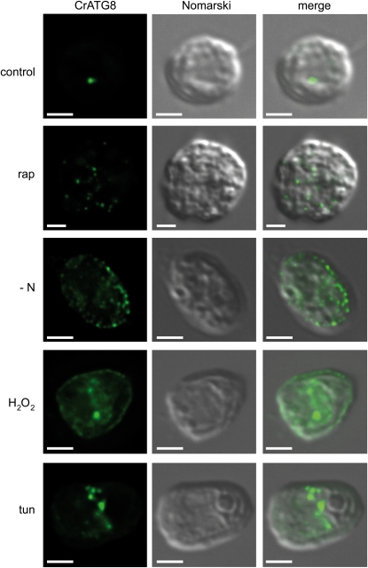
Immunofluorescence (IF) microscopic studies and GFP-ATG8 fusion proteins have been utilized to monitor autophagy in several organisms. We used the anti-CrATG8 antibody to examine the subcellular localization of this protein by IF microscopy in Chlamydomonas cells under different conditions. In cells growing exponentially in acetate-rich medium, CrATG8 was localized to punctate structures and a single spot was detected in most of the cells (Fig. 9). Similarly, the signal of CrATG8 in cells grown in minimal medium was confined to discrete punctae, although the number of these spots was usually higher than in acetate-grown cells, likely due to a higher requirement of autophagy under these conditions (Supplemental Fig. S1). The distribution of ATG8 in the cell has been shown to be modified by the activation of autophagy (Kirisako et al., 1999; Kabeya et al., 2000; Kim et al., 2001). Thus, we investigated whether CrATG8 cellular localization may respond to autophagy induction. Exponentially growing cells were treated with rapamycin or tunicamycin or subjected to nitrogen limitation or oxidative stress to trigger autophagy, and CrATG8 localization was examined by IF microscopy. Treatment of acetate-grown cells with rapamycin resulted in a pronounced detection of CrATG8 in more punctate structures and an increase in cell size (Fig. 9). Rapamycin caused a similar effect on CrATG8 localization in minimal medium, although a diffuse CrATG8 signal could also be detected at the periphery and the anterior end of the cell (Supplemental Fig. S1). Accordingly, this region of the cell has been reported to become vacuolated upon rapamycin treatment under similar experimental conditions (Crespo et al., 2005). Nitrogen depletion also resulted in a strong modification of CrATG8 cellular localization, being detected as multiple small dots all over the cell with a predominant localization around the chloroplast (Fig. 9; Supplemental Fig. S1). Similar to nitrogen starvation, oxidative stress induced by H2O2 increased the overall intensity of the CrATG8 signal in the cell, and some large spots could also be detected (Fig. 9; Supplemental Fig. S1). The effect of tunicamycin on the cellular distribution of CrATG8 differed in some respects from other stresses. First, the signal was remarkably stronger, and second, CrATG8 accumulated at a specific region in the middle of the cell that might correspond to the ER, in agreement with the localized effect of tunicamycin at this compartment (Fig. 9; Supplemental Fig. S1). Taken together, these results indicate that the cellular distribution of CrATG8 strongly depends on the autophagic requirement of the cell.
Figure 9.
IF localization of CrATG8 in Chlamydomonas cells. Cells growing exponentially in acetate medium were subjected to different treatments as follows: cells were treated with rapamycin for 16 h (rap), shifted to nitrogen-free medium for 24 h (−N), treated with 2 mm H2O2 for 8 h to induce oxidative stress, or treated with 5 μg mL−1 tunicamycin (tun) for 8 h to trigger ER stress. As a control, cells were maintained in exponential growth. Cells were collected and processed for IF microscopy analysis as described in “Materials and Methods.” The signal recognized by the affinity-purified anti-CrATG8 antibody is shown in green (CrATG8). The same acquisition time was used in all samples except for tunicamycin-treated cells, where it was reduced to 50% to avoid signal saturation. Bars = 2 μm.
DISCUSSION
The Chlamydomonas genome contains potential homologs to yeast and plant ATG genes, including those involved in the ATG8/ATG12-conjugating systems (Diaz-Troya et al., 2008b), which strongly suggests that Chlamydomonas might recycle intracellular components through autophagic processes. In agreement with this hypothesis, it has been recently shown that autophagosome-like structures are generated as a response to prolonged salt stress in the unicellular green alga Micrasterias denticulata (Affenzeller et al., 2009). However, the molecular features of autophagy have never been characterized in green algae. Here, we report the participation of the Chlamydomonas CrATG8 protein in autophagy and demonstrate that this protein may be used to monitor autophagic processes in this green alga.
Compared with the ATG8 proteins from other organisms, the C-terminal extension downstream of the conserved Gly-120 of CrATG8 is particularly large (Fig. 1A). A comparative analysis of the C-terminal extension of ATG8 proteins from different organisms indicated that the sequence after the conserved Gly completely diverged (Kabeya et al., 2000). In Arabidopsis, two of the nine ATG8 homologs, AtATG8h and AtATG8i, do not possess additional amino acids downstream of the conserved Gly, while the ATG8 protein from other organisms such as rats or the symbiotic fungus Laccaria bicolor have large C-terminal extensions (Kabeya et al., 2000). The C-terminal extension does not seem to be required for the functionality of the protein, since all nine Arabidopsis ATG8s, including AtATG8h and AtATG8i, are active in plant autophagy (Yoshimoto et al., 2004). Unprocessed CrATG8 could not be detected in Chlamydomonas cells (Fig. 4), indicating that similar to what has been described in yeast or plants (Kirisako et al., 2000; Kim et al., 2001), newly synthesized CrATG8 is immediately processed. Whether the C-terminal extension of CrATG8 may play a role in the stability of the protein and/or its processing remains to be investigated.
Functional studies performed in yeast revealed that CrATG8 is able to restore pAPI processing in an atg8 mutant strain under normal growth conditions (Fig. 2), demonstrating the functionality of this protein in yeast ATG8-mediated processes. Yeast complementation assays have also been performed with Arabidopsis and Trypanosoma cruzi ATG8 homologs, and in contrast to CrATG8, these proteins were able to functionally replace yeast ATG8 under starvation conditions only (Ketelaar et al., 2004; Alvarez et al., 2008). Mutation of the conserved Gly residue to Ala in CrATG8 fully abolished pAPI processing, suggesting that despite the presence of the large C-terminal extension, CrATG8 could be processed and lipidated in yeast cells (Fig. 2A). Indeed, in vitro cleavage assays demonstrated that CrATG8 is maturated at Gly-120 by yeast ATG4 (Fig. 3).
Our in vivo and in vitro heterologous studies strongly suggested that CrATG8 performs a similar function to yeast ATG8 in Chlamydomonas. Several lines of evidence support this hypothesis. On the one hand, CrATG8 is processed in Chlamydomonas at the conserved Gly, as revealed by ATG4 proteolytic assays performed in cell-free extracts of Chlamydomonas (Fig. 4). In agreement with findings in plants and T. cruzi, where ATG4 proteases are constitutively expressed regardless of nutrient conditions (Yoshimoto et al., 2004; Alvarez et al., 2008), ATG4 activity was detected in Chlamydomonas cells under optimal and nutrient-limiting growth conditions (Fig. 4; data not shown). On the other hand, CrATG8 levels increased upon autophagy activation. The abundance of plant ATG8s is up-regulated at the protein and mRNA levels in response to autophagy induction by nutrient starvation (Yoshimoto et al., 2004; Contento et al., 2005; Thompson et al., 2005; Chung et al., 2009). Accordingly, a significant increase in CrATG8 protein abundance was detected in Chlamydomonas cells limited for nutrients (Fig. 6). Entry into stationary phase also resulted in the up-regulation of CrATG8 levels (Fig. 5), which links autophagy to cell aging in Chlamydomonas. Autophagy has been shown to play a role in plant senescence. The phenotypic analysis of Arabidopsis atg mutants has led to the hypothesis that autophagy occurs before or during senescence, likely to allow the plant to accomplish efficient nutrient reallocation (Doelling et al., 2002; Hanaoka et al., 2002; Yoshimoto et al., 2004; Thompson et al., 2005; Xiong et al., 2005; Phillips et al., 2008). Similarly, high levels of CrATG8, and by inference autophagy, might be required for the metabolic adaptation of Chlamydomonas cells before entering into the stationary phase. Supporting a role for CrATG8 in the cell cycle, we found that CrATG8 abundance is down-regulated as soon as stationary cells are allowed to enter the log phase (Fig. 6C). Moreover, modified CrATG8 is efficiently delipidated in exponentially growing cells (Fig. 6C), indicating that CrATG8 modification is a reversible process in Chlamydomonas. Deconjugation of ATG8-PE has been demonstrated in yeast and is mediated by the ATG4 protease (Kirisako et al., 2000). It is likely, therefore, that CrATG8 deconjugation is catalyzed by a homologous ATG4 protease in photosynthetic organisms.
A common feature of ATG8 cellular localization in lower and higher eukaryotes seems to be a punctate pattern under optimal growth that is enhanced upon autophagy activation. In unicellular organisms such as yeast or protozoa, ATG8 was localized to tiny dots as well as larger spots distributed throughout the cytoplasm in rich medium (Kirisako et al., 1999; Kim et al., 2001; Alvarez et al., 2008). Nutrient depletion resulted in both organisms in an increase in the number of large spots, reflecting the activation of autophagy and the formation of mature autophagosomes. The cellular distribution of CrATG8 in Chlamydomonas displayed common features with yeast and protozoa ATG8 localization. In acetate-rich medium, CrATG8 signal was restricted to discrete punctate structures that may correspond to PAS. However, autophagy induction by nutrient limitation, oxidative stress, or tunicamycin treatment drastically modified the localization of CrATG8 in the cell. Numerous punctate structures could be detected all over the cell upon nutrient starvation or H2O2 treatment, while a specific stress in the ER caused a massive accumulation of CrATG8 at this cellular compartment (Fig. 9). Using GFP-ATG8 fusion proteins, it has been shown in plant cells that the cellular distribution of this protein is modified upon autophagy activation by nutrient limitation (Yoshimoto et al., 2004; Contento et al., 2005; Thompson et al., 2005; Xiong et al., 2005). Oxidative stress also induces autophagy in plants, since H2O2 or methyl viologen treatment results in increased autophagosome detection (Xiong et al., 2007). It remains to be determined whether ER stress may trigger autophagy in plants as it does in yeast (Yorimitsu et al., 2006) or algae.
Compared with plants, the global picture of CrATG8 cellular localization and hence autophagosome generation in Chlamydomonas seems to be less complex. In plants, it has been proposed that autophagy occurs constantly in significant amounts under optimal growth based on the presence of ATG8s in many rings and punctate structures representing autophagosomes or PAS (Yoshimoto et al., 2004; Thompson et al., 2005). This model is strengthened by the detection of most ATG8s in the pellet fraction in plant cells, an indication of ATG8 association with the autophagosomes (Yoshimoto et al., 2004). In exponentially growing Chlamydomonas cells, mature and modified forms of CrATG8 were also found in association with membrane fractions (Fig. 8A), which may imply the participation of CrATG8 in autophagy or other membrane-trafficking events under optimal growth conditions. Upon autophagy induction, CrATG8 becomes lipidated and binds to membrane, although mature, unmodified CrATG8 could also be detected with membrane fractions (Fig. 8B). The presence of unmodified CrATG8 in membranes is not exclusive to Chlamydomonas, since this phenomenon has also been reported in mammals (Tanida et al., 2004) and plants (Chung et al., 2009).
The TORC1 pathway perceives the nutritional status of the cell and transmits these signals to the autophagic machinery, acting as a negative regulator of autophagy (Noda and Ohsumi, 1998). The molecular mechanisms by which TORC1 inhibits autophagy were elucidated from studies performed in yeast (Noda and Ohsumi, 1998; Kamada et al., 2000; Kabeya et al., 2005), and significant advances in this regulatory pathway were recently reported in insects and mammals (Chang and Neufeld, 2009; Hosokawa et al., 2009; Jung et al., 2009). Components of TORC1, including the TOR kinase, are functionally conserved in plants (Menand et al., 2002; Anderson et al., 2005; Deprost et al., 2005; Mahfouz et al., 2006), although the participation of this signaling pathway in the control of autophagy has not been shown in photosynthetic eukaryotes. The data presented in this study demonstrate that TOR signaling inhibits autophagy in Chlamydomonas. Rapamycin-mediated inhibition of CrTOR resulted in increased CrATG8 modification and a relocalization of CrATG8 in Chlamydomonas cells similar to the one observed under nutrient-limiting conditions (Figs. 7 and 9). Thus, our results assign a prominent role to the TOR pathway in the control of autophagy in photosynthetic organisms. In yeast and animal cells, the ATG1 kinase and its regulatory proteins ATG11, ATG13, and ATG17 transmit the signal from TORC1 to the autophagosome-generating machinery. Whether these proteins may perform a similar function in plants or algae is unknown.
Autophagy has been recently involved in chloroplast degradation and Rubisco mobilization to the vacuole during plant senescence and stress (Ishida et al., 2008; Wada et al., 2009). Rubisco and other stroma-targeted proteins are mobilized from the chloroplast to the vacuole through an ATG-mediated autophagic process, which does not implicate entire chloroplast degradation. In Chlamydomonas, it has been shown that a substantial amount of chloroplast material, including the large subunit of Rubisco and ribosomes, is transferred to a class of vacuoles distinct from contractile vacuoles via protrusion of the outer membrane of the plastid envelope (Park et al., 1999). However, the participation of autophagy in this degradative process has not been demonstrated. The availability of a specific autophagy marker in Chlamydomonas will certainly facilitate the investigation of the role of autophagy in this and other cellular events.
MATERIALS AND METHODS
Strains and Growth Conditions
Chlamydomonas reinhardtii wild-type strain 6C+ was obtained from the laboratory of J.-D. Rochaix (University of Geneva). rap2 and rap2 + M52V E54Q strains were characterized previously (Crespo et al., 2005). Chlamydomonas cells were grown under continuous illumination at 25°C in Tris-acetate phosphate or high-salt minimal medium as described (Harris, 1989). When required, 1.2% bacto agar (Difco) was added to the medium. Saccharomyces cerevisiae wild-type (SEY6210) and atg8 mutant (WPHYD7) strains were obtained from the laboratory of D.J. Klionsky (University of Michigan). Yeast cells were grown in rich or synthetic medium as described previously (Kim et al., 2001). Rapamycin was added to Chlamydomonas to 500 nm from a 1 mg mL−1 stock in 90% ethanol and 10% Tween 20.
DNA and Protein Sequence Alignments
The amino acid Chlamydomonas ATG8 sequence was obtained from the Chlamydomonas database (www.chlamy.org/chlamydb.html). The ATG8 amino acid sequences from other organisms were obtained from the National Center for Biotechnology Information (www.ncbi.nlm.nih.gov). The multiple sequence alignment was performed with the ClustalW2 program (www.ebi.ac.uk/Tools/clustalw2).
Cloning and Protein Purification
The ATG8 coding region from Chlamydomonas (351 bp) was amplified by PCR from a Chlamydomonas cDNA library using the following primers: ATG8For (5′-CCGGCATATGGTTGGCTCCCGACCCCCGACATTC-3′) and ATG8Rev (5′-CCGGCTCGAGTCACAACGCCAGCTCCTCCACAGG-3′), designed to contain a NdeI or XhoI site (underlined), respectively. The resulting product was digested with NdeI and XhoI and inserted into pET28a (Novagen) for expression in Escherichia coli. Expression of the recombinant protein was induced by adding 1 mm isopropyl-β-d-thiogalactopyranoside for 3 h at 37°C. The ATG8 His6-tagged protein was purified from the lysates by two consecutive affinity chromatographies on a nickel matrix from Novagen.
The His6-tagged Gly-to-Ala mutants of ATG8, denoted as G120A, G122A, and G120122A, whose Gly-120, Gly-122, or both were replaced by Ala, respectively, were obtained by PCR using the wild-type version of the ATG8 gene cloned into pET28a and reverse primers including the proper mutation as follows: G120ARev (5′-TCACAACGCCAGCTCCTCCACAGGCAGCTGCAGCTGCTCCCCGGCAGCGAACGTGTT-3′) for the G120A mutant, G122ARev (5′-CCGGCTCGAGTCACAACGCCAGCTCCTCCACAGGCAGCTGCAGCTGCTCCTCCGCGGCAC-3′) for G122A, and DMRev (5′-CCGGCTCGAGTCACAACGCCAGCTCCTCCACAGGCAGCTGCAGCTGCTCCGCGGCAGCGAAC-3′) for G120122A. The His6-tagged Gly-120 truncated mutant, named G120, was similarly obtained by PCR using 5′-CCGGCTCGAGTCAACCGAACGTGTTCTCGC-3′ as the reverse primer. ATG8For was used as the forward primer in PCR to obtain all ATG8 mutant versions. All ATG8 mutants (G120A, G122A, G120122A, and G120) were purified as described for wild-type ATG8.
The ATG4 coding region from S. cerevisiae was amplified by PCR from yeast genomic DNA using the following primers: ATG4For (5′-CCGGCATATGCAGAGGTGGCTACAACTGTG-3′) and ATG4Rev (5′-CCGGCTCGAGCTAGCATTTTTCATCAATAG-3′), designed to contain a NdeI or XhoI site (underlined), respectively. Cloning and protein expression were performed as described above for Chlamydomonas ATG8. Prior to purification, His6-tagged ATG4 was solubilized with 4 m urea due to the presence of the recombinant protein in inclusion bodies. Purification was performed as described for CrATG8.
Yeast Complementation
For expression of CrATG8 in yeast cells, the cDNA of the CrATG8 gene and the mutant version, G120A (whose Gly-120 was substituted by Ala), was cloned into the SpeI site of the p426TEF vector under control of the TEF constitutive promoter (Mumberg et al., 1995). Wild-type and atg8 strains were transformed with empty vector, the pTEF-CrATG8 plasmid, or the pTEF-G120A plasmid and grown in rich or synthetic medium as described (Kim et al., 2001).
Preparation and Analysis of Proteins
Chlamydomonas cells from liquid cultures were collected by centrifugation (4,000g, 5 min), washed once in 50 mm Tris-HCl (pH 7.5) buffer, and resuspended in a minimal volume of the same solution. Cells were lysed by two cycles of slow freezing to −80°C followed by thawing to room temperature. The soluble cell extract was separated from the insoluble fraction by centrifugation (15,000g, 15 min) in a microcentrifuge at 4°C. Yeast extracts from logarithmically growing cells were prepared in lysis buffer (50 mm Tris-HCl, pH 7.5, and 0.1% Triton X-100) by vortexing them 10 times for 1 min each time with glass beads (Sigma-Aldrich). Crude extracts were cleared by centrifugation at 15,000g for 15 min at 4°C. Proteins were quantified with the Coomassie Brilliant Blue dye-binding method as described by the manufacturer (Bio-Rad).
In Vitro ATG4 Cleavage Assays
Recombinant wild-type CrATG8 and the different Gly-to-Ala mutant versions (5 μg) were incubated with different amounts of purified ScATG4 in 100 μL of Tris-buffered saline (TBS) containing 1 mm EDTA and 1 mm 1,4-dithiothreitol. The reaction mixtures were incubated at 30°C for different times and stopped by the addition of Laemmli sample buffer followed by 5 min of boiling. Proteins were resolved on a 15% SDS-PAGE gel and stained with Coomassie Brilliant Blue. For the detection of ATG4 activity in Chlamydomonas, cell-free extracts were prepared as described above and incubated with 10 ng of the different recombinant ATG8 proteins in TBS buffer for 45 min. To determine which protease group Chlamydomonas ATG4 belongs to, proteolytic assays were carried out in the presence of the protease inhibitor iodoacetamide (3 mm) or PMSF (5 mm). The reactions were stopped by the addition of Laemmli sample buffer followed by 5 min of boiling. Proteins were resolved on a 15% SDS-PAGE gel and immunoblotted with the anti-CrATG8 antibody.
Antibody Production and Immunoblot Analysis
The anti-CrATG8 polyclonal antibody was produced by injecting the recombinant wild-type CrATG8 protein into a rabbit using standard immunization protocols. Polyclonal serum was affinity purified by coupling the recombinant protein CrATG8 (12 mg) to a solid resin (Aminolink Plus Immobilization kit; Thermo Scientific) according to the manufacturer's instructions. For immunoblot analyses, total protein extracts that had been subjected to SDS-PAGE were transferred to nitrocellulose membranes (Bio-Rad). Anti-CrATG8 and secondary antibodies were diluted 1:2,000 and 1:10,000, respectively, in phosphate-buffered saline containing 0.1% Tween 20 (Sigma-Aldrich) and 5% milk powder. The ECL-Plus immunoblotting detection system (GE Healthcare) was used to detect the proteins with horseradish peroxidase-conjugated anti-rabbit secondary antibodies. The anti-FKBP12 antibody has been described previously (Crespo et al., 2005). The anti-pAPI antibody was obtained from the laboratory of D.J. Klionsky (University of Michigan). Both antibodies were used at 1:2,000 dilution. The anti-D1 and anti-vacuolar membrane, anti-BiP, and anti-FOX1 antibodies were purchased from Agrisera and diluted to 1:20,000, 1:10,000, 1:2,000, and 1:10,000, respectively.
Subcellular Fractionation and Solubilization of CrATG8
For fractionation assays, Chlamydomonas whole-cell extract was prepared from wild-type cells as described above. After freezing and thawing, samples were centrifuged at 500g for 5 min to remove cell debris. The supernatant was centrifuged at 15,000g for 15 min to generate the soluble extract and the low-speed membrane fraction. The microsomal fraction was obtained by centrifuging the soluble extract in an 80-Ti rotor in a Beckman Coulter ultracentrifuge at 100,000g for 2 h at 4°C. For CrATG8 solubilization, the membrane pellet fraction was resuspended in extraction buffer plus 1% deoxycholate and incubated on ice for 1 h. Samples were then centrifuged at 100,000g for 2 h to separate soluble from insoluble material. PlpD treatment was performed by incubating the membrane fraction at 37°C for 1 h with Streptomyces chromofuscus PlpD (Sigma), and reactions were stopped by the addition of SDS-PAGE sample buffer. The different fractions were processed and subjected to western-blot analysis with the anti-CrATG8 antibody. Suc density gradient analysis was performed as described previously (Diaz-Troya et al., 2008a).
Fluorescence Microscopy
Wild-type Chlamydomonas cells were fixed and stained for IF microscopy as described previously (Cole et al., 1998; Diaz-Troya et al., 2008a). The primary antibody used was rabbit polyclonal anti-CrATG8 (1:500). For signal detection, a fluorescein isothiocyanate-labeled goat anti-rabbit antibody (1:500; Sigma) was used. Preparations were photographed on a DM6000B microscope (Leica) with an ORCA-ER camera (Hamamatsu) and processed with the Leica Application Suite Advanced Fluorescence software package (Leica). Deconvolution analysis of images was performed with the same software. For the comparative analysis of the fluorescein isothiocyanate signal from different samples, the same acquisition time was fixed except for tunicamycin-treated cells, where it was reduced to 50% to avoid signal saturation.
Supplemental Data
The following materials are available in the online version of this article.
Supplemental Figure S1. IF localization of CrATG8.
Supplementary Material
Acknowledgments
We thank Daniel J. Klionsky for the generous gift of wild-type and atg8 yeast strains and the anti-pAPI antibody.
References
- Affenzeller MJ, Darehshouri A, Andosch A, Lutz C, Lutz-Meindl U. (2009) Salt stress-induced cell death in the unicellular green alga Micrasterias denticulata. J Exp Bot 60: 939–954 [DOI] [PMC free article] [PubMed] [Google Scholar]
- Alvarez VE, Kosec G, Sant'Anna C, Turk V, Cazzulo JJ, Turk B. (2008) Autophagy is involved in nutritional stress response and differentiation in Trypanosoma cruzi. J Biol Chem 283: 3454–3464 [DOI] [PubMed] [Google Scholar]
- Anderson GH, Veit B, Hanson MR. (2005) The Arabidopsis AtRaptor genes are essential for post-embryonic plant growth. BMC Biol 3: 12. [DOI] [PMC free article] [PubMed] [Google Scholar]
- Bassham DC. (2007) Plant autophagy: more than a starvation response. Curr Opin Plant Biol 10: 587–593 [DOI] [PubMed] [Google Scholar]
- Bassham DC. (2009) Function and regulation of macroautophagy in plants. Biochim Biophys Acta 1793: 1397–1403 [DOI] [PubMed] [Google Scholar]
- Bassham DC, Laporte M, Marty F, Moriyasu Y, Ohsumi Y, Olsen LJ, Yoshimoto K. (2006) Autophagy in development and stress responses of plants. Autophagy 2: 2–11 [DOI] [PubMed] [Google Scholar]
- Cebollero E, Reggiori F. (2009) Regulation of autophagy in yeast Saccharomyces cerevisiae. Biochim Biophys Acta 1793: 1413–1421 [DOI] [PubMed] [Google Scholar]
- Chang YY, Neufeld TP. (2009) An Atg1/Atg13 complex with multiple roles in TOR-mediated autophagy regulation. Mol Biol Cell 20: 2004–2014 [DOI] [PMC free article] [PubMed] [Google Scholar]
- Chung T, Suttangkakul A, Vierstra RD. (2009) The ATG autophagic conjugation system in maize: ATG transcripts and abundance of the ATG8-lipid adduct are regulated by development and nutrient availability. Plant Physiol 149: 220–234 [DOI] [PMC free article] [PubMed] [Google Scholar]
- Cole DG, Diener DR, Himelblau AL, Beech PL, Fuster JC, Rosenbaum JL. (1998) Chlamydomonas kinesin-II-dependent intraflagellar transport (IFT): IFT particles contain proteins required for ciliary assembly in Caenorhabditis elegans sensory neurons. J Cell Biol 141: 993–1008 [DOI] [PMC free article] [PubMed] [Google Scholar]
- Contento AL, Xiong Y, Bassham DC. (2005) Visualization of autophagy in Arabidopsis using the fluorescent dye monodansylcadaverine and a GFP-AtATG8e fusion protein. Plant J 42: 598–608 [DOI] [PubMed] [Google Scholar]
- Crespo JL, Diaz-Troya S, Florencio FJ. (2005) Inhibition of target of rapamycin signaling by rapamycin in the unicellular green alga Chlamydomonas reinhardtii. Plant Physiol 139: 1736–1749 [DOI] [PMC free article] [PubMed] [Google Scholar]
- Deprost D, Truong HN, Robaglia C, Meyer C. (2005) An Arabidopsis homolog of RAPTOR/KOG1 is essential for early embryo development. Biochem Biophys Res Commun 326: 844–850 [DOI] [PubMed] [Google Scholar]
- Diaz-Troya S, Florencio FJ, Crespo JL. (2008a) Target of rapamycin and LST8 proteins associate with membranes from the endoplasmic reticulum in the unicellular green alga Chlamydomonas reinhardtii. Eukaryot Cell 7: 212–222 [DOI] [PMC free article] [PubMed] [Google Scholar]
- Diaz-Troya S, Perez-Perez ME, Florencio FJ, Crespo JL. (2008b) The role of TOR in autophagy regulation from yeast to plants and mammals. Autophagy 4: 851–865 [DOI] [PubMed] [Google Scholar]
- Doelling JH, Walker JM, Friedman EM, Thompson AR, Vierstra RD. (2002) The APG8/12-activating enzyme APG7 is required for proper nutrient recycling and senescence in Arabidopsis thaliana. J Biol Chem 277: 33105–33114 [DOI] [PubMed] [Google Scholar]
- Fujiki Y, Yoshimoto K, Ohsumi Y. (2007) An Arabidopsis homolog of yeast ATG6/VPS30 is essential for pollen germination. Plant Physiol 143: 1132–1139 [DOI] [PMC free article] [PubMed] [Google Scholar]
- Fujioka Y, Noda NN, Fujii K, Yoshimoto K, Ohsumi Y, Inagaki F. (2008) In vitro reconstitution of plant Atg8 and Atg12 conjugation systems essential for autophagy. J Biol Chem 283: 1921–1928 [DOI] [PubMed] [Google Scholar]
- Fujita N, Hayashi-Nishino M, Fukumoto H, Omori H, Yamamoto A, Noda T, Yoshimori T. (2008) An Atg4B mutant hampers the lipidation of LC3 paralogues and causes defects in autophagosome closure. Mol Biol Cell 19: 4651–4659 [DOI] [PMC free article] [PubMed] [Google Scholar]
- Geng J, Klionsky DJ. (2008) The Atg8 and Atg12 ubiquitin-like conjugation systems in macroautophagy. ‘Protein modifications: beyond the usual suspects’ review series. EMBO Rep 9: 859–864 [DOI] [PMC free article] [PubMed] [Google Scholar]
- Hanada T, Noda NN, Satomi Y, Ichimura Y, Fujioka Y, Takao T, Inagaki F, Ohsumi Y. (2007) The Atg12-Atg5 conjugate has a novel E3-like activity for protein lipidation in autophagy. J Biol Chem 282: 37298–37302 [DOI] [PubMed] [Google Scholar]
- Hanaoka H, Noda T, Shirano Y, Kato T, Hayashi H, Shibata D, Tabata S, Ohsumi Y. (2002) Leaf senescence and starvation-induced chlorosis are accelerated by the disruption of an Arabidopsis autophagy gene. Plant Physiol 129: 1181–1193 [DOI] [PMC free article] [PubMed] [Google Scholar]
- Harris EH. (1989) The Chlamydomonas Sourcebook. Academic Press, San Diego [Google Scholar]
- Harris EH. (2001) Chlamydomonas as a model organism. Annu Rev Plant Physiol Plant Mol Biol 52: 363–406 [DOI] [PubMed] [Google Scholar]
- Hofius D, Schultz-Larsen T, Joensen J, Tsitsigiannis DI, Petersen NH, Mattsson O, Jorgensen LB, Jones JD, Mundy J, Petersen M. (2009) Autophagic components contribute to hypersensitive cell death in Arabidopsis. Cell 137: 773–783 [DOI] [PubMed] [Google Scholar]
- Hosokawa N, Hara T, Kaizuka T, Kishi C, Takamura A, Miura Y, Iemura S, Natsume T, Takehana K, Yamada N, et al. (2009) Nutrient-dependent mTORC1 association with the ULK1-Atg13-FIP200 complex required for autophagy. Mol Biol Cell 20: 1981–1991 [DOI] [PMC free article] [PubMed] [Google Scholar]
- Huang WP, Scott SV, Kim J, Klionsky DJ. (2000) The itinerary of a vesicle component, Aut7p/Cvt5p, terminates in the yeast vacuole via the autophagy/Cvt pathways. J Biol Chem 275: 5845–5851 [DOI] [PubMed] [Google Scholar]
- Ichimura Y, Kirisako T, Takao T, Satomi Y, Shimonishi Y, Ishihara N, Mizushima N, Tanida I, Kominami E, Ohsumi M, et al. (2000) A ubiquitin-like system mediates protein lipidation. Nature 408: 488–492 [DOI] [PubMed] [Google Scholar]
- Ishida H, Yoshimoto K, Izumi M, Reisen D, Yano Y, Makino A, Ohsumi Y, Hanson MR, Mae T. (2008) Mobilization of Rubisco and stroma-localized fluorescent proteins of chloroplasts to the vacuole by an ATG gene-dependent autophagic process. Plant Physiol 148: 142–155 [DOI] [PMC free article] [PubMed] [Google Scholar]
- Jung CH, Jun CB, Ro SH, Kim YM, Otto NM, Cao J, Kundu M, Kim DH. (2009) ULK-Atg13-FIP200 complexes mediate mTOR signaling to the autophagy machinery. Mol Biol Cell 20: 1992–2003 [DOI] [PMC free article] [PubMed] [Google Scholar]
- Kabeya Y, Kamada Y, Baba M, Takikawa H, Sasaki M, Ohsumi Y. (2005) Atg17 functions in cooperation with Atg1 and Atg13 in yeast autophagy. Mol Biol Cell 16: 2544–2553 [DOI] [PMC free article] [PubMed] [Google Scholar]
- Kabeya Y, Mizushima N, Ueno T, Yamamoto A, Kirisako T, Noda T, Kominami E, Ohsumi Y, Yoshimori T. (2000) LC3, a mammalian homologue of yeast Apg8p, is localized in autophagosome membranes after processing. EMBO J 19: 5720–5728 [DOI] [PMC free article] [PubMed] [Google Scholar]
- Kamada Y, Funakoshi T, Shintani T, Nagano K, Ohsumi M, Ohsumi Y. (2000) Tor-mediated induction of autophagy via an Apg1 protein kinase complex. J Cell Biol 150: 1507–1513 [DOI] [PMC free article] [PubMed] [Google Scholar]
- Ketelaar T, Voss C, Dimmock SA, Thumm M, Hussey PJ. (2004) Arabidopsis homologues of the autophagy protein Atg8 are a novel family of microtubule binding proteins. FEBS Lett 567: 302–306 [DOI] [PubMed] [Google Scholar]
- Kim J, Huang WP, Klionsky DJ. (2001) Membrane recruitment of Aut7p in the autophagy and cytoplasm to vacuole targeting pathways requires Aut1p, Aut2p, and the autophagy conjugation complex. J Cell Biol 152: 51–64 [DOI] [PMC free article] [PubMed] [Google Scholar]
- Kirisako T, Baba M, Ishihara N, Miyazawa K, Ohsumi M, Yoshimori T, Noda T, Ohsumi Y. (1999) Formation process of autophagosome is traced with Apg8/Aut7p in yeast. J Cell Biol 147: 435–446 [DOI] [PMC free article] [PubMed] [Google Scholar]
- Kirisako T, Ichimura Y, Okada H, Kabeya Y, Mizushima N, Yoshimori T, Ohsumi M, Takao T, Noda T, Ohsumi Y. (2000) The reversible modification regulates the membrane-binding state of Apg8/Aut7 essential for autophagy and the cytoplasm to vacuole targeting pathway. J Cell Biol 151: 263–276 [DOI] [PMC free article] [PubMed] [Google Scholar]
- Liu Y, Schiff M, Czymmek K, Talloczy Z, Levine B, Dinesh-Kumar SP. (2005) Autophagy regulates programmed cell death during the plant innate immune response. Cell 121: 567–577 [DOI] [PubMed] [Google Scholar]
- Mahfouz MM, Kim S, Delauney AJ, Verma DP. (2006) Arabidopsis TARGET OF RAPAMYCIN interacts with RAPTOR, which regulates the activity of S6 kinase in response to osmotic stress signals. Plant Cell 18: 477–490 [DOI] [PMC free article] [PubMed] [Google Scholar]
- Meijer WH, van der Klei IJ, Veenhuis M, Kiel JA. (2007) ATG genes involved in non-selective autophagy are conserved from yeast to man, but the selective Cvt and pexophagy pathways also require organism-specific genes. Autophagy 3: 106–116 [DOI] [PubMed] [Google Scholar]
- Menand B, Desnos T, Nussaume L, Berger F, Bouchez D, Meyer C, Robaglia C. (2002) Expression and disruption of the Arabidopsis TOR (target of rapamycin) gene. Proc Natl Acad Sci USA 99: 6422–6427 [DOI] [PMC free article] [PubMed] [Google Scholar]
- Mizushima N, Noda T, Yoshimori T, Tanaka Y, Ishii T, George MD, Klionsky DJ, Ohsumi M, Ohsumi Y. (1998) A protein conjugation system essential for autophagy. Nature 395: 395–398 [DOI] [PubMed] [Google Scholar]
- Mumberg D, Muller R, Funk M. (1995) Yeast vectors for the controlled expression of heterologous proteins in different genetic backgrounds. Gene 156: 119–122 [DOI] [PubMed] [Google Scholar]
- Nakatogawa H, Ichimura Y, Ohsumi Y. (2007) Atg8, a ubiquitin-like protein required for autophagosome formation, mediates membrane tethering and hemifusion. Cell 130: 165–178 [DOI] [PubMed] [Google Scholar]
- Nakatogawa H, Suzuki K, Kamada Y, Ohsumi Y. (2009) Dynamics and diversity in autophagy mechanisms: lessons from yeast. Nat Rev Mol Cell Biol 10: 458–467 [DOI] [PubMed] [Google Scholar]
- Noda T, Ohsumi Y. (1998) Tor, a phosphatidylinositol kinase homologue, controls autophagy in yeast. J Biol Chem 273: 3963–3966 [DOI] [PubMed] [Google Scholar]
- Park HEL, Roberson RW, Hoober JK. (1999) Transfer of proteins from the chloroplast to vacuoles in Chlamydomonas reinhardtii (Chlorophyta): a pathway for degradation. J Phycol 35: 528–538 [Google Scholar]
- Phillips AR, Suttangkakul A, Vierstra RD. (2008) The ATG12-conjugating enzyme ATG10 is essential for autophagic vesicle formation in Arabidopsis thaliana. Genetics 178: 1339–1353 [DOI] [PMC free article] [PubMed] [Google Scholar]
- Tanida I, Sou Y, Ezaki J, Minematsu-Ikeguchi N, Ueno T, Kominami E. (2004) HsAtg4B/HsApg4B/Autophagin-1 cleaves the carboxyl termini of three human Atg8 homologues and delipidates microtubule-associated protein light chain 3- and GABAA receptor-associated protein-phospholipid conjugates. J Biol Chem 279: 36268–36276 [DOI] [PubMed] [Google Scholar]
- Thompson AR, Doelling JH, Suttangkakul A, Vierstra RD. (2005) Autophagic nutrient recycling in Arabidopsis directed by the ATG8 and ATG12 conjugation pathways. Plant Physiol 138: 2097–2110 [DOI] [PMC free article] [PubMed] [Google Scholar]
- Vierstra RD. (2009) The ubiquitin-26S proteasome system at the nexus of plant biology. Nat Rev Mol Cell Biol 10: 385–397 [DOI] [PubMed] [Google Scholar]
- Wada S, Ishida H, Izumi M, Yoshimoto K, Ohsumi Y, Mae T, Makino A. (2009) Autophagy plays a role in chloroplast degradation during senescence in individually darkened leaves. Plant Physiol 149: 885–893 [DOI] [PMC free article] [PubMed] [Google Scholar]
- Xie Z, Klionsky DJ. (2007) Autophagosome formation: core machinery and adaptations. Nat Cell Biol 9: 1102–1109 [DOI] [PubMed] [Google Scholar]
- Xie Z, Nair U, Klionsky DJ. (2008) Atg8 controls phagophore expansion during autophagosome formation. Mol Biol Cell 19: 3290–3298 [DOI] [PMC free article] [PubMed] [Google Scholar]
- Xiong Y, Contento AL, Bassham DC. (2005) AtATG18a is required for the formation of autophagosomes during nutrient stress and senescence in Arabidopsis thaliana. Plant J 42: 535–546 [DOI] [PubMed] [Google Scholar]
- Xiong Y, Contento AL, Nguyen PQ, Bassham DC. (2007) Degradation of oxidized proteins by autophagy during oxidative stress in Arabidopsis. Plant Physiol 143: 291–299 [DOI] [PMC free article] [PubMed] [Google Scholar]
- Yorimitsu T, Nair U, Yang Z, Klionsky DJ. (2006) Endoplasmic reticulum stress triggers autophagy. J Biol Chem 281: 30299–30304 [DOI] [PMC free article] [PubMed] [Google Scholar]
- Yoshimoto K, Hanaoka H, Sato S, Kato T, Tabata S, Noda T, Ohsumi Y. (2004) Processing of ATG8s, ubiquitin-like proteins, and their deconjugation by ATG4s are essential for plant autophagy. Plant Cell 16: 2967–2983 [DOI] [PMC free article] [PubMed] [Google Scholar]
Associated Data
This section collects any data citations, data availability statements, or supplementary materials included in this article.


