

Abstract
BACKGROUND:
The propensity of Ascaris lumbricoides to wander leads to varied surgical complications in the abdomen. Wandering A lumbricoides may sometimes reach the vermiform appendix and its presence there may remain silent or incite pathology. Our aim was to study ascariadial appendicitis.
METHODS:
Over a period of 3 years, we identified children who were found to have appendiceal ascariasis during surgery for different intestinal complications due to ascariasis. We studied the relationship between ascariasis and its lodgement inside the vermiform appendix in these patients. No preoperative diagnosis was made in this series.
RESULTS:
We found 11 patients with appendiceal ascariasis. It was incidentally found that 8/11 (72.7%) patients had worms inside their vermiform appendix but not appendicitis, whereas the remaining three patients (27.2%) were found to have Ascaris-associated appendicitis. The characteristic finding in Ascaris-infested vermiform appendix was that the worm is positioned with its head at the base and its tail at the tip of the appendix.
CONCLUSION:
Migration of A lumbrocoides inside the vermiform appendix is an incidental finding and tends to pursue a silent course in most patients. Only rarely does the presence of Ascaris inside the vermiform appendix cause appendicitis.
Ascaris lumbricoides is rarely seen in the vermiform appendix although they are seen in the intestines of individuals in tropical countries. Ascaris-associated appendicitis is a form of wandering ascariasis and is usually the sequelae of a high intestinal worm load.1,2 Ascaris can be found in the normal appendix but may also be associated with appendicitis. We studied the clinical and pathological sequelae of the migration of Ascaris to the appendix.
METHODS
Between May 2005 and May 2008, we identified children who were found to have appendiceal ascariasis during surgery for different intestinal complications due to ascariasis at SMHS Hospital. Age, sex, clinical features, operative diagnosis, and pathological findings confirmed by histological examination were recorded for patients whose vermiform appendix showed the presence of Ascaris.
RESULTS
During a three-year period, a total of 11 patients with appendiceal ascariasis were encountered. Ten (91%) of these 11 patients were male and one was female (9%) (Table 1). Worms were found in the appendix during the course of surgery for ascaridial intestinal obstruction. Intestinal obstruction by worms was observed in seven patients (63.6%), adhesion obstruction (not related to Ascaris) in one patient (9%), appendicitis in two patients (18%), and peritonitis in one patient (9%).
Table 1.
Patient characteristics and pathology of appendix.
| Patient number | Age (years) | Sex | Symptoms | Preoperative diagnosis | Number of worms in the appendix | Treatment | Appendicitis |
|---|---|---|---|---|---|---|---|
| 1 | 4 | F | Abdominal pain, fever, vomiting, worms in the stool | Peritonitis | (5 worms in the peritoneal cavity) | Appendectomy | Yes |
| 2 | 4 | M | Abdominal pain, fever, vomiting of worms, constipation | Intestinal obstruction by worms | 1 | Enterotomy, appendectomy | No |
| 3 | 4 | M | Abdominal pain, vomiting of worms, worms in the stool | Intestinal obstruction by worms | 1 | Kneading, appendectomy | No |
| 4 | 5 | M | Abdominal pain, constipation | Intestinal obstruction by worms | 1 | Kneading, appendectomy | No |
| 5 | 5 | M | Abdominal pain, vomiting of worms, worms in the stool, diarrhea | Intestinal obstruction by worms | 3 | Kneading, appendectomy | No |
| 6 | 6 | M | Abdominal pain, fever, vomiting of worms, constipation | Intestinal obstruction by worms | 1 | Enterotomy, appendectomy | No |
| 7 | 7 | M | Abdominal pain, constipation | Intestinal obstruction by worms | 1 | Kneading, appendectomy | No |
| 8 | 8 | M | Right lower abdominal pain, fever, diarrhea | Appendicitis | 1 | Appendectomy | Yes |
| 9 | 8 | M | Abdominal pain, vomiting, constipation | Intestinal obstruction by worms | 1 | Kneading, appendectomy | No |
| 10 | 11 | M | Right lower abdominal pain, fever, worms in the stool | Appendicitis | 1 | Appendectomy | Yes |
| 11 | 12 | M | Abdominal pain, vomiting, constipation | Intestinal obstruction by adhesion | 1 | Adhesionolysis, appendectomy | No |
Worms were found incidentally in the appendix of five patients (45.4%) who had kneading of worms, in two patients (18%) who had enterotomy for worms (Figures 1–4), during appendectomy for appendicitis for two patients (18%) (Figures 5 and 6), during the course of incidental appendectomy in one patient (9%) who had adhesional intestinal obstruction, and in one patient (9%) who had appendectomy for perforated appendix. The number of worms in the appendix ranged from one to three worms. A single worm was found in the vermiform appendix in nine patients (81.8%) and three worms were found in the vermiform appendix in one patient (9%). A characteristic finding was that worms in the appendix had their heads at the base and their tails at the tip of the appendix. One patient (9%) had five worms in the peritoneal cavity after perforation of the appendix. Only three patients (27.2%) had histopathologically documented evidence of Ascaris-associated appendicitis while the other eight cases were found to have normal appendix on histopathological examination (Figure 7). Laparotomy revealed worms in the appendix of three patients (27.2%) through the appendiceal wall. Live worms were observed whenever a diagnosis was made intraoperatively.
Figure 1.
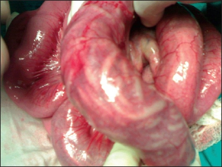
Long impacted worm bolus with transerosal visbility in a child who had incidental finding of worm inside appendix.
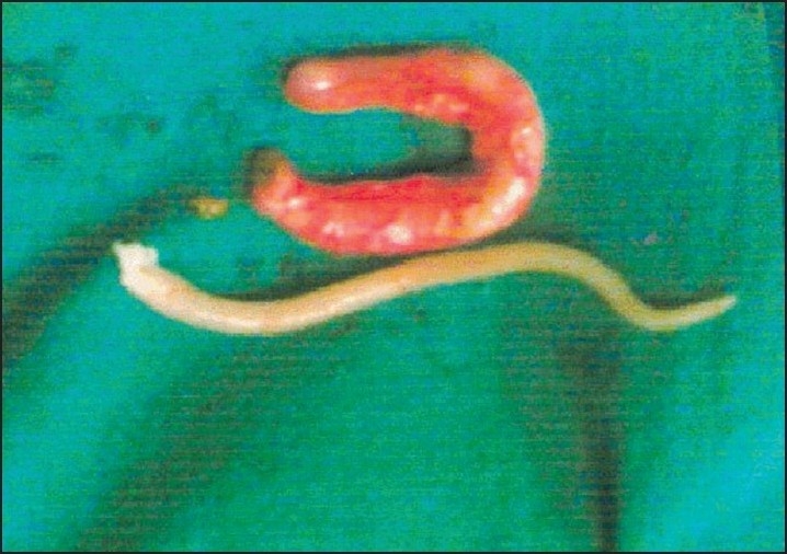
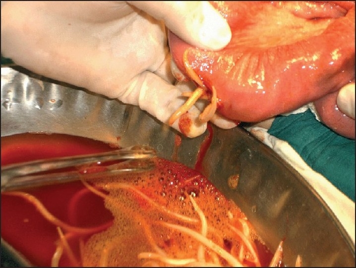
Figure 4.
Second worm being removed through above normal appendix.Worm lying with tail end at tip of appendix, held at tail end and being removed.
Figure 5.
Ascaris lumbricoides with head end at base of appendix in a grossly inflammed appendix which had features of ascaridial appendicitis on histopathology.
Figure 6.
Ascaris lumbrocoides which was lying with tail end lying at the tip and head end at base of grossly inflammed vermiform appendix.
Figure 7.
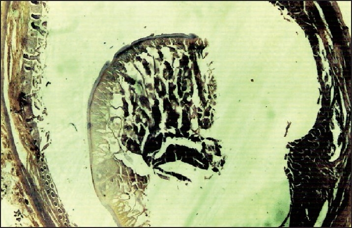
Cross section of vermiform appendix having Ascaris lumbrocoides in lumen; no features of appendicitis can be seen.
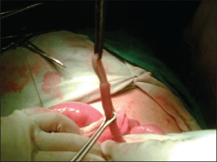
Figure 2.
Enterotomy being done for impacted worm bolus in a child.
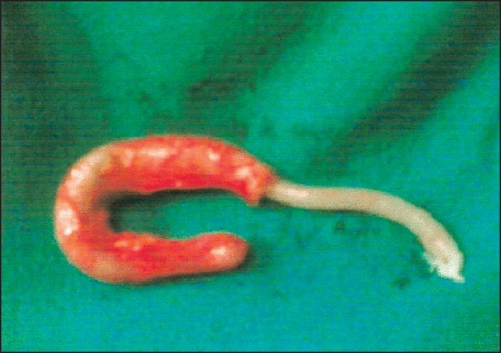
Figure 3.
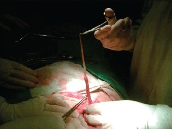
Ascaris lumbricoides being removed through tip of vermiform appendix in grossly normal appendix which has no evidence of any appendicitis at histopathology
DISCUSSION
Surgical manifestations of abdominal ascariasis are varied and are attributed to the wandering nature of Ascaris lumbricoides. The preoperative diagnosis of this condition continues to remain difficult, although the parasite can sometimes be observed inside the lumen during micropathological examination. Appendicitis due to the migration of Ascaris lumbrocoides into the appendix is still debatable because the symptoms of this migration may simulate appendicitis, but rarely cause it.3,4 The hypothesis that Ascaris lumbricoides is a major cause of appendicitis in children has been disproved.5
In Ascaris infestations associated with a normal appendix, Ascaris lodges in the appendix and comes and goes on its own accounting for the intermittent pain observed sometimes in children with high worm load. During the kneading of the worms, this high intestinal worm load coupled with a competent illeaocecal valve can sometimes provide a high load of worms in the cecum. This leads to the entry of the worms into the lumen of the appendix to escape the kneading. A competent ileocecal valve prevents the worms from escaping through the retrograde route. An incompetent ileocecal valve with proximal worm bolus obstruction may force the worm to travel again towards the cecum. This further contributes to the worm load in the cecum and in an attempt to seek natural orifices, the worms may enter the vermiform appendix. This type of Ascaris-associated normal appendix occurs in the wide-lumen, free-lying appendix. More than one worm can be seen in the lumen even when there are no grossly or microscopically visible features of appendicitis. An inflamed appendix can contain worms inside its lumen although it is debatable whether the worms caused the inflammation or whether they migrated to an already inflamed appendix. However, the presence of live worms and the associated pathology of the appendix do not favor the hypothesis that the worms cause appendicitis. Also, the presence of Ascaris inside the inflamed appendix favors the hypothesis that Ascaris has an affinity for pathological tissue. The wandering nature of Ascaris lumbricodes makes these worms seek openings just as they do in the perforated appendix wherein they reach the perforation site and lodge freely in the peritoneal cavity.
One of the characteristic findings of this study was that the worms were seen in the appendix with their heads at the base and their tail ends at the tip end of the appendix, which might lead to the frequent escape of worms from the appendix. Ascaris can be removed through the distal tip of the appendix when more than one worm is seen inside the appendix. It is to be stressed that complete removal of worms from the appendix is to be done when only a portion of the worm is lying inside the appendix and part of it is inside the cecum to avoid necrosis of the portion inside the appendicular stump, which may lead to fecal fistula. Our observations support the direct evidence of the presence of Ascaris in the vermiform appendix in contrast to reports of indirect evidence of migration of the worms into the appendix due to the presence of Ascaris lumbricoides eggs lodged in the appendix without any features of appendicitis.6
In conclusion, Ascaris lumbrocoides is rarely found in the appendix and its presence there is rarely associated with appendicitis. Worms in the appendix can be extracted through the distal tip of the appendix. Ascaris position themselves with their heads directed towards the base and their tail ends at the tip of the vermiform appendix.
REFERENCES
- 1.Khuroo MS. Ascariasis. Gastroenterol Clin North Am. 1996;25:553–77. doi: 10.1016/s0889-8553(05)70263-6. [DOI] [PubMed] [Google Scholar]
- 2.Misra SP, Dwivedi M, Misra V, Singh PA, Agarwal VK. Acute appendicitis caused by Ascaris lumbricoides is an uncommon variant of a common disease. J Clin Ultrasound. 1999;27:96–7. doi: 10.1002/(sici)1097-0096(199902)27:2<96::aid-jcu10>3.0.co;2-a. [DOI] [PubMed] [Google Scholar]
- 3.Aydin O. Incidental parasitic infestations in surgically removed appendices: A retrospective analysis. Diagn Pathol. 2007;2:16. doi: 10.1186/1746-1596-2-16. [DOI] [PMC free article] [PubMed] [Google Scholar]
- 4.Yildirim S, Nursal TZ, Tarim A, Kayaselcuk F, Noyan T. A rare cause of acute appendicitis: parasitic infection. Scand J Infect Dis. 2005;37:757–9. doi: 10.1080/00365540510012161. [DOI] [PubMed] [Google Scholar]
- 5.Dorfman S, Cardozo J, Dorfman D, Del Villar A. The role of parasites in acute appendicitis of pediatric patients. Invest Clin. 2003;44:337–40. [PubMed] [Google Scholar]
- 6.Zoguéreh DD, Lemaétre X, Ikoli JF, Delmont J, Chamlian A, Mandaba JL, et al. Acute appendicitis at the National university Hospital in Bangui, Central African Republic: epidemiologic, clinical, paraclinical and therapeutic aspects. Sante. 2001;11:117–25. [PubMed] [Google Scholar]