Abstract
Aggressive periodontitis is characterized by a rapid and severe periodontal destruction in young systemically healthy subjects. A greater prevalence is reported in Africans and African descendent groups than in Caucasians and Hispanics. We first fine mapped the interval 1q24.2 to 1q31.3 suggested as containing an aggressive periodontitis locus. Three hundred and eighty-nine subjects from 55 pedigrees were studied. Saliva samples were collected from all subjects, and DNA was extracted. Twenty-one single nucleotide polymorphisms were selected and analyzed by standard polymerase chain reaction using TaqMan chemistry. Non-parametric linkage and transmission distortion analyses were performed. Although linkage results were negative, statistically significant association between two markers, rs1935881 and rs1342913, in the FAM5C gene and aggressive periodontitis (p = 0.03) was found. Haplotype analysis showed an association between aggressive periodontitis and the haplotype A-G (rs1935881-rs1342913; p = 0.009). Sequence analysis of FAM5C coding regions did not disclose any mutations, but two variants in conserved intronic regions of FAM5C, rs57694932 and rs10494634, were found. However, these two variants are not associated with aggressive periodontitis. Secondly, we investigated the pattern of FAM5C expression in aggressive periodontitis lesions and its possible correlations with inflammatory/immunological factors and pathogens commonly associated with periodontal diseases. FAM5C mRNA expression was significantly higher in diseased versus healthy sites, and was found to be correlated to the IL-1β, IL-17A, IL-4 and RANKL mRNA levels. No correlations were found between FAM5C levels and the presence and load of red complex periodontopathogens or Aggregatibacter actinomycetemcomitans. This study provides evidence that FAM5C contributes to aggressive periodontitis.
Introduction
Aggressive periodontitis is characterized by a rapid and severe periodontal destruction in young systemically healthy subjects, and can be subdivided into localized and generalized forms according to the extension of the periodontal destruction [1]. Epidemiological surveys have shown that the prevalence of aggressive periodontitis varies among ethnic groups, regions and countries, and may range from 0.1% to 15% [2], [3]. A greater prevalence is reported in Africans and African descendent groups than in Caucasians and Hispanics [4], [5].
There are many reports in the literature describing families with multiple aggressive periodontitis affected individuals, suggesting familial aggregation [6]–[8]. Several research groups have used segregation analysis to determine the likely mode of inheritance for this trait. The patterns of disease in these families have led investigators to postulate both dominant and recessive modes of Mendelian inheritance for aggressive periodontitis [9]–[11]. Segregation analysis that included the families in the present study suggested an excessive disease transmission from heterozygous parents. This model provides support for the hypothesis that a few loci, each one with relatively small effects, contribute to aggressive periodontitis, with or without interaction with environmental factors [12].
Candidate gene approaches have been used to study aggressive periodontitis, but the results so far are very diverse and conflicting [13], [14]. A case-control genome wide association study suggested a role for GLT6D1 in aggressive periodontitis in Germans [15]. One linkage study in African American families [16] showed that aggressive periodontitis is linked to the marker D1S492, located on chromosome 1q. A susceptibility locus for aggressive periodontitis was determined between the markers D1S196 and D1S533. This region of chromosome 1 (from base pair 165,770,752 to base pair 192,424,848) includes the cytogenetic regions from 1q24.2 to 1q31.3. In this study, we first investigated this chromosomal region for genetic variants that contribute to aggressive periodontitis in a clinically well-characterized group of families, several of African descent (Table 1), segregating this condition. The hypothesis of this study is that genetic variation located between 1q24.2 to 1q31.3 contributes to aggressive periodontitis. Since the present genetic studies provide evidence that FAM5C gene contributes to aggressive periodontitis, we also investigated the pattern of FAM5C expression in periodontal lesions and its possible correlations with inflammatory/immunological factors and pathogens commonly associated with periodontal diseases in a second population presenting aggressive periodontitis, compared to periodontally-healthy controls.
Table 1. Ethnic background of the families and number of individuals by affection status and gender in 55 families with at least a proband affected with aggressive periodontitis and average age of the probands.
| Family Characteristic | N (%) |
| African descent families | 38 (69%) |
| Affected individuals | 132 (34%) |
| Probands | 55 |
| Male | 18 |
| Female | 37 |
| Average age of the probands (minimum-maximum) | 31.1 years (16–40 years) |
| Relatives | 77 |
| Male | 26 |
| Female | 51 |
| Unaffected individuals | 193 (49.6%) |
| Male | 90 |
| Female | 103 |
| Unknown | 64 (16.4%) |
| Male | 36 |
| Female | 28 |
| Total | 389 (100%) |
Results
Genetic results
All markers studies (Table 2) were in Hardy-Weinberg equilibrium (data not shown). Non-parametric linkage analysis showed no linkage between genetic markers in 1q24.2-1q31.3 and aggressive periodontitis (Table 3 [48]). Association could be seen between aggressive periodontitis and markers in FAM5C, rs1935881 and rs1342913. Both the A allele (common allele) of marker rs1935881 and the G allele (rare allele) of marker rs1342913 were observed to be over-transmitted among cases (p = 0.03 for both, complete results in Table S3). The results of PLINK also suggested an association between aggressive periodontitis and the same marker alleles: most common allele A of marker rs1935881 (OR = 0.50, 95% CI 0.15–1.66, p = 0.07) and rare allele G of marker rs1342913 (OR = 3.2, 95% CI 1.17–8.73, p = 0.03). No linkage disequilibrium was apparent between these two markers (Table S4). Haplotype analysis also showed an association between the haplotype A–G (rs1935881-rs1342913; p = 0.009) and aggressive periodontitis (Table 4). Additional haplotypes including these two markers also had suggestive association results (Table S5).
Table 2. Genetic variants studied.
| SNP | Position | Gene | Region | Change |
| rs366839 | 187,825,289 | ---- | ---- | AG |
| rs463228 | 187,828,998 | ---- | ---- | AG |
| rs2208921 | 187,947,000 | ---- | ---- | AG |
| rs12132519 | 187,997,671 | ---- | ---- | AG |
| rs1935885 | 188,314,448 | ---- | ---- | AG |
| rs1935881* | 188,333,009 | FAM5C | 3′ | GA |
| rs35296429* | 188,333,533 | FAM5C | 3′ | -/A |
| rs1053081* | 188,333,608 | FAM5C | 3′ | AG |
| rs35481069* | 188,334,216 | FAM5C | exon 8 | AC (K619T) |
| rs34739035* | 188,334,578 | FAM5C | exon 8 | AC (K498N) |
| rs34098782* | 188,334,831 | FAM5C | exon 8 | GA (G414D) |
| rs10800889 | 188,341,501 | FAM5C | intron | AG |
| rs1342913 | 188,387,648 | FAM5C | intron | AG |
| rs4633293 | 188,461,532 | FAM5C | intron | AG |
| rs12140456 | 188,479,488 | FAM5C | intron | CG |
| rs57694932# | 188,705,935 | FAM5C | intron | AG |
| rs10494634# | 188,706,091 | FAM5C | intron | AT |
| rs61818811* | 188,713,220 | FAM5C | 5′ | AC |
| rs1377924 | 189,425,718 | ---- | ---- | CG |
| rs2061018 | 189,429,170 | ---- | ---- | AT |
| rs7526348 | 190,390,334 | ---- | ---- | AG |
| rs1175111 | 190,472,655 | ---- | ---- | AG |
| rs1175152 | 190,479,859 | ---- | ---- | AG |
FAM5C = family with similarity 5, member C;
* Variants included after the first association results;
# Variants found during sequencing of highly conserved intronic regions.
Table 3. Results of non-parametric linkage analysis.
| Marker | Position (cM) | Z | p value | Delta | Logarithm of Odds § | p value |
| * | Minimum | −4.96 | 1 | −0.188 | −1.64 | 1 |
| Maximum | 9.6 | 0 | 0.707 | 7.71 | 0 | |
| rs366839 | 42.129 | −0.17 | 0.6 | −0.061 | −0.01 | 0.6 |
| rs463228 | 42.130 | −0.17 | 0.6 | −0.061 | −0.01 | 0.6 |
| rs2208921 | 42.168 | −0.09 | 0.5 | −0.031 | 0 | 0.5 |
| rs12132519 | 42.184 | −0.06 | 0.5 | −0.021 | 0 | 0.5 |
| rs1935885 | 42.284 | −0.07 | 0.5 | −0.024 | 0 | 0.5 |
| rs10800889 | 42.293 | −0.1 | 0.5 | −0.033 | 0 | 0.5 |
| rs1342913 | 42.307 | 0.28 | 0.4 | 0.085 | 0.02 | 0.4 |
| rs4633293 | 42.331 | 0.43 | 0.3 | 0.134 | 0.06 | 0.3 |
| rs12140456 | 42.337 | 0.43 | 0.3 | 0.134 | 0.06 | 0.3 |
| rs1377924 | 42.630 | 0.28 | 0.4 | 0.099 | 0.03 | 0.4 |
| rs2061018 | 42.632 | 0.43 | 0.3 | 0.151 | 0.06 | 0.3 |
| rs7526348 | 42.906 | 0.11 | 0.5 | 0.038 | 0 | 0.4 |
| rs1175111 | 42.927 | 0.1 | 0.5 | 0.035 | 0 | 0.5 |
| rs1175152 | 42.929 | 0.1 | 0.5 | 0.034 | 0 | 0.5 |
* The first two lines indicate the maximum possible scores for this dataset. These are followed by analysis results at each location: cM position, Z score, p-value assuming normal approximation, delta [48], logarithm of odds score [48], and p-value [48].
§ Positive non-parametric logarithm of odds score indicates excess allele sharing among affected individuals. A negative non-parametric logarithm of odds score indicates less than expected allele sharing among these groups of individuals.
Table 4. Haplotype results for two, three and four-marker windows in FAM5C.
| Haplotype Analysis Results | |||||
| FAM5C | rs1935881 | rs1342913 | rs57694932 | rs10494634 | Haplotype |
| Frequency | |||||
| p value | 0.03 | 0.03 | 0.36 | 0.94 | f |
| Sliding Windows | |||||
| 2 windows | 0.009 | 0.326 | |||
| 0.03 | 0.647 | ||||
| 0.07 | 0.009 | ||||
| 3 windows | 0.02 | 0.109 | |||
| 0.05 | 0.107 | ||||
| 4 windows | 0.08 | 0.110 | |||
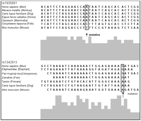
The rs1935881 wild type allele A is conserved in several mammals, while the G allele of rs1342913 is conserved back to zebrafish (Figure 1). The TRANSFAC program predicted the presence of transcription factors in the binding-sites of rs1935881 and rs1342913 (Table S6).
Figure 1. Multispecies sequence comparisons.
Multispecies sequence comparison of variant sites (indicated by arrows) in FAM5C associated with aggressive periodontitis.
Sequencing of FAM5C coding regions did not disclose any etiologic mutations. Two variants were found in highly conserved intronic regions of FAM5C gene: rs57694932 and rs10494634. TRANSFAC predicted changes in binding site affinity with these variants (Table S6). These two variants were genotyped in the entire population but they did not show an association with aggressive peridontitis (Tables 3 and S7). They are in moderate linkage disequilibrium (D' = 0.655) with the two markers associated with aggressive periodontitis (rs1935881 and rs1342913), which suggests they do not explain the association observed.
The genome wide scan of the two large families not linked to chromosome 1 yielded suggestive results for an association with markers in chromosomes 2q21.2-q37.3, 3p24.2-p24.1, 5p15.2-q33.3, 6p12.3-q12, and 18q12.3q21.2 (p = 0.0009 for all associated markers in these loci; Table 5).
Table 5. List of markers associated with aggressive periodontitis in the genome wide scan analysis.
| Chromosome 2 | Chromosome 3 | Chromosome 5 | Chromosome 6 | Chromosome 18 |
| rs13402622 | rs9833191 | rs2578619 | rs1480617 | rs2085796 |
| rs13388210 | rs4858608 | rs2548552 | rs1525354 | rs1865555 |
| rs12052971 | rs3935025 | rs1379544 | rs10948618 | rs633667 |
| rs10177619 | rs2196427 | rs7705454 | rs4715425 | rs11082925 |
| rs6755528 | rs3951794 | rs1368329 | rs9382239 | rs12457182 |
| rs6437372 | rs6801153 | rs11167472 | rs513041 | rs9320010 |
| rs708078 | rs12635000 | rs919221 | rs2478878 | rs188918 |
| rs10173407 | rs6774513 | rs2548554 | rs9381981 | rs17800754 |
| rs13029625 | rs1382305 | rs9357777 | rs10502903 | |
| rs908265 | rs919222 | rs11963528 | rs9965852 | |
| rs6431472 | rs10066281 | rs7764904 | rs8085750 | |
| rs6741220 | rs2614119 | rs2753070 | rs1623892 | |
| rs1996286 | rs17113771 | rs2677024 | rs1800640 | |
| rs4312490 | rs10070224 | rs824383 | rs2919451 | |
| rs6749707 | rs2244960 | rs9296812 | rs17785419 | |
| rs10460245 | rs13181236 | rs659446 | rs8085360 | |
| rs4571012 | rs6579746 | rs17625497 | rs1787614 | |
| rs2060127 | rs9688110 | rs2268855 | rs12606093 | |
| rs4675792 | rs7715047 | rs12662737 | rs6507852 | |
| rs7577417 | rs3097779 | rs2297985 | rs9947627 | |
| rs13016717 | rs2910263 | rs2677023 | rs17749350 | |
| rs2645778 | rs1025260 | rs9381454 | rs748317 | |
| rs4663247 | rs286958 | rs9474972 | rs4613156 | |
| rs4398270 | rs4566790 | rs4094394 | rs1787292 | |
| rs1822882 | rs10038971 | rs6929426 | rs7235757 | |
| rs10166257 | rs10796 | rs1723527 | rs9965625 | |
| rs16845023 | rs1461240 | rs1779758 | rs323118 | |
| rs13009175 | rs778825 | rs2969931 | ||
| rs13032395 | rs699113 | rs9946886 | ||
| rs184586 | rs9952398 | |||
| rs2244964 | rs1800639 | |||
| rs10064971 | rs9965170 | |||
| rs10052410 | rs8097738 | |||
| rs6883565 | rs628531 | |||
| rs890832 | rs1504504 | |||
| rs2548553 | rs17707448 | |||
| rs6874995 | rs627697 | |||
| rs6875111 | rs1787613 | |||
| rs11741184 | rs1787606 | |||
| rs4702684 | rs669350 | |||
| rs2662532 | rs12456253 | |||
| rs183495 | rs9304344 | |||
| rs2292267 | rs533064 | |||
| rs1442076 | ||||
| rs12457104 |
Gene expression results
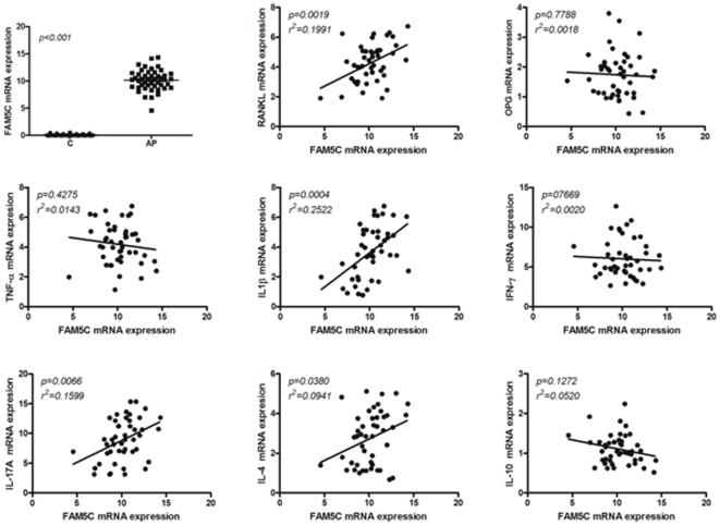
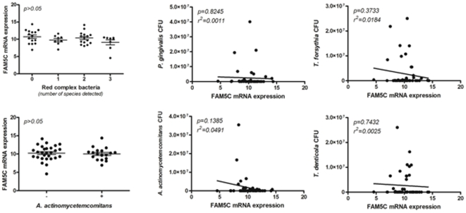
In order to support the potential association of FAM5C with aggressive periodontitis pathogenesis, we next investigated its expression in diseased versus healthy tissues. Our data demonstrate (Figure 2) that FAM5C expression was significantly higher in diseased tissues (p<0.001). In addition, FAM5C mRNA levels were positively correlated with IL-1β (p = 0.0004, r2 = 0.2522), IL-17A (p = 0.0066, r2 = 0.1599), IL-4 (p = 0.0380, r2 = 0.0941), and RANKL (p = 0.0019, r2 = 0.1991) expression, while no correlations were found with TNF-α (p = 0.4275, r2 = 0.0143), IFN-γ (p = 07669, r2 = 0.0020), IL-10 (p = 0.1272, r2 = 0.0520), and OPG (p = 0.7788, r2 = 0.0018). FAM5C mRNA levels were not associated with the presence or load of red complex periodontal pathogens or Aggregatibacter actinomycetemcomitans (p>0.05; Figure 3).
Figure 2. Summary of gene expression results.
FAM5C expression is significantly higher in diseased tissues. In addition, FAM5C mRNA levels were positively correlated with IL-1β, IL-17A, IL-4, and RANKL expression, while no correlations were found with TNF-α, IFN-γ, IL-10, and OPG. C = periodontally-healthy controls; AP = aggressive periodontitis cases.
Figure 3. Summary of bacterial DNA quantification results.
For red complex bacteria, “0” indicates sites with no bacteria, “1” indicates the detection of one species, “2” indicates the detection of two species, and “3” indicates the detection of three species. FAM5C mRNA levels were not associated with the presence or load of red complex periodontopathogens or Aggregatibacter actinomycetemcomitans.
Discussion
Aggressive periodontitis is a group of infrequent types of periodontal diseases with rapid attachment loss and bone destruction initiated at a young age. Though a variety of factors, such as microbial, environmental, behavioral and systemic disease, are suggested to influence the risk of aggressive periodontitis, an individual genetic profile is a crucial factor influencing their systemic or host response-related risk [17], [18]. This is the first report that provides evidence of an association between variation in FAM5C and aggressive periodontitis. Our work supports the initial findings of linkage [16] between chromosome 1q and aggressive periodontitis.
The family-based study design that we used is robust to problems resulting from population admixture or stratification [19]. Brazil is a trihybrid population of Native Indians, Caucasians with Portuguese ancestry and Africans [20]. The last National Research for Sample of Domiciles census in Rio de Janeiro revealed that in this city 53.4% are white, 46.1% are black, and 0.5% are Asian or Amerindian [21]. Table 1 describes additional demographic variables of the families studied.
We found evidence of association between aggressive periodontitis and FAM5C, but not linkage. Since marker allele-disease association and linkage between a disease locus and a marker locus are two different events, linkage without evidence of association and association without evidence of linkage are possible observations [22]. In linkage analysis, we take advantage of the process of forming new allelic combinations (recombination) to identify loci that are linked to the disease. One can argue that these alleles are necessary for the disease to happen. However, an association can exist if the disease-causing variants are in linkage disequilibrium with the associated marker/locus. An association can also exist if the associated genetic marker is a susceptibility locus that increases the probability of developing the disease. By themselves, these alleles are not sufficient for disease manifestation. If the linkage disequilibrium hypothesis is correct, there will be evidence for linkage. If the susceptibility locus hypothesis is correct, there may be strong evidence against linkage [22].
The FAM5C gene (NM199051-1, Gene ID:339479) is located on chromosome 1q31.1, comprises eight exons and encodes a protein of 766 amino acids named FAM5C (family with sequence similarity 5, member C; aliases BRINP3, DBCCR1L, RP11-445K1.1). FAM5C was originally identified in the mouse brain as a gene that is induced by bone morphogenic protein and retinoic acid signaling [23]. Importantly, FAM5C is localized in the mitochondria and that over-expression of this molecule leads to increased proliferation, migration, and invasion of non-tumorogenic pituitary cells [24], a phenotype relevant to the cellular changes of smooth muscle cells that are associated with the formation and vulnerability of an atherosclerotic plaque [25], [26]. FAM5C alleles are also implicated in the risk of myocardial infarction [27]. Through complex signaling cascades, mitochondria have the ability to activate multiple pathways that modulate both cell proliferation and, inversely, promote cell arrest and programmed cell death [28], all phenomena relevant in the pathogenesis of periodontal diseases.
Our exploratory genome wide scan analysis unveiled new candidate loci for aggressive periodontitis. The regions on chromosomes 2, 3, 5, 6, and 18 included many associated markers (Table 5) and spanned over large segments, and included several hundred genes but fine-mapping approaches such as the one used in this study can considerably reduce the time and cost effort to study these loci. Out of the most studied genes in aggressive periodontitis [IL1-A and IL1-B (2q14), IL-4 (5q31.1), IL-10 (1q31-q32), FcγRIIa, FcγRIIb, and FcγRIIIb (1q23), and TNFA (6p21.3)], IL-4 and TNFA map in the intervals with suggestive association results. IL-10 maps in the interval analyzed in the present studied (1q24.2 to 1q31.3) and FcγRIIa, FcγRIIb, and FcγRIIIb are just outside of it.
Interestingly, this preliminary genome wide scan analysis did not suggest linkage to 9q34.3. This locus was recently shown to be associated with aggressive periodontitis in Germans [15]. Since the families studied here are from a distinct geographic location, it is possible that the role of GLT6D1 in 9q34.3 in these families is less pronounced. Future investigations in our study population include replication of the German genome wide scan finding.
Since literature data is scarce to suggest a mechanism linking FAM5C to the pathogenesis of aggressive periodontitis, we next investigated its pattern of expression in periodontal lesion and possible correlations with inflammatory/immunological and microbial factors classically associated with the periodontitis outcome. FAM5C expression was found to be significantly higher in disease tissues, and to present a slight but significant correlation with IL-1β, IL-17A, IL-4 and RANKL expressions (Figure 2). The pro-inflammatory cytokine IL-1β has been classically associated with inflammatory cell influx and osteoclastogenesis in the periodontal environment [29], and a similar role for IL-17A was recently suggested [30]. Interestingly, both cytokines are positive regulators of RANKL expression, the master regulator of osteoclasts differentiation and activation, which is thought to account for alveolar bone loss throughout the periodontal disease process [31]. Conversely, IL-4 was described as an inhibitor of RANKL expression, but in certain conditions may increase osteoclast activity [32]. While some studies suggest a possible destructive role for IL-4 in both chronic and aggressive periodontitis [33], [34], other studies suggest that this cytokine has a protective role against tissue destruction [35], [36]. Therefore, it is possible to suppose that FAM5C may somehow modulate/interfere in cytokine network in diseased periodontal tissues, and consequently impact disease outcome. Interestingly, while destructive cytokine expression have been linked to the presence of classic periodontopathogens [33], FAM5C mRNA levels were not associated with the presence or load of red complex periodontopathogens or Aggregatibacter actinomycetemcomitans, reinforcing the putative strong genetic control of its expression in periodontal tissues.
In summary, this study provides evidence that variation in FAM5C might contribute to aggressive periodontitis, and that the markers rs1935881 and rs1342913 are candidate functional variants (based on multispecies nucleotide sequence comparisons and electronic transcription binding site predictions - Figure 1 and Table S6) or are in linkage disequilibrium with still unknown disease-predisposing alleles. Future work will investigate if expression profiles of FAM5C are associated with genetic variation in the gene.
Materials and Methods
Subjects (Genetic Studies)
Three hundred and seventy-one subjects from 54 pedigrees (75 nuclear mother-father-affected child) were recruited at the Periodontology Department at the Rio de Janeiro State University (Rio de Janeiro, RJ, Brazil), and UNIGRANRIO (Duque de Caxias, RJ, Brazil) (Figure S1). One additional family was recruited at Guarulhos University (Guarulhos, SP, Brazil) and included father, mother and sixteen offspring (Figure S2). All subjects were of Brazilian descent. The protocol for the study was reviewed and approved by the Ethics Committee of the Rio de Janeiro State University, Guarulhos University, and University of Pittsburgh, and written informed consent was obtained from all individuals prior any research activity. Aggressive periodontitis were diagnosed according to the 1999 international classification of periodontal diseases [1] and positive individuals were assigned as affected. If individuals were edentulous and reported having lost all their teeth at young age (before 35 years), for no obvious reasons such as trauma or extensive cavities, this was recognized as a potential indicator that they started as an aggressive periodontitis case and we also designated them as affected. In addition, the following information was collected by the same examiner from all probands and family members: affection status, gender, age, family relationship and ethnicity, cigarette smoking habits, current medications taken and general health status. In addition, clinical data (pocket probing depth and clinical attachment level) and radiological examinations were collected from all participants. Individuals with co-existing morbidities (e.g. diabetes) or smokers were not defined as affected to minimize the risk of inadvertently including chronic periodontitis in the analysis.
Isolation of genomic DNA
Saliva samples were collected from all of the 389 individuals with Oragene™ DNA Self-Collection Kit (DNA Genotek Inc., Kanata, ON, Canada). The DNA was extracted using the protocol for manual purification of DNA from 0.5 mL of Oragene™/saliva. The DNA integrity was checked and quantified using the absolute quantification in real-time PCR as suggested by Applied Biosystems (Foster City, CA, USA).
Selection of single nucleotide polymorphisms (SNPs)
The region between markers D1S196 and D1S533 on chromosome 1(1q24.2-1q31.3), covering about 26 million base pairs, was studied using the data from the International HapMap Project [37] and the University of California Santa Cruz Genome Bioinformatics, and viewed through the software Haploview [38]. Based on pairwise linkage disequilibrium, haplotype block structures, and structure of genes, we identified the 14 most informative single nucleotide polymorphisms in the region (Table 2).
Genotyping
Polymerase chain reactions [39] with TaqMan chemistry (Applied Biosystems, Foster City, CA, USA) [40] held in total 3 µL/reaction were used for genotyping all selected markers in a PTC-225 tetrad thermocycler (Peltier Thermal Cycler, Bio-Rad Life Sciences, Corston, UK).
Subjects (Gene expression studies)
One hundred and three subjects (57 healthy controls and 46 presenting aggressive periodontitis) were recruited at the Department of Periodontics, University of Ribeirão Preto Dental School (UNAERP). All subjects were of Brazilian descent. The protocol for the study was reviewed and approved by the Ethics Committee of the UNAERP and written informed consent was obtained from all individuals prior any research activity. All subjects were diagnosed as described above for genetic analysis.
Gene expression analysis
One biopsy of gingival tissue of each periodontally-healthy subjects (N = 57) were taken from sites that showed no bleeding on probing, probing depth smaller than three millimeters, and clinical attachment loss smaller than one millimeter during surgical procedures due to esthetics, orthodontic or prosthetic reasons. Samples included junctional epithelium, gingival crevicular epithelium and connective gingival tissue. One biopsy of gingival tissue from each aggressive periodontitis patients (N = 46) were taken from the gingival margin to the bottom of the gingival pocket of affected sites, and included junctional epithelium, periodontal pocket epithelium, and connective gingival or granulation tissue. These samples were collected during surgical therapy of the sites that exhibited persistent bleeding on probing and increased probing depth three to four weeks after the basic periodontal therapy (non-responsive sites), as previously described [41]. The extraction of total RNA from periodontal tissue samples was performed with Trizol reagent (Invitrogen, Carlsbad, CA, USA), and the cDNA synthesis was accomplished as previously described [41]. Real-Time-PCR mRNA experiments were performed in a MiniOpticon system (BioRad, Hercules, CA, USA), using SybrGreen MasterMix (Invitrogen, Carlsbad, CA, USA), using 2.5 ng of cDNA in each reaction and primers previously described [41]. Calculations for determining the relative levels of gene expression were made from triplicate measurements of the target gene, with normalization to β-actin in the sample, using the cycle threshold (Ct) method and the 2ΔΔct equation, as previously detailed [41].
Bacterial DNA quantification
In order to allow the detection of Porphyromonas gingivalis, Tannerella forsythia, Treponema denticola, and Aggregatibacter actinomycetemcomitans, periodontal crevice/pocket biofilm samples were collected with sterile paper point ISO #40 from the same site biopsied previously to the surgical procedure [41]. Bacterial DNAs were extracted from plaque samples using the DNA Purification System (Promega, Madison, WI, USA). RealTime-PCR mRNA or DNA analyses were performed in a MiniOpticon system (BioRad, Hercules, CA, USA), using SybrGreen MasterMix (Invitrogen, Carlsbad, CA, USA), using 5 ng of DNA in each reaction and the primers previously described [41]. The positivity to bacteria detection and the bacterial counts in each sample were determined based on the comparison with a standard curve comprised by specific bacterial DNA (109 to 10−2 bacteria) and negative controls [41]. The sensibility range of bacteria detection and quantification of our real time-PCR technique was of 101 to 108 bacteria to each of the four periodontal pathogens tested.
Statistical analysis
Calculations of linkage disequilibrium were computed with the Graphical Overview of Linkage Disequilibrium (GOLD) software [42] for both the squared correlation coefficient (r2) and Lewontin's standardized disequilibrium coefficient (D'). The program Rutgers Map Interpolator (www.compgen.rutgers.edu/map-interpolator/) was used to convert the physical position of the 14 markers from base pairs to centiMorgans. Non-parametric linkage analysis was performed with the program Merlin [43], [44]. Alleles and haplotypes were tested for association with aggressive periodontitis with the programs Family-Based Association Test (FBAT) [45], [46] and PLINK version 1.05 [47]. To generate odds ratios, the most common allele was used as reference. In the analysis, only probands and relatives with aggressive periodontitis were considered as affected individuals, while relatives who could not be definitely diagnosed with aggressive periodontitis were considered as unaffected individuals (including healthy individuals and individuals with chronic periodontitis). Data was analyzed with and without the family recruited in the Guarulhos University.
Analyses regarding gene expression were performed with t test or by ANOVA, followed by Tukey's test. Multiple logistic and linear regression analyses were performed to evaluate possible associations between the expression of FAM5C and inflammatory/immunological and microbial factors. Values of p<0.05 were considered statistically significant.
Follow up experiments after preliminary results
Increasing genotyping density
After the first genotyping results and association analysis, seven additional markers were chosen in the proximity of the rs1342913 marker. The same criteria described previously were used to select these additional markers (Table 2). Genotypes were generated and analyzed as described above.
FAM5C sequencing
The coding regions, including the exon-intron boundaries of FAM5C, were sequenced in eleven unrelated individuals carrying two copies of the haplotype A–G of rs1935881-rs1342913 (nine diagnosed with aggressive periodontitis and two unaffected relatives – Figure S1). As a positive control for good DNA quality, one sample from the Centre D'Étude du Polymorphisme Humain - Fondation Jean Dausset (obtained through Coriell Institute for Medical Research, Camden, NJ, USA) was also sequenced. This sample originated from an anonymous healthy individual. The FASTA sequences of FAM5C exons were obtained based on data from the Ensemble Genome Browser (www.ensembl.org). Primer3 (version 0.4.0) (www.primer3.sourceforge.net) was used to design primers covering each exon and exon-intron boundary. FAM5C has 8 exons (Figure S3). Primer sequences and polymerase chain reaction conditions are available as Supporting Document (Table S1).
Since no etiologic variants were identified in FAM5C coding regions, five highly conserved FAM5C intronic sequences were identified in the University of California Santa Cruz Genome Bioinformatics database (www.genome.ucsc.edu) and sequenced (Table S2). Two single nucleotide variants were identified in the conserved regions. These two variants (Table 2) were genotyped in all samples and data was analyzed as described above.
Bioinformatic analysis
The program ENDEAVOUR [49] was used to perform gene prioritization in the selected region based on genes already described in the literature as associated with the target disease. A list of 10 genes previously described [14] as showing evidence of involvement with periodontitis in humans was used. Secondly, we used the program TRANSFAC ® 7.0 Public 2005 (www.gene-regulation.com) in order to assess the likely transcription factors binding to the sites of the variants associated with aggressive periodontitis in this study. Finally, the BLAST function (Basic Local Alignment Search Tool) of NCBI (National Center for Biotechnology Information, www.ncbi.nlm.nih.gov) was used to make sequence comparisons between humans and other species in selected nucleotide sequences.
Genome Wide Scan
The family recruited in the Guarulhos University (Figure S2) was not associated with markers in 1q (data not shown) and we decided to investigate if this family, in addition to pedigree 24 (Figure S1), would yield the identification of additional contributing loci to aggressive periodontitis, since we previously showed that more than one loci may contribute to the disease [12]. Genome wide genotyping was performed with the GeneChip 500K arrays (Affymetrix, Santa Clara, CA, USA) at the Genomics and Proteomics Core Laboratories, University of Pittsburgh. In brief, two aliquots of 250 ng of DNA each are digested with NspI and StyI, respectively, an adaptor is ligated and molecules are then fragmented and labeled. At this stage each enzyme preparation is hybridized to the corresponding array. Samples were processed in 96-well plate format; each plate carried a positive and a negative control, up to the hybridization step. A total of 443,816 markers were genotyped. Data was analyzed using the PLINK software.
Supporting Information
Families recruited in Rio de Janeiro, Brazil. Black color indicates affected individuals. White color indicates unaffected individuals. Arrows indicate proband. Blue color indicates individuals who could not be examined.
(3.99 MB TIF)
Family recruited in the Guarulhos University, Brazil. Black color indicates affected individuals. White color indicates unaffected individuals. Arrow indicates proband.
(0.66 MB TIF)
FAM5C localization in chromosome 1q. Schematic representation of chromosome 1 (top). In the middle is the linkage disequilibrium plot generated for chromosomal region 1q31 including the FAM5C gene. Below is the schematic representation of the FAM5C gene: boxes represent exons, lines connecting boxes are introns. Blue boxes represent untranslated regions and red boxes represent coding regions. The horizontal arrow (bottom) indicates direction of gene.
(0.57 MB TIF)
FAM5C primer sequences and polymerase chain reaction (PCR) conditions.
(0.06 MB DOC)
FAM5C highly conserved intronic region primer sequences and polymerase chain reaction (PCR) conditions.
(0.04 MB DOC)
Association* results between aggressive periodontitis and genetic variation in 1q24.2-1q31.3. *Family-Based Association Test (FBAT).
(0.08 MB DOC)
Linkage disequilibrium between markers genotyped in the study. * r2 is above diagonal; D' is below diagonal.
(0.05 MB PDF)
Hsplotype results for four-marker windows across the region studied. * f indicates haplotype frequencies.
(0.07 MB PDF)
Predicted transcription binding sites for studied markers.
(0.06 MB DOC)
Acknowledgments
The authors are indebted to the subjects that participated in this study. We thank Ariadne Letra, Wendy Carricato, and Melissa Carp for all their contributions during this work. Flavia M. Carvalho conducted this work while at the Department of Periodontology, School of Dentistry, Rio de Janeiro State University, Rio de Janeiro, RJ, Brazil. This paper is partly based on a thesis submitted to the graduate faculty, State University of Rio de Janeiro, in partial fulfillment of the requirements for the PhD degree (for FMC).
Footnotes
Competing Interests: The authors have declared that no competing interests exist.
Funding: This work was supported by the Coordenacao de Aperfeiacoamento de Pessoal de Nivel Superior [CAPES/BEX 094308-8 to F.M.C.; http://www.capes.gov.br/]; and the National Institutes of Health [K99-DE018413 to R.M.; http://www.nih.gov/]. The funders had no role in study design, data collection and analysis, decision to publish, or preparation of the manuscript.
References
- 1.Armitage GC. Development of a classification system for periodontal diseases and conditions. Ann Periodontol. 1999;4:1–6. doi: 10.1902/annals.1999.4.1.1. [DOI] [PubMed] [Google Scholar]
- 2.Albandar JM, Brown LJ, Genco RJ, Loe H. Clinical classification of periodontitis in adolescents and young adults. J Clin Periodontol. 1997;68:545–555. doi: 10.1902/jop.1997.68.6.545. [DOI] [PubMed] [Google Scholar]
- 3.Tinoco EM, Beldi MI, Loureiro CA, Lana M, Campedelli F, et al. Localized juvenile periodontitis and Actinobacillus actinomycetemcomitans in a Brazilian population. Eur J Oral Sci. 1997;105:9–14. doi: 10.1111/j.1600-0722.1997.tb00174.x. [DOI] [PubMed] [Google Scholar]
- 4.Löe H, Brown LJ. Early onset periodontitis in the United States of America. J Periodontol. 1991;62:608–616. doi: 10.1902/jop.1991.62.10.608. [DOI] [PubMed] [Google Scholar]
- 5.Albandar JM, Tinoco EM. Global epidemiology of periodontal diseases in children and young persons. Periodontol 2000. 2002;29:153–176. doi: 10.1034/j.1600-0757.2002.290108.x. [DOI] [PubMed] [Google Scholar]
- 6.van der Velden U, Abbas F, Armand S, de Graaff J, Timmerman MF, et al. The effect of sibling relationship on the periodontal condition. J Clin Periodontol. 1993;20:683–690. doi: 10.1111/j.1600-051x.1993.tb00716.x. [DOI] [PubMed] [Google Scholar]
- 7.Novak MJ, Novak KF. Early-onset periodontitis. Curr Opin Periodontol. 1996;3:45–58. [PubMed] [Google Scholar]
- 8.Tinoco EM, Sivakumar M, Preus HR. The distribution and transmission of Actinobacillus actinomycetemcomitans in families with localized juvenile periodontitis. J Clin Periodontol. 1998;25:99–105. doi: 10.1111/j.1600-051x.1998.tb02415.x. [DOI] [PubMed] [Google Scholar]
- 9.Boughman JA, Halloran SL, Roulston D, Schwartz S, Suzuki JB, et al. An autosomal-dominant form of juvenile periodontitis: its localization to chromosome 4 and linkage to dentinogenesis imperfecta and Gc. J Craniofac Genet Dev Biol. 1986;6:341–350. [PubMed] [Google Scholar]
- 10.Hart TC, Marazita ML, McCanna KM, Schenkein HA, Diehl SR. Reevaluation of the chromosome 4q candidate region for early onset periodontitis. Hum Genet. 1993;91:416–422. doi: 10.1007/BF00217764. [DOI] [PubMed] [Google Scholar]
- 11.Marazita ML, Burmeister JA, Gunsolley JC, Koertge TE, Lake K, et al. Evidence for autosomal dominant inheritance and race-specific heterogeneity in early-onset periodontitis. J Periodontol. 1994;65:623–630. doi: 10.1902/jop.1994.65.6.623. [DOI] [PubMed] [Google Scholar]
- 12.Carvalho FM, Tinoco EM, Govil M, Marazita ML, Vieira AR. Aggressive periodontitis is likely influenced by a few small effect genes. J Clin Periodontol. 2009;36:468–473. doi: 10.1111/j.1600-051X.2009.01410.x. [DOI] [PMC free article] [PubMed] [Google Scholar]
- 13.Brett PM, Zygogianni P, Griffiths GS, Tomaz M, Parkar M, et al. Functional gene polymorphisms in aggressive and chronic periodontitis. J Dent Res. 2005;84:1149–1153. doi: 10.1177/154405910508401211. [DOI] [PubMed] [Google Scholar]
- 14.Covani U, Marconcini S, Giacomelli L, Sivozhelevov V, Barone A, et al. Bioinformatic prediction of leader genes in human periodontitis. J Periodontol. 2008;79:1974–1983. doi: 10.1902/jop.2008.080062. [DOI] [PubMed] [Google Scholar]
- 15.Schaefer AS, Richter GM, Nothnagel M, Manke T, Dommisch H, et al. GLT6D1 as a susceptibility locus for periodontitis. Hum Mol Genet. 2010;19:553–562. doi: 10.1093/hmg/ddp508. [DOI] [PubMed] [Google Scholar]
- 16.Li Y, Xu L, Hasturk H, Kantarci A, DePalma SR, et al. Localized aggressive periodontitis is linked to human chromosome 1q25. Hum Genet. 2004;114:291–297. doi: 10.1007/s00439-003-1065-7. [DOI] [PubMed] [Google Scholar]
- 17.Kinane DF, Shiba H, Hart TC. The genetic basis of periodontitis. Periodontol 2000. 2005;39:91–117. doi: 10.1111/j.1600-0757.2005.00118.x. [DOI] [PubMed] [Google Scholar]
- 18.Meng H, Xu L, Li Q, Han J, Zhao Y. Determinants of host susceptibility in aggressive periodontitis. Periodontol 2000. 2007;43:133–159. doi: 10.1111/j.1600-0757.2006.00204.x. [DOI] [PubMed] [Google Scholar]
- 19.Laird NM, Horvath S, Xu X. Implementing a unified approach to family-based tests of association. Genet Epidemiol. 2000;(Suppl 1):S36–S42. doi: 10.1002/1098-2272(2000)19:1+<::AID-GEPI6>3.0.CO;2-M. [DOI] [PubMed] [Google Scholar]
- 20.Alves-Silva J, Silva MS, Guimarães PE, Ferreira AC, Bandelt HJ, et al. The ancestry of Brazilian mtDNA lineages. Am J Hum Genet. 2000;67:444–461. doi: 10.1086/303004. [DOI] [PMC free article] [PubMed] [Google Scholar]
- 21.Instituto Brasileiro de Geografia e Estatística. Síntese de indicadores sociais 2008. Rio de Janeiro, Brazil 2008 [Google Scholar]
- 22.Greenberg DA, Doneshka P. Partitioned association-linkage test: distinguishing “necessary” from “susceptibility” loci. Genet Epidemiol. 1998;13:243–252. doi: 10.1002/(SICI)1098-2272(1996)13:3<243::AID-GEPI2>3.0.CO;2-7. [DOI] [PubMed] [Google Scholar]
- 23.Kawano H, Nakatani T, Mori T, Ueno S, Fukaya M, et al. Identification and characterization of novel developmentally regulated neural-specific proteins, BRINP family. Mol Brain Res. 2004;125:60–75. doi: 10.1016/j.molbrainres.2004.04.001. [DOI] [PubMed] [Google Scholar]
- 24.Shorts-Cary L, Xu M, Ertel J, Kleinschmidt-Demasters BK, Lillehei K, et al. Bone morphogenetic protein and retinoic acid-inducible neural specific protein-3 is expressed in gonadotrope cell pituitary adenomas and induces proliferation, migration, and invasion. Endocrinology. 2007;148:967–975. doi: 10.1210/en.2006-0905. [DOI] [PubMed] [Google Scholar]
- 25.Clarke MCH, Figg N, Maguire JJ, Davenport AP, Goddard M, et al. Apoptosis of vascular smooth muscle cells induces features of plaque vulnerability in atherosclerosis. Nature Medicine. 2006;12:1075–1080. doi: 10.1038/nm1459. [DOI] [PubMed] [Google Scholar]
- 26.Watkins H, Farrall M. Genetic susceptibility to coronary artery disease: from promise to progress. Nat Rev Genet. 2006;7:163–173. doi: 10.1038/nrg1805. [DOI] [PubMed] [Google Scholar]
- 27.Connelly JJ, Shah SH, Doss JF, Gadson S, Nelson S, et al. Genetic and functional association of FAM5C with myocardial infarction. BMC Med Genet. 2008;9:33–47. doi: 10.1186/1471-2350-9-33. [DOI] [PMC free article] [PubMed] [Google Scholar]
- 28.McBride HM, Neuspiel M, Wasiak S. More than just a powerhouse. Curr Biol. 2006;16:R551–R560. doi: 10.1016/j.cub.2006.06.054. [DOI] [PubMed] [Google Scholar]
- 29.Graves D. Cytokines that promote periodontal tissue destruction. J Periodontol. 2008;79:1585–1591. doi: 10.1902/jop.2008.080183. [DOI] [PubMed] [Google Scholar]
- 30.Cardoso CR, Garlet GP, Crippa GE, Rosa AL, Júnior WM, et al. Evidence of the presence of T helper type 17 cells in chronic lesions of human periodontal disease. Oral Microbiol Immunol. 2009;24:1–6. doi: 10.1111/j.1399-302X.2008.00463.x. [DOI] [PubMed] [Google Scholar]
- 31.Jin Q, Cirelli JA, Park CH, Sugai JV, Taba M, Jr, et al. RANKL inhibition through osteoprotegerin blocks bone loss in experimental periodontitis. J Periodontol. 2007;78:1300–1308. doi: 10.1902/jop.2007.070073. [DOI] [PMC free article] [PubMed] [Google Scholar]
- 32.Yu M, Moreno JL, Stains JP, Keegan AD. Complex regulation of tartrate-resistant acid phosphatase (TRAP) expression by interleukin 4 (IL-4): IL-4 indirectly suppresses receptor activator of NF-kappaB ligand (RANKL)-mediated TRAP expression but modestly induces its expression directly. J Biol Chem. 2009;284:32968–32979. doi: 10.1074/jbc.M109.001016. [DOI] [PMC free article] [PubMed] [Google Scholar]
- 33.Gemmell E, Yamazaki K, Seymour GJ. Destructive periodontitis lesions are determined by the nature of the lymphocytic response. Crit Rev Oral Biol Med. 2002;13:17–34. doi: 10.1177/154411130201300104. [DOI] [PubMed] [Google Scholar]
- 34.Gonzales JR, Gröger S, Haley G, Bödeker RH, Meyle J. The interleukin-4 -34TT and -590TT genotype is correlated with increased expression and protein production in aggressive periodontitis. Mol Immunol. 2010;47:701–705. doi: 10.1016/j.molimm.2009.10.025. [DOI] [PubMed] [Google Scholar]
- 35.Eastcott JW, Yamashita K, Taubman MA, Harada Y, Smith DJ. Adoptive transfer of cloned T helper cells ameliorates periodontal disease in nude rats. Oral Microbiol Immunol. 1994;9:284–289. doi: 10.1111/j.1399-302x.1994.tb00072.x. [DOI] [PubMed] [Google Scholar]
- 36.Garlet GP, Martins W, Jr, Fonseca BA, Ferreira BR, Silva JS. Matrix metalloproteinases, their physiological inhibitors and osteoclast factors are differentially regulated by the cytokine profile in human periodontal disease. J Clin Periodontol. 2004;31:671–679. doi: 10.1111/j.1600-051X.2004.00545.x. [DOI] [PubMed] [Google Scholar]
- 37.The International HapMap Consortium. The international HapMap project. Nature. 2003;426:789–796. doi: 10.1038/nature02168. [DOI] [PubMed] [Google Scholar]
- 38.Barrett JC, Fry B, Maller J, Daly MJ. Haploview: analysis and visualization of LD and haplotype maps. Bioinformatics. 2005;21:263–265. doi: 10.1093/bioinformatics/bth457. [DOI] [PubMed] [Google Scholar]
- 39.Saiki RK, Scharf S, Faloona F, Mullis KB, Horn GT, et al. Enzymatic amplification of beta-globin genomic sequences and restriction site analysis for diagnosis of sickle cell anemia. Science. 1985;230:1350–1354. doi: 10.1126/science.2999980. [DOI] [PubMed] [Google Scholar]
- 40.Ranade K, Chang MS, Ting CT, Pei D, Hsiao CF, et al. High-throughput genotyping with single nucleotide polymorphisms. Genome Res. 2001;11:1262–1268. doi: 10.1101/gr.157801. [DOI] [PMC free article] [PubMed] [Google Scholar]
- 41.Repeke CE, Trombone AP, Ferreira SB, Jr, Cardoso CR, Silveira EM, et al. Strong and persistent microbial and inflammatory stimuli overcome the genetic predisposition to higher matrix metalloproteinase-1 (MMP-1) expression: a mechanistic explanation for the lack of association of MMP1-1607 single-nucleotide polymorphism genotypes with MMP-1 expression in chronic periodontitis lesions. J Clin Periodontol. 2009;36:726–738. doi: 10.1111/j.1600-051X.2009.01447.x. [DOI] [PubMed] [Google Scholar]
- 42.Abecasis GR, Cookson WO. GOLD - Graphical overview of linkage disequilibrium. Bioinformatics. 2000;16:182–183. doi: 10.1093/bioinformatics/16.2.182. [DOI] [PubMed] [Google Scholar]
- 43.Abecasis GR, Cherney SS, Cookson WO, Cardon LR. Merlin-rapid analysis of dense genetic maps using sparse gene flow trees. Nat Genet. 2002;30:97–101. doi: 10.1038/ng786. [DOI] [PubMed] [Google Scholar]
- 44.Wigginton JE, Abecasis GR. PEDSTATS: descriptive statistics, graphics and quality assessment for gene mapping data. Bioinformatics. 2005;21:3345–3447. doi: 10.1093/bioinformatics/bti529. [DOI] [PubMed] [Google Scholar]
- 45.Horvath S, Xu X, Laird NM. The family based association test method: strategies for studying general genotype-phenotype associations. Eur J Hum Genet. 2001;9:301–306. doi: 10.1038/sj.ejhg.5200625. [DOI] [PubMed] [Google Scholar]
- 46.Horvath S, Xu X, Lake SL, Silverman EK, Weiss ST, et al. Family-based tests for associating haplotypes with general phenotype data: application to asthma genetics. Genet Epidemiol. 2004;26:61–69. doi: 10.1002/gepi.10295. [DOI] [PubMed] [Google Scholar]
- 47.Purcell S, Neale B, Todd-Brown K, Thomas L, Ferreira MAR, et al. PLINK: a tool set for whole-genome association and population-based linkage analysis. Am J Hum Genet. 2007;81:559–575. doi: 10.1086/519795. [DOI] [PMC free article] [PubMed] [Google Scholar]
- 48.Kong A, Cox NJ. Allele-sharing models: LOD scores and accurate linkage tests. Am J Hum Genet. 1997;61:1179–1188. doi: 10.1086/301592. [DOI] [PMC free article] [PubMed] [Google Scholar]
- 49.Tranchevent LC, Barriot R, Yu S, van Vooren S, van Loo P, et al. ENDEAVOUR update: a web resource for gene priorization in multiple species. Nucleic Acids Res. 2008;36:377–384. doi: 10.1093/nar/gkn325. [DOI] [PMC free article] [PubMed] [Google Scholar]
Associated Data
This section collects any data citations, data availability statements, or supplementary materials included in this article.
Supplementary Materials
Families recruited in Rio de Janeiro, Brazil. Black color indicates affected individuals. White color indicates unaffected individuals. Arrows indicate proband. Blue color indicates individuals who could not be examined.
(3.99 MB TIF)
Family recruited in the Guarulhos University, Brazil. Black color indicates affected individuals. White color indicates unaffected individuals. Arrow indicates proband.
(0.66 MB TIF)
FAM5C localization in chromosome 1q. Schematic representation of chromosome 1 (top). In the middle is the linkage disequilibrium plot generated for chromosomal region 1q31 including the FAM5C gene. Below is the schematic representation of the FAM5C gene: boxes represent exons, lines connecting boxes are introns. Blue boxes represent untranslated regions and red boxes represent coding regions. The horizontal arrow (bottom) indicates direction of gene.
(0.57 MB TIF)
FAM5C primer sequences and polymerase chain reaction (PCR) conditions.
(0.06 MB DOC)
FAM5C highly conserved intronic region primer sequences and polymerase chain reaction (PCR) conditions.
(0.04 MB DOC)
Association* results between aggressive periodontitis and genetic variation in 1q24.2-1q31.3. *Family-Based Association Test (FBAT).
(0.08 MB DOC)
Linkage disequilibrium between markers genotyped in the study. * r2 is above diagonal; D' is below diagonal.
(0.05 MB PDF)
Hsplotype results for four-marker windows across the region studied. * f indicates haplotype frequencies.
(0.07 MB PDF)
Predicted transcription binding sites for studied markers.
(0.06 MB DOC)


