Abstract
WEB SITE FEATURE
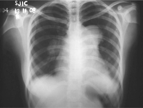
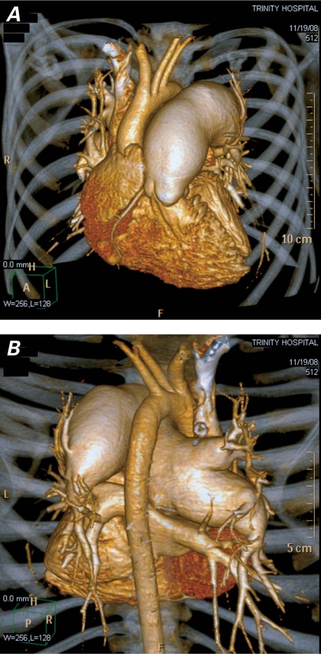
A 19-year-old woman with a 2-year history of progressive dyspnea, dull aching chest pain, and hoarseness was referred to our institution for clinical evaluation. Her general physical examination was normal. Auscultation revealed a palpable systolic impulse in the 2nd left anterior intercostal space and a grade 2/6 systolic murmur at the lower left sternal border. Her chest radiograph showed cardiomegaly, together with a hugely dilated main pulmonary artery (PA) and bilateral PA enlargement (Fig. 1) with peripheral pruning. Electrocardiography suggested right ventricular (RV) hypertrophy with strain. Later, transthoracic echocardiography showed dilated right atrium and RV with aneurysmally dilated main PA (4.6 cm) and right (3.4 cm) and left (4 cm) PA branches (Fig. 2A). The pulmonary valve was normal. She had mild tricuspid regurgitation with pulmonary hypertension (PA systolic pressure, 80 mmHg) and RV dysfunction. The left-sided chambers were normal, with normal left ventricular function. There was no echocardiographic evidence of congenital or valvular heart disease. In order to evaluate the lesions of the pulmonary vasculature, 64-slice computed tomographic angiography was performed; it confirmed the aneurysm, which affected the main PA (4.8 cm) and bilateral branches (right PA, 4.4 cm; left PA, 4.6 cm), with blunting of the lobar, segmental, and sub-segmental PA radicles (Fig. 3). The bilateral peripheral lung fields were mosaic patterned and hypoperfused, with marked attenuation of the computed tomographic image, possibly due to trapped air secondary to pressure on the bronchi by the aneurysm (Fig. 2B). There was no evidence of thrombus within the pulmonary vasculature.
Fig. 1 Chest radiograph (posteroanterior view) shows cardiomegaly, dilated main pulmonary artery, and right pulmonary artery with peripheral pruning.
Fig. 2 A) Transthoracic echocardiography (basal short-axis view) shows dilated main pulmonary artery (MPA), right pulmonary artery (RPA), and left pulmonary artery (LPA). B) Multislice contrast computed tomographic scan of the thorax shows dilated pulmonary artery and compression of the bilateral bronchi due to pressure.
Real-time motion image of Fig. 2A is available at www.texasheart.org/journal.
Fig. 3 Multislice computed tomographic scans (volume-rendered images) show dilated main pulmonary artery and its branches. A) Anterior view. B) Posterior view, showing also the peripheral pruning.
The patient was investigated for causes of the pulmonary hypertension that possibly were associated with other clinical conditions. It was decided to administer a calcium-channel blocker, an anticoagulant, and sildenafil citrate. There was dramatic improvement in her signs and symptoms upon follow-up.
Comment
Pulmonary artery aneurysm—defined as PA dilation greater than 4 cm1—is a rare condition, regardless of cause. It may develop as a result of congenital heart disease, infection, vasculitis, arteriovenous communication, trauma, or connective-tissue disease.2 The most common cause (in approximately 50% of cases) is congenital heart disease productive of pulmonary hypertension. We present a highly unusual phenomenon: giant pulmonary artery aneurysm secondary to pulmonary hypertension in a young woman.
Chest radiography can provide the initial clue indicative of PA aneurysm. Echocardiography is useful both for confirmation of diagnosis and for ascertainment of possible cause. Multislice computed tomography of the thorax provides excellent 3-dimensional images that show the relationships of mediastinal and thoracic structures to the aneurysm.
Supplementary Material
Footnotes
Address for reprints: Nagaraja Moorthy, MD, Department of Cardiology, Sri Jayadeva Institute of Cardiovascular Sciences and Research, 9th block Jayanagar, Bangalore – 560069, Karnataka, India
E-mail: drnagaraj_moorthy@yahoo.com
References
- 1.Chetty KG, McGovern J, Mahutte CK. Hilar mass in a patient with chest pain. Chest 1996;109(6):1643–4. [DOI] [PubMed]
- 2.Mann P, Seriki D, Dodds PA. Embolism of an idiopathic pulmonary artery aneurysm. Heart 2002;87(2):135. [DOI] [PMC free article] [PubMed]
Associated Data
This section collects any data citations, data availability statements, or supplementary materials included in this article.


