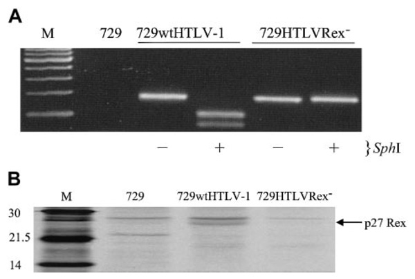
Figure 3. 729HTLVRex− stable cell line contains the Rex mutation and does not produce detectable Rex.
(A) HTLV-1 genome fragment containing the Rex start codon was amplified by PCR from genomic DNA of 729wtHTLV-1 and 729HTLVRex− cells. PCR-amplified product was incubated in the presence (+) or absence (−) of SphI, separated on 2% agarose gel, and visualized by ethidium bromide staining. (B) Rex-1 immunoprecipitation. 729, 729wtHTLV-1, and 729HTLVRex− cells were metabolically labeled with [35S] methionine-cysteine. Cell lysates were immunoprecipitated with polyclonal antibody against the HTLV-1 Rex in the presence of protein A–sepharose. The sizes (in daltons, indicated on the left) were determined by comparison to protein markers.

