Abstract
Listerial brain abscesses are rare, and are found mostly in patients with underlying hematological malignancies or solid-organ transplants. A case of a patient with Crohn’s disease and multiple brain abscesses involving the left cerebellum and right sylvian fissure is described. The Gram stain and histopathology of the cerebellar abscess revealed Gram-positive, beaded rods suggestive of Nocardia. However, on culture, Listeria monocytogenes was identified. Listeria may appear Gram-variable and has been misidentified as streptococci, enterococci and diphtheroids. The present case is the first reported case of L monocytogenes resembling Nocardia on both microbiological and histopathological assessment. Reported cases of listerial brain abscesses are sporadic, while the current case was part of a nationwide listerial outbreak linked to consumption of contaminated deli meats. Broad antimicrobial therapy (including antilisterial coverage) in immunosuppressed patients presenting with brain abscess is crucial, until cultures confirm the identification of the organism.
Keywords: Brain abscess, Cerebellum, Listeria, Listeria monocytogenes, Nocardia
Abstract
Les abcès cérébraux à Listeria sont rares, et on les observe surtout chez des patients ayant des tumeurs hématologiques malignes ou une greffe d’organe. Est décrit le cas d’un patient ayant la maladie de Crohn, de multiples abcès cérébraux dans le cervelet gauche et une scissure latérale droite. La coloration de Gram et l’histopathologie de l’abcès cérébelleux ont révélé des bacilles en chapelet Gram positif évocateurs d’une nocardiose. Cependant, à la culture, on a dépisté une Listeria monocytogenes. La Listeria peut sembler Gram variable et être prise à tort pour des streptocoques, des entérocoques ou des diphthéroïdes. Le présent cas est le premier cas déclaré de L monocytogenes évocateur d’une nocardiose à la fois à l’évaluation microbiologique et histopathologique. Les cas déclarés d’abcès cérébraux à Listeria sont sporadiques, tandis que le cas à l’étude faisait partie d’une flambée nationale de Listeria liée à la consommation de charcuterie contaminée. Il est essentiel d’amorcer une antibiothérapie à large spectre (incluant les antilistérioses) chez les patients immunosupprimés ayant un abcès cérébral, jusqu’à ce que les cultures confirment de quel organisme il s’agit.
Listeria monocytogenes is a saprophytic organism present in soil and on plants (1). This Gram-positive bacterium is psychrotropic, which makes it an ideal food-borne pathogen because it readily grows in refrigerated foods. Human exposure occurs as a result of consumption of contaminated dairy, vegetable, fish, poultry or meat products (2). Most human infections occur sporadically; however, numerous outbreaks have also occurred as a result of consumption of contaminated coleslaw, milk and soft cheeses (3–5). There have only been a few nosocomial outbreaks reported: in Costa Rica, infants were bathed with mineral oil contaminated with L monocytogenes (6), and in the United States, a large multistate outbreak of listeriosis due to contaminated deli meats occurred, in which 18% of outbreak patients may have been served contaminated meat in health care institutions (7).
L monocytogenes is an intracellular pathogen with the ability to infect adjacent cells with little exposure to the extracellular environment. In this way, it can escape humoral immunity, leaving cell-mediated immunity as the main defense against infection. As a result, L monocytogenes is more commonly implicated as a serious infectious agent in patient populations with impaired cell-mediated immunity such as pregnant women, organ transplant recipients and the elderly. Mortality rates for invasive listerial infection can exceed 30% in high-risk individuals (8). Because it can be confused with corynebacteria or other organisms that are commonly considered to be contaminants, Listeria can be overlooked in the microbiology laboratory. Therefore, a high level of suspicion and correct identification is crucial.
CASE PRESENTATION
A 51-year-old woman with long-standing Crohn’s disease with a remote history of colectomy with ileostomy was admitted to a peripheral hospital on June 8, 2008, with symptoms of nausea, diarrhea, pain and bleeding around her stoma site. She received oral prednisone 20 mg daily as treatment for possible Crohn’s exacerbation, while being maintained on her regular immunosuppressive regimen of azathioprine. In addition to oral prednisone, she was also receiving metronidazole and steroid enemas per stoma. She had not received any anti-tumour necrosis factor agents. Her symptoms improved somewhat, but she continued to experience nausea and pain around the stoma. In the third week of hospitalization, the patient experienced an episode of drowsiness and delirium, which was attributed to opiate medications and resolved with dose reduction. Around this time, she developed recurrent headaches and memory difficulties. During the sixth week of hospitalization, she developed blurry vision, weakness of her left arm and ataxia, leading to a fall. She was immediately transferred to a referral centre on July 18, 2008, for further management of her bowel disease and investigation of her new neurological symptoms.
On admission to the tertiary care hospital, the patient’s blood pressure was 117/77 mmHg and her heart rate was 68 beats/min. She had a single episode of low-grade fever of 38.2°C. She was alert and oriented with no obvious dysarthria. There was no nuchal rigidity. She had a full range of extraocular movements, with no nystagmus or diplopia. Her visual fields were intact, and her optic fundi appeared normal. The patient’s tone and sensory functions were also intact; muscle strength of the upper and lower extremities was within normal limits. Specifically, despite a subjective feeling of loss of strength, there was no left arm weakness detected on examination. Her deep tendon reflexes were 2+ and symmetrical. Plantar responses were downgoing. However, she had marked left-sided dysdiadochokinesia and dysmetria. She had an unsteady gait and complained of nausea. The rest of her physical examination was unremarkable.
Her white blood cell count was 5.1×10⁹/L (neutrophils 3.8×10⁹/L and monocytes 0.2×10⁹/L), platelet count and hemoglobin level were 227×109/L and 124 g/L, respectively. Her liver enzymes were normal, with the exception of lactate dehydrogenase at 350 U/L (reference range 90 U/L to 210 U/L). Her erythrocyte sedimentation rate was 8 mm/h and albumin level was 37 g/L (reference range 34 g/L to 50 g/L). The patient’s blood cultures were negative. A contrast computed tomography scan of the head revealed a 2.4 cm ring-enhancing lesion in the medial aspect of the left cerebellar hemisphere with adjacent edema. There was also a smaller lesion in the apex of the right sylvian fissure (Figure 1). Magnetic resonance imaging later confirmed these findings, in addition to the presence of tiny satellite lesions with adjacent edema, mostly seen in the right sylvian fissure region (Figure 2). Lumbar puncture was deferred because of concerns about the patient’s increased intracranial pressure.
Figure 1).
Computed tomography scans of the head (with contrast) demonstrating a 2.4 cm ring-enhancing mass within the medial aspect of the left cerebellar hemisphere, with adjacent edema and moderate mass effect of the right cerebellar hemisphere (A), and showing a small lesion in the right sylvian fissure (B)
Figure 2).
Magnetic resonance images of the head (axial fluid-attenuated inversion recovery) demonstrating left cerebellar abscess with surrounding edema (A), and right sylvian fissure abscess with surrounding foci of satellite edema representing adjacent small abscesses (B)
The patient had a unique exposure history. She and her husband hunt and eat game animals, mostly ungulates, but had also recently skinned and eaten a cougar killed on their property. They always serve their meat cooked. They breed Yorkshire terriers and have an adult cat at home. Our patient recalled eating soft cheeses three months before presenting to hospital, and also remembers being served deli meats while in hospital.
The differential diagnosis included pyogenic brain abscesses, Toxoplasma gondii and lymphoma. Her immunocompromised state and various exposures led to the consideration of other possible etiological agents including the following: Nocardia species, Mycobacterium tuberculosis, Cryptococcus species, Aspergillus species and L monocytogenes. She was started on broad antibacterial coverage including meropenem, vancomycin and pyrimethamine-sulfadiazine pending biopsy diagnosis.
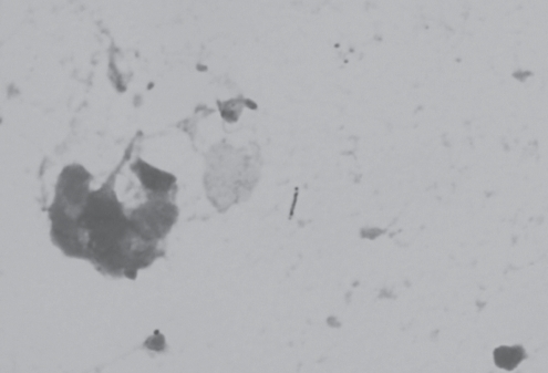
The patient underwent neurosurgical biopsy of the cerebellar mass. Gram stain revealed two or more polymorphs and nonbranching, Gram-positive rods, which appeared beaded (Figure 3). The presence of Gram-positive bacilli in the form of beaded rods led to the possibility of Nocardia species or Mycobacterium species. Similarly, small, delicate, Gram-positive, filamentous, rod-like structures consistent with Nocardia were seen on histopathology. Acid-fast and modified acid-fast stains were negative. After two days of growth in thioglycollate broth, turbid streaks were seen. When plated, the colonies appeared creamy, white and moist on blood agar. Wet mount revealed tumbling motility at room temperature. Biochemical tests included positive catalase, bile esculin and hippurate hydrolysis. The organism exhibited salt tolerance; however, the CAMP test with a beta-lysin-producing Staphylococcus aureus strain was negative. L monocytogenes was identified by MicroScan panel (Siemens United Kingdom) and confirmed by the Canadian National Microbiology Laboratory (Winnipeg, Manitoba). On diagnosis of listerial brain abscess, the patient’s antibiotic regimen was changed to ampicillin and gentamicin.
Figure 3).
Listeria monocytogenes from the cerebellar biopsy specimen. Gram stain showing a beaded Gram-positive rod located in the centre of the figure (original magnification ×1000)
During the remainder of her hospitalization, the patient’s word finding ability improved, but she had persistent left-sided dysmetria, ataxia and her short-term memory remained impaired. She completed a three-month course of intravenous ampicillin at a dose of 2 mg every 4 h in hospital. On repeat magnetic resonance imaging of the head on October 20, 2008, there was no evidence of fluid to suggest ongoing abscesses and there was an improvement in surrounding vasogenic edema. She was subsequently discharged to a rehabilitation facility to improve her balance and mobility.
DISCUSSION
Listeria brain abscesses reported in the literature are mostly sporadic, with no definite source identified (9). Potential exposures for our patient include consumption of game meat, ingestion of soft cheeses as an outpatient, and luncheon meats while in hospital. Listerial meningoencephalitis in a cougar has been reported in the veterinary literature (10), but does not appear to be a common phenomenon. Given a mean incubation period of 31 days (range 11 to 70 days) for invasive listeriosis (5), we rejected soft cheeses as a potential source for the patient’s infection. Interestingly, there were an increased number of listeriosis cases reported to the Public Health Agency of Canada in July 2008 and, in August 2008, a nationwide listeriosis outbreak was declared (11). The outbreak was linked to consumption of contaminated deli meats from a single manufacturer. Because our patient was served deli meat sandwiches in hospital from the same manufacturer, this was thought to be the most likely source of her infection.
According to standard laboratory surveillance procedures, cases of listeriosis nationwide are referred to the National Microbiology Laboratory for molecular subtyping by pulsed-field gel electrophoresis (PFGE). This internationally standardized whole-genome restriction fragment length polymorphism technique is a part of the PulseNet Canada program (12,13), which applies PFGE in real time to cases of listeriosis to link geographically dispersed cases and outbreaks to potential sources. The present case had the same rare PFGE pattern as the national outbreak strain and L monocytogenes recovered from the implicated deli meat products (Figure 4). Therefore, our case met the case definition criteria for being part of the nationwide listeriosis outbreak.
Figure 4).

AscI pulsed-field gel electrophoresis (PFGE) profile of Listeria monocytogenes from case patient compared with national listeriosis outbreak strain. A AscI PFGE pattern of case. B AscI PFGE pattern of outbreak strain. The case and the outbreak strain also had indistinguishable PFGE patterns using a second enzyme ApaI (data not shown)
L monocytogenes is a facultative anaerobic bacillus that grows well on blood agar, producing an incomplete zone of beta-hemolysis. Because listeriae may appear Gram-variable, misidentification as diphtheroids, streptococci or enterococci have been reported (14). In the present case, the organism in both the microbiology laboratory and on histopathology appeared as a Gram-positive, beaded rod and was initially identified as being most consistent with Nocardia species, although Mycobacterium species was also considered. This is particularly troublesome because it can be difficult to distinguish listerial and tuberculous meningitis clinically because both could have a subacute course, and propensity to cause basal meningitis with, at times, predominance of monocytes in the cerebrospinal fluid (15). While Listeria can have a variable appearance on Gram stain, we are unaware of other cases in which Listeria has shown this particular microscopic appearance. We hypothesize that antibiotic therapy preceding collection of the specimen may have been responsible for the unusual morphology of this organism.
Listeria is an uncommon cause of brain abscess; however, brain abscesses account for approximately 10% of listerial central nervous system (CNS) infections – commonly associated with bacteremia (16). In a 2001 review of 39 cases of listerial brain abscesses, Eckburg at al (17) found that the majority of patients had underlying hematological malignancies or were solid organ transplant recipients. In a follow-up review by Cone et al (9), 10 of the 40 cases of supratentorial listerial brain abscesses had multiple areas of involvement. Patients with multiple brain abscesses were more likely to have suppressed immunity than those with a solitary abscess. In addition, corticosteroid use was found to be a predisposing factor for having two or more brain abscesses, but Crohn’s disease was not identified as a risk factor. We found only one previous case report of a patient with both acute myeloid leukemia and Crohn’s disease who developed a listerial brain abscess (17), leading us to believe that it is primarily the immunosuppressive regimen our patient was treated with that led to her infection. While azathioprine alone can depress T lymphocyte function, it is likely that the use of prednisone is the more important risk factor (14).
There are no large trials comparing the various treatment modalities for Listeria. Recommended treatment regimens are based on in vitro susceptibilities, animal studies and clinical experience. Ampicillin is the drug of choice for listeriosis. It is recommended that gentamicin be added for synergy, especially when treating Listeria bacteremia in patients with impaired T lymphocyte immunity and in all cases of meningitis. The organism also demonstrates susceptibility to sulpha drugs in vitro and in vivo. In a small study (18) of severe cases of L monocytogenes meningoencephalitis, the combination of trimethoprim-sulfamethoxazole with ampicillin has shown greater efficacy in decreasing morbidity and mortality than the ampicillin-aminoglycoside combination. This observation raises the possibility that the use of trimethoprim-sulfamethoxazole for Pneumocystis carinii prophylaxis in patients on chronic steroids could be an effective method for preventing listeriosis. Patients with brain abscess require treatment for at least six weeks and should be followed up by serial neuroimaging until the abscesses are resolved. Mortality for listerial CNS abscesses was reported to be 40% compared with 17% for other types of brain abscesses (9).
Due to the severity of listerial illness, its predilection for the CNS and high mortality rate, clinicians should have a high index of suspicion in susceptible patients presenting with CNS infection. Because Listeria can often be misidentified on Gram stain, it is important to have a high suspicion for it in the laboratory and to continue with antilisterial therapy until the identification of the organism can be confirmed by culture.
REFERENCES
- 1.Weis J, Seeliger HP. Incidence of Listeria monocytogenes in nature. Appl Microbiol. 1975;30:29–32. doi: 10.1128/am.30.1.29-32.1975. [DOI] [PMC free article] [PubMed] [Google Scholar]
- 2.Farber JM, Peterkin PI. Listeria monocytogenes, a food-borne pathogen. Microbiol Rev. 1991;55:476–511. doi: 10.1128/mr.55.3.476-511.1991. [DOI] [PMC free article] [PubMed] [Google Scholar]
- 3.Schlech WF, Lavigne PM, Bortolussi RA, et al. Epidemic listeriosis – evidence for transmission by food. N Engl J Med. 1983;308:203–6. doi: 10.1056/NEJM198301273080407. [DOI] [PubMed] [Google Scholar]
- 4.Fleming DW, Cochi SL, MacDonald KL, et al. Pasteurized milk as a vehicle of infection in an outbreak of listeriosis. N Engl J Med. 1985;312:404–7. doi: 10.1056/NEJM198502143120704. [DOI] [PubMed] [Google Scholar]
- 5.Linnan MJ, Mascola L, Lou XD, et al. Epidemic listeriosis associated with Mexican-style cheese. N Engl J Med. 1988;319:823–8. doi: 10.1056/NEJM198809293191303. [DOI] [PubMed] [Google Scholar]
- 6.Schuchat A, Lizano C, Broome CV, Swaminathan B, Kim C, Winn K. Outbreak of neonatal listeriosis associated with mineral oil. Pediatr Infect Dis J. 1991;10:183–9. doi: 10.1097/00006454-199103000-00003. [DOI] [PubMed] [Google Scholar]
- 7.Gottlieb SL, Newbern EC, Griffin PM, et al. Multistate outbreak of Listeriosis linked to turkey deli meat and subsequent changes in US regulatory policy. Clin Infect Dis. 2006;42:29–36. doi: 10.1086/498113. [DOI] [PubMed] [Google Scholar]
- 8.Ramaswamy V, Cresence VM, Rejitha JS, et al. Listeria – review of epidemiology and pathogenesis. J Microbiol Immunol Infect. 2007;40:4–13. [PubMed] [Google Scholar]
- 9.Cone LA, Leung MM, Byrd RG, Annunziata GM, Lam RY, Herman BK. Multiple cerebral abscesses because of Listeria monocytogenes: Three case reports and a literature review of supratentorial listerial brain abscess(es) Surg Neurol. 2003;59:320–8. doi: 10.1016/s0090-3019(03)00056-9. [DOI] [PubMed] [Google Scholar]
- 10.Langohr IM, Ramos-Vara JA, Wu CC, Froderman SF. Listeric meningoencephalomyelitis in a cougar (Felis concolor): Characterization by histopathologic, immunohistochemical, and molecular methods. Vet Pathol. 2006;43:381–3. doi: 10.1354/vp.43-3-381. [DOI] [PubMed] [Google Scholar]
- 11.Lessons Learned: The Public Health Agency of Canada’s Response to the 2008 Listeriosis Outbreak. <http://www.phac-aspc.gc.ca/fs-sa/listeria/2008-intro-lessons-lecons-eng.php> (Accessed on February 2, 2010).
- 12.Graves LM, Swaminathan B. PulseNet standardized protocol for subtyping Listeria monocytogenes by macrorestriction and pulsed-field gel electrophoresis. Int J Food Microbiol. 2001;65:55–62. doi: 10.1016/s0168-1605(00)00501-8. [DOI] [PubMed] [Google Scholar]
- 13.Barrett TJ, Gerner-Smidt P, Swaminathan B. Interpretation of pulsed-field gel electrophoresis patterns in foodborne disease investigations and surveillance. Foodborne Pathog Dis. 2006;3:20–31. doi: 10.1089/fpd.2006.3.20. [DOI] [PubMed] [Google Scholar]
- 14.Nieman RE, Lorber B. Listeriosis in adults: A changing pattern. Report of eight cases and review of the literature, 1968–1978. Rev Infect Dis. 1980;2:207–27. doi: 10.1093/clinids/2.2.207. [DOI] [PubMed] [Google Scholar]
- 15.Larsson S, Cronberg S, Winblad S. Clinical aspects on 64 cases of juvenile and adult listeriosis in Sweden. Acta Med Scand. 1978;204:503–8. doi: 10.1111/j.0954-6820.1978.tb08480.x. [DOI] [PubMed] [Google Scholar]
- 16.Lorber B. Listeriosis. Clin Infect Dis. 1997;24:1–9. doi: 10.1093/clinids/24.1.1. [DOI] [PubMed] [Google Scholar]
- 17.Eckburg PB, Montoya JG, Vosti KL. Brain abscess due to Listeria monocytogenes: Five cases and a review of the literature. Medicine (Baltimore) 2001;80:223–35. doi: 10.1097/00005792-200107000-00001. [DOI] [PubMed] [Google Scholar]
- 18.Merle-Melet M, Dossou-Gbete L, Maurer P, et al. Is amoxicillincotrimoxazole the most appropriate antibiotic regimen for Listeria meningoencephalitis? Review of 22 cases and the literature. J Infect. 1996;33:79–85. doi: 10.1016/s0163-4453(96)92929-1. [DOI] [PubMed] [Google Scholar]


