

Abstract
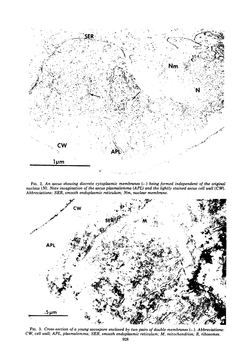
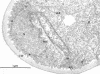
Observations with the electron microscope of the early stages of ascospore formation in Nematospora coryli Peglion reveal the following sequence of events. Partial dissociation of the nuclear membrane is followed by the appearance of unit membranes. Each unit membrane gives rise to two pairs of double membranes delimiting the ascospores from the epiplasm of the ascus. Enlarged mitochondria which have a granular matrix and limited cristae development are also regularly seen.
Full text
PDF


Images in this article

Selected References
These references are in PubMed. This may not be the complete list of references from this article.
- CONTI S. F., NAYLOR H. B. Electron microscopy of ultrathin sections of Schizosaccharomyces octosporus. III. Ascosporogenesis, ascospore structure, and germination. J Bacteriol. 1960 Mar;79:417–425. doi: 10.1128/jb.79.3.417-425.1960. [DOI] [PMC free article] [PubMed] [Google Scholar]
- HASHIMOTO T., GERHARDT P., CONTI S. F., NAYLOR H. B. Studies on the fine structure of microorganisms. V. Morphogenesis of nuclear and membrane structures during ascospore formation in yeast. J Biophys Biochem Cytol. 1960 Apr;7:305–310. doi: 10.1083/jcb.7.2.305. [DOI] [PMC free article] [PubMed] [Google Scholar]
- KELLENBERGER E., RYTER A., SECHAUD J. Electron microscope study of DNA-containing plasms. II. Vegetative and mature phage DNA as compared with normal bacterial nucleoids in different physiological states. J Biophys Biochem Cytol. 1958 Nov 25;4(6):671–678. doi: 10.1083/jcb.4.6.671. [DOI] [PMC free article] [PubMed] [Google Scholar]
- MARQUARDT H. ELEKTRONENOPTISCHE UNTERSUCHUNGEN UEBER DIE ASCOSPORENBILDUNG BEI SACCHAROMYCES CEREVISIAE UNTER CYTOLOGISCHEM UND CYTOGENETISCHEM ASPEKT. Arch Mikrobiol. 1963 Sep 16;46:308–320. [PubMed] [Google Scholar]
- Spurr A. R. A low-viscosity epoxy resin embedding medium for electron microscopy. J Ultrastruct Res. 1969 Jan;26(1):31–43. doi: 10.1016/s0022-5320(69)90033-1. [DOI] [PubMed] [Google Scholar]