



Abstract
Arterial restenosis associated with intimal hyperplasia is the major cause of long-term failure of vascular interventions. Endothelium injury and the proliferation and migration of smooth muscle cells (SMC) are key events in the development of intimal hyperplasia. The objectives of this study were to develop an ex vivo artery injury model for studying endothelial cell (EC) migration and to compare it with an in vitro co-culture arterial wall injury model in terms of the effect of flow on EC migration and its effect on SMC migration and proliferation. Our results demonstrated that shear flow improves reendothelialization in the injured area by promoting EC migration. The migration distance of ECs is much smaller in the arteries than in an in vitro cell culture model (3.57 ± 1.29 mm vs. 5.2 ± 1.4 cm, p< 0.001). SMC proliferation was significantly less in the EC intact and reendothelialization areas than in the EC denuded areas indicating that reendothelialization suppresses SMC proliferation. Our models provide a new approach to study techniques to enhance endothelium healing.
Keywords: Organ culture, cell culture, reendothelialization, cell proliferation, cell migration, porcine, artery
Introduction
Angioplasty and arterial stenting are the most frequently performed cardiovascular interventions for the treatment of occlusive atherosclerotic artery disease.24 However, the use of balloon catheters and stents is associated with injury to the arterial wall, especially to the endothelium.11, 32, 34 The endothelium is critical in preventing thrombosis, regulating mass transportation, and controlling underlying arterial smooth muscle cell (SMC) growth and function.6, 23, 35 Loss of the endothelium can lead to thrombosis and intimal hyperplasia, featured by migration of SMCs into the intima and SMC proliferation. Restenosis due to intimal hyperplasia at the stent site occurs in approximately 20% of patients2, 35 and remains a challenging problem.26, 33, 41 On the other hand, restoration of the endothelium can inhibit the migration and proliferation of SMC and thereby limit intimal hyperplasia.30, 40, 42 Therefore, accelerating the rate of reendothelialization at injured areas is an alternative strategy to limit SMC proliferation and arterial restenosis without the use of cytotoxic or cytostatic agents such as those used in drug-eluting stents.25, 29, 35
Migration of endothelial cells (EC) from the adjacent arterial lumen surface is the primary source of EC to re-populate the injured surface area.18, 28, 35 Extensive studies using animal models and cell culture models have shown that increased shear stress significantly influences EC morphology, function, and migration. 1, 19, 37 Further understanding of EC migration to the injured area is important in developing new strategies to promote EC migration. To this end, it is necessary to develop an effective model to evaluate the effect of various factors on the migration of EC into an injured area. Artery organ culture models have been proposed as alternative models that combine the advantages of excellent hemodynamic and environmental control and native wall structure for studying vascular biology and wall remodeling. 4, 8, 9, 13, 15, 38 Arterial structure and vasomotor function are well maintained under physiologic flow in the organ culture models.3, 8, 15, 21 Here we explore the use of the organ culture model to study EC migration.
The objectives of this study were to develop an ex vivo artery injury model for studying EC migration and to compare it with an in vitro co-culture arterial wall injury model in terms of the effect of flow on EC migration and its effect on SMC migration and proliferation. The comparison would allow us to validate the model and evaluate the differences in EC and SMC migration between in vitro and natural arteries.
Methods
Artery preparation
Porcine common carotid arteries were harvested from farm pigs (250-300 lb) at a local slaughterhouse. Arteries were rinsed with sterilized PBS (Dulbecco's Phosphate buffered saline, Sigma; supplemented with 1% antibiotic-antimycotic solution, Sigma) and transported to the laboratory in ice cold PBS. Segments of 3-5 cm in length without any leaks were prepared for ex vivo organ culture.16 The total preparation time between artery harvesting and placing in the ex-vivo organ culture system is 3 to 5 hours.
Creation of endothelium denudation band in arteries
In order to measure EC migration to the injured area in arteries, an 8 mm EC denudation band was created in the middle of the arterial segments using a retractable injury device (Fig. 1a). The retractable injury device, which was custom-made in our lab, consisted of metal wires that can be deployed from a thin metal tube and partially retracted back into the metal tube. The tube's outer diameter (2.5 mm) was much smaller than the inner diameter of the arteries (4.2 mm) and therefore can be inserted into the arteries without causing significant damage to the intima. The tube was connected to an adjustable stopper to control the axial position of its tip in an artery. Arterial segments were mounted onto the stainless steel cannulae in the vessel chambers in their in vivo length using sterile surgical sutures. The injury device with the metal wires retracted in the metal tube was inserted from the distal end of the arteries through the distal cannula. After positioning the tip at the middle of the arteries (half length from the distal end), the wires were deployed to denude the endothelium and created an 8 mm band at the middle of the arteries (Fig. 1b). The wires were then retracted back into the metal tube and the device was gently pulled out of the arteries.
Figure 1.


Photographs of the retractable injury device (a) and the lumen of an artery stained with 0.5% Evans blue dye (b) illustrating a band of the EC injured area in the middle of the artery created by the retractable device. Schematics of ex-vivo artery organ culture system (c) and in-vitro co-culture system (d)
Artery organ culture
After the creation of the EC denudation band, arteries were maintained using our ex vivo perfusion organ culture system (Fig. 1c) which has been described in detail previously.15, 16 The organ culture system was placed in incubators at 37°C and aerated with a mixture of air and 5% CO2. The perfusion to the arteries was in the same direction of in vivo blood flow (from proximal to distal) and was driven by a peristaltic pump. The perfusion and bath media consisted of Dulbecco's Modified Eagle's Medium (DMEM, Sigma), supplemented with sodium bicarbonate (3.7 g/L, Sigma), L-glutamine (2 mM, Sigma), calf serum (10%, Sigma), hepes buffer solution (25 mM, Gibco), antibiotic-antimycotic solution (1%, Sigma) and dextran (50 g/L; Sigma). Bromodeoxyuridine (BrdU, at a dose of 5 mg/L, Sigma) was also added to the perfusion medium at the beginning of the experiment to label the nuclei of proliferating cells. The perfusion flow and pressure were gradually increased to the designed flow rate, pressure, and pulse frequency and monitored using a computer.
All arteries were maintained under the same axial stretch ratio (λz=1.5), mean pressure (100 mmHg), and pulse amplitude (20 mmHg). One group of arteries were maintained under normal arterial flow of 160 ml/min to generate a normal arterial shear stress of ∼1.5 Pa to the endothelium (arterial shear group) while the other group of arteries were cultured under a low flow of 16 ml/min to generate a low shear stress of ∼0.2 Pa (low shear group). The mean wall shear stress was determined from the flow rates using Poiseuille flow equation 20
| (1) |
where r is the lumen radius of the vessel, μ is the viscosity of the medium (4 cP), and Q is the mean flow rate. Our measurements showed that when the pulsatile pressure oscillated between 80 and 120 mmHg, the flow rate oscillated from 156 to 165 ml/min (a 5.4% change) and the vessel diameter oscillated about 2% from their mean values.
Measurement of endothelial cell migration in arteries
After 7 days of organ culture, the arteries were harvested from the vessel chamber and cut longitudinally using a surgical blade. The arterial specimens were pinned down in their in vivo stretch ratio (i.e. at the same lengths as in organ culture) and stained with 0.5% Evans blue dye (MP Biomedicals) to visualize EC denuded area. Then the specimens were fixed with 10% buffered formalin for two hours, stained with Hoechst 33258 (1 mg/L, Molecular Probes), and mounted onto concavity glass slides. The EC denuded areas were identified under a fluorescent microscope (10× objective) and consecutive pictures were taken from the upstream border to the downstream border. Then, the band width of the EC denuded area was measured at three to five locations along the circumference and averaged for each artery. The migration distance was then computed from the reduction of the band width of the EC denuded area.
Histology
After measuring the migration distance, each specimen was divided into three longitudinal strips and processed for paraffin embedding and longitudinal sectioning. Consecutive 5 μm sections were cut and processed for hematoxylin and eosin staining, alpha-actin smooth muscle immunostaining, and anti-BrdU immunostaining for light and fluorescent microscopy as described below.
Anti-BrdU immunostaining and cell counting
Cell proliferation in the arterial sections was detected using an anti-BrdU staining kit (Labeling and Detection Kit I, Roche Diagnostics) and counterstained with Hoechst 33258 as described previously.10, 17 First, BrdU-positive cells were counted at two locations in the originally EC intact area and three locations in the EC denuded area (not re-endothelialized) for each longitudinal section to evaluate the effect of EC denudation on SMC proliferation. Then, BrdU-positive cells were counted at locations of 1 mm intervals in the re-endothelialized area to evaluate the effect of EC migration on SMC proliferation.
Alpha-actin immunohistochemistry staining
To identify SMC within EC intact and denuded area, arterial sections were stained with anti-smooth muscle alpha-actin following a standard immunostaining protocol.7 Briefly, arterial microsections were deparaffined and processed with blocking agent (labeled streptavidin biotin, LSAB Kit, DAKO) for an hour at room temperature. Then the sections were incubated with mouse anti-actin, alpha smooth muscle primary antibody (Clone 1A4A at 1:1000 dilution buffer, Sigma) for 1 hour at room temperature, followed by peroxidase blocking solution (LSAB Kit, DAKO) for 10 minutes and biotinylated horse anti-mouse IgG (LSAB Kit, DAKO) for 30 minutes at room temperature. The sections were then incubated with RP-Streptavidin diluted in HRP-streptavidin dilution buffer for 30 minute (DAB kit, Vector) to detect the alpha smooth muscle actin.
Measurement of smooth muscle cell area, aspect ratio, shape index, and orientation angle
After the alpha-actin staining, smooth muscle cell morphology in the EC intact area and EC denuded area was examined using a light microscope and photographed. The area, aspect ratio, perimeter, and major axis orientation angle of SMCs close to the intima were measured using Image-Pro® Plus 4.5 software. The aspect ratio and shape index of the SMC were calculated by the following equations: 22
| (2) |
The aspect ratio is one for a circle and infinity for a line while shape index is one for a circle and zero for a line. A larger aspect ratio and smaller shape index indicate a more elongated shape.
Media thickness measurement
Wall thickness was measured using the alpha-actin stained longitudinal sections. Media thickness was measured in both EC intact and EC denuded areas. For each area, measurements were made at ten different locations and averaged to represent the medial thickness in that area of the artery.
In vitro co-culture injury model
In parallel to the ex vivo organ culture model, an in vitro EC-SMC co-culture injury model was developed for comparison using EC and SMC isolated from porcine carotid arteries. Artery segments were opened longitudinally using small iris scissors and pinned to a sterile wax sheet. ECs were obtained using a scalpel blade to scrape along the luminal surface. The scraped ECs were placed in culture medium, centrifuged, re-suspended in culture medium, seeded into culture flasks, and maintained in culture until use. The identification and purity of the endothelial cell cultures was verified by the uptake of diI-acetylated LDL (Biomedical Technologies, Stoughton, MA). On the other hand, the EC-removed, adventitia-striped arterial media was dissected, diced into 1-2 mm pieces, and individually pressed on to a culture surface to facilitate SMC outgrowth. SMCs were then removed by trypsinization, rinsed, and reseeded in culture medium into culture flasks and maintained in culture until use.
The SMC-EC co-culture model was then created using a protocol modified from our previous model.36 Specifically, SMCs were mixed into 10 ml neutralized collagen I (5×10⁵ SMC/ml) and poured into a 10 cm × 14 cm × 1.2 cm polyester frame with a solid flat polyester film base to a depth of 1.2 mm. This seeded gel was then placed in a culture dish to culture for 48 h. A 10 cm ×14 cm × 600 μm porous polyester mesh (5 μm nominal pores) was then layered on the surface of the SMC gel. A polyester spacer of the same dimensions as the underlying frame was positioned on top of the mesh to hold the mesh in place and to serve as a spacer for construction of the flow chamber. With 50% of the surface (7 cm in length across the whole width of 10 cm) protected by a solid polyester film, ECs (∼5×10⁵/cm2) were seeded on the remaining half of the surface. This construct was then returned to the incubator to allow the ECs to attain confluence within the seeded area. After 24h, the protective shield was removed to simulate endothelial injury and this assembly was then placed in a parallel plate flow chamber (Fig. 1d) for exposure to either low flow that generated a low shear stress of 0.2 Pa (low shear group) or normal flow that generated a normal arterial level shear stress of 1.6 Pa (arterial shear group) for up to 7 days. The shear stress was calculated by 36
| (3) |
where b is the width of the chamber, h is the height, μ is the viscosity of the medium, and Q is the time-averaged volume flow rate.
Measurement of cell migration and proliferation in co-culture model
After the 7-day exposure to the shear flows, the assemblies were removed and fixed with 4% formalin. Anti-smooth muscle alpha actin coupled with a Texas red conjugated mouse anti-IgG was used to identify SMC that had migrated through the porous membrane to the top (“intimal”) layer. These migrating SMCs were counted along the entire top surface which was divided into 7 consecutive sections (2 cm in length by 10 cm in width) along the flow direction.
After quantifying SMC migration, the ECs on the porous polyester films were stained using Giemsa staining. The EC migration distance into the “wounded area” was measured under a microscope using a micrometer. Cell proliferation was detected by immunostaining the gel with anti-proliferating cell nuclear antigen (anti-PCNA) coupled with an FITC conjugated mouse anti-IgG. The numbers of proliferating SMC per view field were counted microscopically at 5 consecutive sections along the length of the gel (each section covers an area of 3 cm length by 10 cm width and the last section was 2 cm by 10 cm).
Statistical analysis
All values of measurements were presented as the mean ± SD for each group. Statistical significance between means was determined using the Student's t-test and one-way ANOVA test followed by Tukey's multiple pairwise comparison test. The significance level was set as a p value less than 0.05.
Results
Endothelium denudation
EC denudation was confirmed by both light and fluorescent microscopic examination in four arteries right after the denudation process. First, Evans blue dye staining showed a clear dark blue band at the middle of the artery (Fig. 1b). Secondly, Hoechst staining showed an almost complete denudation of EC at the middle of the artery. The denudation band width averaged 8.36 ± 0.65 mm. In addition, Verhoff elastin stain confirmed the integrity of internal elastic laminar in both normal and EC denuded arteries after 7 days in organ culture (Figure 2). On the other hand, for the co-culture assemblies, an EC denuded area of 7 cm in width was generated to allow EC migration upon removal of the protective film as described above.
Figure 2.

Micrographs of arterial longitudinal sections at EC intact (a) and EC denuded (b) areas after being cultured under low flow conditions for 7 days. The internal elastic lamina is intact in both areas. Verhoff stain, scale: 10μm.
Flow effects on endothelial cell migration
In both the ex vivo arteries and in vitro co-culture model, the EC migration distance was significantly higher under a normal arterial shear level than under a low shear level. In arteries exposed to low shear, the width of the EC denudation area was only slightly decreased after completion of the flow regimen compared to the width of this zone measured in freshly denuded arteries (7.42 ± 0.87 mm vs. 8.37 ± 0.65 mm, n = 4, p=NS, Figure 3, panel a). However, in arteries exposed to arterial shear, the width of the EC-free area was significantly narrowed compared to that of the freshly denuded arteries (4.80 ± 1.29 vs. 8.37 ± 0.65 mm, p < 0.01) or the low shear group (4.80 ± 1.29 vs. 7.42 ± 0.87 mm, p<0.05). This decreased width corresponded to an EC migration distance of 3.57 ± 1.29 mm over 7 days under the arterial shear stress level. Similarly, endothelial migration in the co-culture model was also significantly increased under arterial shear compared to low shear (5.2 ± 1.4 vs. 1.3 ± 0.5 cm, p <0.001) over 7 day (Figure 3, panel b). Thus, the endothelial migration rate was increased in response to arterial shear stress levels compared to low shear stress in both the ex vivo arterial and in vitro arterial wall models, although the increase observed in the in vitro model was an order of magnitude larger.
Figure 3.

Top: Comparison of the width of endothelium denuded area in arteries (mean ± SD, n = 4) after being cultured for 7 days under low and normal arterial shear stress (at low shear stress of 0.2 Pa and arterial shear stress of 1.5 Pa, respectively). ** p<0.01 versus Fresh, # p<0.05 versus Low Shear.
Bottom: Comparison of the migration distances of EC in the in vitro co-culture model after being cultured for 7 days under flows at a low shear stress of 0.2 Pa and an arterial shear stress of 1.6 Pa, respectively, ***p<0.001.
Effect of endothelial cell migration on existing endothelial cell proliferation
EC proliferation, as measured by the BrdU incorporation, was significantly higher in the EC intact area upstream of the EC denuded area than in areas of intact endothelium in arteries without EC denudation that were cultured under the same hemodynamic conditions in our previous study 22 (8.16 ± 3.04 vs. 18.99 ± 8.23 %, n=4,4, p <0.05, Figure 4). These results indicate that EC migration is associated with increased EC proliferation.
Figure 4.

Comparison of BrdU index (mean ± SD) in the intima in the EC intact area upstream EC denuded area and in normal arteries without EC denudation after 7 days in organ culture. * p<0.05.
Effects of flow and endothelial cell denudation on smooth muscle cell proliferation
In arteries, SMC proliferation was significantly higher in EC denuded areas than in EC intact areas (Figure 5, top panels) regardless of their exposure to either low or arterial shear stress levels. SMC proliferation was lower under arterial shear than under low shear (Figure 5 bottom panels).
Figure 5.
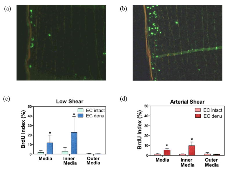
Top panels: Fluorescent micrographs of BrdU-stained arterial longitudinal sections of an EC intact area (a) and an EC denuded area (b) in an artery after being cultured under arterial shear condition for 7 days. Bottom panels: Comparisons of BrdU indices in the EC intact and EC denuded areas of arteries cultured under low shear and arterial shear conditions for 7 days (mean ± SD, n = 4, *p<0.05).
In addition, the media thickness also increased in EC denuded areas compared to the media thickness in the neighboring EC intact areas (Figure 6). The average thickness was 309.35 ± 50.89 μm in EC intact areas, but was significantly increased to 412.83 ± 59.62 μm (n=4, p<0.05) in EC denuded areas.
Figure 6.

Upper panels: Smooth muscle alpha-actin staining of: a) an upstream EC intact area, b) an EC denuded area, and c) a downstream EC intact area. Bottom panel: Comparison of the medial wall thickness between EC intact and EC denuded areas (mean ± SD, n = 4, *p<0.05)
Endothelialization suppressed SMC proliferation
SMC proliferation was significantly reduced in areas covered with migrating ECs in both EC injured arteries (Figure 7) and the co-culture arterial wall model (Figures 8). In arteries, the SMC proliferation rates in areas fully covered with migrating ECs (located 1- 3 mm from the edge of the initial injury band) were similar to those in the EC intact areas. The SMC proliferation rate increased in the transition areas with incomplete EC coverage which were located 4-6 mm from the original wound edge. In contrast, SMC proliferation rate in areas that remain un-endothelialized (no EC, located 6-8 mm from the edge of the original injury band) was significantly higher than in the EC intact areas (5.45 ± 1.8 % vs. 1.4 ± 0.86 %, p<0.001, Figure 7). Similarly, in the co-culture arterial wall model, SMC proliferation was also suppressed by endothelialization and arterial shear stress. Specifically, SMC proliferation rates were significantly decreased in areas covered by migrating EC under arterial shear conditions compared to either the pre-endothelialized areas (under either low or arterial shear conditions) or the areas not covered by EC that were exposed to low shear stress (p < 0.001, Figure 8). The SMC proliferation rates were increased by almost 20-fold in areas not covered by EC and exposed low shear conditions compared to EC covered areas exposed to arterial shear conditions. The increase is approximately 2-fold compared to the pre-endothelialized areas exposed to low shear stress. In addition, SMC proliferation was lower under arterial shear than under low shear in EC denuded areas in both the artery injury model and co-culture model,.
Figure 7.

Distribution of medial SMC proliferation (BrdU index, mean ± SD, n = 4) along the flow direction in arteries cultured under arterial shear for 7 days. Locations 1-3: fully reendothelialized areas, 4-6: incomplete reendothlialization areas, and noEC: un-endothelialized areas. *** p<0.001 versus EC intact.
Figure 8.

Distribution of the number of proliferating SMC per section (3 cm in length by 10cm in width) along the flow direction in EC-SMC co-culture model after cultured for 7 days under low shear and arterial shear conditions. Dotted lines with arrows indicate the ranges the EC migrated. n = 4, * p < 0.05, ** p<0.005 versus Arterial Shear.
The effects of flow and reendothelialization on SMC migration
In injured arteries, the medial SMCs underneath the EC denuded area exhibited characteristics associated with increased migration. Specifically, the SMCs in the first sub-layer underneath the internal elastic lamina (IEL) in the EC denuded area were larger and more elongated along the radial direction (towards the lumen) as compared to SMCs underneath the EC covered area (Figure 9, top panels). The average crosss-sectional area of SMCs was significantly increased in EC denuded areas compared to SMCs in the EC intact areas (51.52 ± 11.09 vs. 115.22 ± 20.38 μm2, n=4, p <0.005, Figure 9, middle left). The aspect ratio of SMC was significantly higher in EC denuded areas than in the EC intact areas (2.02 ± 0.37 vs 1.43 ± 0.13, n=4, p <0.05, Figure 9, middle right), while the shape index was significantly reduced (0.55 ± 0.04 vs 0.66 ± 0.03, n=4, p <0.005). The major axis of the elongated SMC was angled close to 90 degrees from the vertical line (94.36 ± 12.11°). These shape changes along with SMC proliferation and medial thickening are the initial stages associated with the development of intimal hyperplasia.
Figure 9.

Top: Smooth muscle alpha-actin staining at EC intact (a) and EC denuded (b) areas after being cultured under arterial shear conditions for 7 days. Scale: 10μm. Middle: Comparisons of areas and aspect ratios (c and d, respectively) of SMCs near the lumen in the EC intact and EC denuded areas (mean ± SD, n = 4, *p<0.05). Bottom: Distributions of the number of migrated SMC per section (2 cm in length by 10 cm in width) along the flow direction in the co-culture model under low shear and arterial shear conditions. n = 3, * p < 0.05, ** p<0.005 versus Arterial Shear.
In the co-culture model, SMCs migrate through the mesh pores into the subendothelial space or onto the unendothelialized flow surface. In 7 consecutive areas along the length of the mesh (in the same direction of the flow), the number of SMCs migrated through the mesh pores was significantly smaller (p<0.005) in endothelialized areas exposed to arterial shear stress compared to both endothelialized and unendothelialized areas exposed to low shear stress (Figure 9, bottom). Furthermore, it was noted that even though all 7 areas of the mesh were endothelialized by the end of the arterial shear stress exposure regimen, this endothelialization was a progressive phenomenon that occurred over 7 days. This progression likely accounts for the somewhat higher rate of SMC migration in the distal areas which became endothelialized at later times.
Discussion
In summary, we developed an ex vivo injured artery model and a parallel in vitro EC-SMC co-culture arterial wall model to investigate the migration of EC under shear flows. Our results demonstrated that flow-generated wall shear stress accelerated EC migration in arteries and in the cell co-culture model. Endothelial cell coverage suppressed the migration and proliferation of the underlying SMCs.
The observed enhanced effect of shear stress on EC migration in our models is similar to what we and others have observed for EC migration in vitro.1, 19, 37 Our data is consistent with the literature in showing that EC denudation promotes the proliferation of the underlying SMCs.12, 14, 18, 43 EC denudation also led to SMC migration to the lumen surface of the artery and medial wall thickening, which are the early signs of restenosis and intimal hyperplasia.41 Furthermore, our data also showed that reendothelialization significantly reduced SMC proliferation. These results support the validity of our models.
The artery injury model using ex vivo organ culture with perfusion provides an excellent model for studying cell proliferation and migration in arteries. This model maintains the natural extracellular matrix (ECM) and mechanical stress environment of vascular cells while providing independent and consistent control of the pressure and flow. Thus, this model has the advantage of specificity in determining the effects of individual factors, especially the mechanical factors, although it lacks the hormone and nerve control as compared to in vivo. By changing flow rate while maintaining other mechanical parameters (pressure, axial stretch etc) unchanged, this model allows us to specify the observed differences that resulted from the changes in flow alone. Therefore. this model provides a tool for better understanding of the EC migration process and a test bed to develop various techniques to promote EC migration and reduce SMC proliferation, as well as a tool to investigate cellular and molecular mechanisms governing these responses. Experimental manipulations such as those using genetically modified smooth muscle cells, endothelial cells, or pharmacologic agents can be evaluated on the basis that the results will likely be relevant and applicable to in vivo physiology or pathophysiology.
In this study, EC proliferation was measured by counting the BrdU positive cells on the intimal surface. While SMCs may migrate into the intima, the likelihood is very low in the EC intact area where the measurement was made. Also note that we used the Poiseuille flow equation to estimate the mean wall shear stress. Due to the pulsatility of the flow, the actual wall shear stress oscillated with time and that caused errors in the estimation. The Womersley number of the perfusion flow was estimated to be 4.6 which was the same as in normal carotid or femoral arteries and thus the effect of pulsatility was expected to be relatively small.31, 44 In addition, our measurement showed that the flow rate oscillated between 156 to 165 ml/min, so the flow had a very large steady component (a mean flow rate of 160 ml/min) that was much higher than the oscillation component (5 ml/min). Therefore, we expect that the error in the shear stress estimation was relatively small.
Comparison of ex vivo artery model and in vitro co-culture model
By comparing the ex vivo and in vitro models, we demonstrated that flow has similar effects on EC migration in vitro and in arteries ex vivo. EC migration depends on the shear force, cell-ECM interaction, signal pathways, and cytoskeletal changes.5 While the shear stresses were qualitatively similar in the artery and co-culture models, the EC-ECM interactions most likely were different. This difference could be the reason for the observed difference in the migration distance. The slow EC migration in the arteries may indicate a stronger EC-ECM binding in the natural arterial structure than the EC binding to the polymer substrate in the co-culture model. These results also indicated that while the two models may yield quantitatively different results with respect to the endothelial cell migration rate, they yield similar qualitative results with respect to the influence of flow-generated shear stress on endothelial cell migration and the influence of endothelium on suppressing SMC proliferation and migration. This is particularly evident with respect to SMC proliferation in areas covered by endothelium exposed to arterial level shear stress.
Clinical relevance
Arterial restenosis associated with thrombosis and intimal hyperplasia at sites of arterial bypass surgery, balloon angioplasty, or stent placement remains a challenging clinical problem.26, 33 Endothelial cell injury during these procedures triggers thrombosis and inflammatory responses followed by SMC proliferation and migration as well as extracellular matrix deposition. Though we used porcine artery and vascular cells, the model can be used to study human arteries and human vascular cells. Previous studies demonstrated that human vascular cells migrated in a similar pattern under shear flow36, indicating that our results on porcine cells are relevant to clinical applications. In addition, previous studies have demonstrated that endothelium in an arterial shear environment exhibits an anti-inflammatory phenotype characterized by down-regulation of inflammatory genes such as monocyte chemotactic protein-1 (MCP-1) and vascular cell adhesion molecule-1 (VCAM-1).27, 39 Thus, rapid restoration of endothelium in an arterial shear stress environment inhibits monocyte-macrophage recruitment to these sites.27 Macrophages, via the release of multiple cytokines, are known to play a key role in promoting intimal hyperplasia. These sequences of events may lead to restenosis26, 41 due to either acute thrombotic occlusion or, more frequently, SMC-associated intimal hyperplasia. Anti-proliferative drugs such as Sirolimus (rapamycin) have been used systemically or delivered locally using drug eluting stents to suppress SMC proliferation. These techniques have generated good results but have associated risks of delayed thrombosis requiring long-term anti-thrombotic therapy.29 Promoting rapid reendothelization either by stent-based agents or stent surface modifications has been proposed as an alternative treatment strategy.25, 35 Our results demonstrated that re-endothelialization associated with EC migration inhibits underlying SMC proliferation and migration.
These studies were solely focused on the influence of reendothelialization from adjacent intact endothelium at arterial injury sites on proliferation and migration of arterial wall smooth muscle cells. Recent evidence indicates that circulating endothelial progenitor cells may also contribute to this critical healing process.45 The relative contribution of these two mechanisms for reendothelialization at injured revascularization sites remains to be determined. Based on previous studies of monocyte adherence under different levels of shear stress in this laboratory and others,27, 39 it seems likely that adherence of circulating progenitor cells under arterial shear stress levels would be reduced compared to adherence under low shear conditions. Nevertheless, our results confirmed that higher levels of shear stress increase the migration rate of functional arterial endothelial cells from adjacent upstream areas of intact endothelium. Future and ongoing studies are directed toward using both the ex vivo and in vitro arterial wall models to evaluate strategies to increase the rate of post-injury endothelial healing.
Acknowledgments
This work was supported by the Advanced Research Program from the Texas Higher Educational Coordinating Board through grant # 003659-0014-2006. It was also partially supported by an MBRS-SCORE pilot grant from the National Institute of Health through grant # S06GM008194 and grant # 0602834 from the National Science Foundation. The authors thank the Granzins at New Braunfels and Wiatrek at Poth, TX for generously providing the arteries for this work. The authors also thank Mr. Kurtis Johnson for his help in this work and Ms. Patricia Navarro for proofreading the manuscript.
References
- 1.Albuquerque ML, Waters CM, Savla U, Schnaper HW, Flozak AS. Shear stress enhances human endothelial cell wound closure in vitro. Am J Physiol. 2000;279(1):H293–302. doi: 10.1152/ajpheart.2000.279.1.H293. [DOI] [PubMed] [Google Scholar]
- 2.Bailey SR. Endovascular stents: update on stents in practice. J Long Term Eff Med Implants. 2000;10(1-2):7–18. doi: 10.1615/jlongtermeffmedimplants.v10.i12.40. [DOI] [PubMed] [Google Scholar]
- 3.Bardy N, Karillon GJ, Merval R, Samuel JL, Tedgui A. Differential effects of pressure and flow on DNA and protein synthesis and on fibronectin expression by arteries in a novel organ culture system. Circ Res. 1995;77(4):684–94. doi: 10.1161/01.res.77.4.684. [DOI] [PubMed] [Google Scholar]
- 4.Chesler NC, Ku DN, Galis ZS. Transmural pressure induces matrix-degrading activity in porcine arteries ex vivo. Am J Physiol. 1999;277(5 Pt 2):H2002–9. doi: 10.1152/ajpheart.1999.277.5.H2002. [DOI] [PubMed] [Google Scholar]
- 5.Chien S, Li S, Shiu YT, Li YS. Molecular basis of mechanical modulation of endothelial cell migration. Front Biosci. 2005;10:1985–2000. doi: 10.2741/1673. [DOI] [PubMed] [Google Scholar]
- 6.Chien S. Mechanotransduction and endothelial cell homeostasis: the wisdom of the cell. Am J Physiol Heart Circ Physiol. 2007;292(3):H1209–24. doi: 10.1152/ajpheart.01047.2006. [DOI] [PubMed] [Google Scholar]
- 7.Christen T, Bochaton-Piallat ML, Neuville P, Rensen S, Redard M, van Eys G, Gabbiani G. Cultured Porcine Coronary Artery Smooth Muscle Cells : A New Model With Advanced Differentiation. Circ Res. 1999;85(1):99–107. doi: 10.1161/01.res.85.1.99. [DOI] [PubMed] [Google Scholar]
- 8.Clerin V, Gusic RJ, O'Brien J, Kirshbom PM, Myung RJ, Gaynor JW, Gooch KJ. Mechanical environment, donor age, and presence of endothelium interact to modulate porcine artery viability ex vivo. Ann Biomed Eng. 2002;30(9):1117–27. doi: 10.1114/1.1519262. [DOI] [PubMed] [Google Scholar]
- 9.Conklin BS, Zhong DS, Zhao W, Lin PH, Chen C. Shear stress regulates occludin and VEGF expression in porcine arterial endothelial cells. J Surg Res. 2002;102(1):13–21. doi: 10.1006/jsre.2001.6295. [DOI] [PubMed] [Google Scholar]
- 10.Davis NP, Han HC, Wayman B, Vito R. Sustained axial loading lengthens arteries in organ culture. Ann Biomed Eng. 2005;33(7):867–77. doi: 10.1007/s10439-005-3488-x. [DOI] [PubMed] [Google Scholar]
- 11.Edelman ER, Rogers C. Pathobiologic responses to stenting. Am J Cardiol. 1998;81(7A):4E–6E. doi: 10.1016/s0002-9149(98)00189-1. [DOI] [PubMed] [Google Scholar]
- 12.El Hamamsy I, Stevens LM, Vanhoutte PM, Perrault LP. Injury of the coronary endothelium at implantation increases endothelial dysfunction and intimal hyperplasia after heart transplantation. J Heart Lung Transplant. 2005;24(3):251–258. doi: 10.1016/j.healun.2003.12.012. [DOI] [PubMed] [Google Scholar]
- 13.Gleason RL, Wilson E, Humphrey JD. Biaxial biomechanical adaptations of mouse carotid arteries cultured at altered axial extension. J Biomech. 2006 doi: 10.1016/j.jbiomech.2006.03.018. [DOI] [PubMed] [Google Scholar]
- 14.Guerin P, Rondeau F, Grimandi G, Heymann MF, Heymann D, Pillet P, Al Habash O, Loirand G, Pacaud P, Crochet D. Neointimal hyperplasia after stenting in a human mammary artery organ culture. J Vasc Res. 2004;41(1):46–53. doi: 10.1159/000076245. [DOI] [PubMed] [Google Scholar]
- 15.Han HC, Ku DN. Contractile responses in arteries subjected to hypertensive pressure in seven-day organ culture. Ann Biomed Eng. 2001;29(6):467–75. doi: 10.1114/1.1376391. [DOI] [PubMed] [Google Scholar]
- 16.Han HC, Ku DN, Vito RP. Arterial wall adaptation under elevated longitudinal stretch in organ culture. Ann Biomed Eng. 2003;31(4):403–11. doi: 10.1114/1.1561291. [DOI] [PubMed] [Google Scholar]
- 17.Han HC, Marita S, Ku DN. Changes of opening angle in hypertensive and hypotensive arteries in 3-day organ culture. J Biomech. 2006;39(13):2410–8. doi: 10.1016/j.jbiomech.2005.08.003. [DOI] [PubMed] [Google Scholar]
- 18.Haudenschild CC, Schwartz SM. Endothelial regeneration. II. Restitution of endothelial continuity. Lab Invest. 1979;41(5):407–18. [PubMed] [Google Scholar]
- 19.Hsu PP, Li S, Li YS, Usami S, Ratcliffe A, Wang X, Chien S. Effects of flow patterns on endothelial cell migration into a zone of mechanical denudation. Biochem Biophys Res Commun. 2001;285(3):751–9. doi: 10.1006/bbrc.2001.5221. [DOI] [PubMed] [Google Scholar]
- 20.Ku DN. Blood Flow in Arteries. Ann Rev Fluid Mech. 1997;29(1):399–434. [Google Scholar]
- 21.Labadie RF, Antaki JF, Williams JL, Katyal S, Ligush J, Watkins SC, Pham SM, Borovetz HS. Pulsatile perfusion system for ex vivo investigation of biochemical pathways in intact vascular tissue. Am J Physiol. 1996;270(2 Pt 2):H760–8. doi: 10.1152/ajpheart.1996.270.2.H760. [DOI] [PubMed] [Google Scholar]
- 22.Lee YU, Drury-Stewart D, Vito RP, Han HC. Morphologic adaptation of arterial endothelial cells to longitudinal stretch in organ culture. J Biomech. 2008;41(15):3274–7. doi: 10.1016/j.jbiomech.2008.08.016. [DOI] [PMC free article] [PubMed] [Google Scholar]
- 23.Lehoux S, Tedgui A. Cellular mechanics and gene expression in blood vessels. J Biomech. 2003;36(5):631–43. doi: 10.1016/s0021-9290(02)00441-4. [DOI] [PubMed] [Google Scholar]
- 24.Lloyd-Jones D, Adams R, Carnethon M, De Simone G, Ferguson TB, Flegal K, Ford E, Furie K, Go A, Greenlund K, Haase N, Hailpern S, Ho M, Howard V, Kissela B, Kittner S, Lackland D, Lisabeth L, Marelli A, McDermott M, Meigs J, Mozaffarian D, Nichol G, O'Donnell C, Roger V, Rosamond W, Sacco R, Sorlie P, Stafford R, Steinberger J, Thom T, Wasserthiel-Smoller S, Wong N, Wylie-Rosett J, Hong Y. Heart disease and stroke statistics--2009 update: a report from the American Heart Association Statistics Committee and Stroke Statistics Subcommittee. Circulation. 2009;119(3):e21–181. doi: 10.1161/CIRCULATIONAHA.108.191261. [DOI] [PubMed] [Google Scholar]
- 25.Losordo DW, Isner JM, Diaz-Sandoval LJ. Endothelial recovery: the next target in restenosis prevention. Circulation. 2003;107(21):2635–7. doi: 10.1161/01.CIR.0000071083.31270.C3. [DOI] [PubMed] [Google Scholar]
- 26.Mitra AK, Agrawal DK. In stent restenosis: bane of the stent era. J Clin Pathol. 2006;59(3):232–9. doi: 10.1136/jcp.2005.025742. [DOI] [PMC free article] [PubMed] [Google Scholar]
- 27.Mohan S, Mohan N, Valente AJ, Sprague EA. Regulation of low shear flow-induced HAEC VCAM-1 expression and monocyte adhesion. Am J Physiol Cell Physiol. 1999;276(5):C1100–1107. doi: 10.1152/ajpcell.1999.276.5.C1100. [DOI] [PubMed] [Google Scholar]
- 28.Ong AT, Aoki J, Kutryk MJ, Serruys PW. How to accelerate the endothelialization of stents. Arch Mal Coeur Vaiss. 2005;98(2):123–6. [PubMed] [Google Scholar]
- 29.Palmaz JC. Intravascular stents in the last and the next 10 years. J Endovasc Ther. 2004;11 2:II200–206. doi: 10.1177/15266028040110S617. [DOI] [PubMed] [Google Scholar]
- 30.Redmond EM, Cullen JP, Cahill PA, Sitzmann JV, Stefansson S, Lawrence DA, Okada SS. Endothelial cells inhibit flow-induced smooth muscle cell migration: role of plasminogen activator inhibitor-1. Circulation. 2001;103(4):597–603. doi: 10.1161/01.cir.103.4.597. [DOI] [PubMed] [Google Scholar]
- 31.Rhee K, Tarbell JM. A study of the wall shear rate distribution near the end-to-end anastomosis of a rigid graft and a compliant artery. J Biomech. 1994;27(3):329–38. doi: 10.1016/0021-9290(94)90009-4. [DOI] [PubMed] [Google Scholar]
- 32.Schwartz RS, Henry TD. Pathophysiology of coronary artery restenosis. Rev Cardiovasc Med. 2002;3 5:S4–9. [PubMed] [Google Scholar]
- 33.Scott NA. Restenosis following implantation of bare metal coronary stents: Pathophysiology and pathways involved in the vascular response to injury. Advanced Drug Delivery Reviews. 2006;58(3):358–376. doi: 10.1016/j.addr.2006.01.015. [DOI] [PubMed] [Google Scholar]
- 34.Soyombo AA, Angelini GD, Newby AC. Neointima formation is promoted by surgical preparation and inhibited by cyclic nucleotides in human saphenous vein organ cultures. J Thorac Cardiovasc Surg. 1995;109(1):2–12. doi: 10.1016/S0022-5223(95)70415-9. [DOI] [PubMed] [Google Scholar]
- 35.Sprague EA. Endothelial and Smooth Muscle Cell Injury in Contrived Models and Natural Disease. In: Bishop SP, Kerns WD, editors. Comprehensive Toxicology. Pergamon; Oxford, United Kingdom: 1997. pp. 213–240. [Google Scholar]
- 36.Sprague EA, Luo J, Palmaz JC. Human aortic endothelial cell migration onto stent surfaces under static and flow conditions. J Vasc Interv Radiol. 1997;8(1 Pt 1):83–92. doi: 10.1016/s1051-0443(97)70521-9. [DOI] [PubMed] [Google Scholar]
- 37.Sprague EA, Luo J, Palmaz JC. Endothelial cell migration onto metal stent surfaces under static and flow conditions. J Long Term Eff Med Implants. 2000;10(1-2):97–110. [PubMed] [Google Scholar]
- 38.Vorp DA, Peters DG, Webster MW. Gene expression is altered in perfused arterial segments exposed to cyclic flexure ex vivo. Ann Biomed Eng. 1999;27(3):366–71. doi: 10.1114/1.158. [DOI] [PubMed] [Google Scholar]
- 39.Walpola PL, Gotlieb AI, Cybulsky MI, Langille BL. Expression of ICAM-1 and VCAM-1 and Monocyte Adherence in Arteries Exposed to Altered Shear Stress. Arterioscler Thromb Vasc Biol. 1995;15(1):2–10. doi: 10.1161/01.atv.15.1.2. [DOI] [PubMed] [Google Scholar]
- 40.Walter DH, Cejna M, Diaz-Sandoval L, Willis S, Kirkwood L, Stratford PW, Tietz AB, Kirchmair R, Silver M, Curry C, Wecker A, Yoon YS, Heidenreich R, Hanley A, Kearney M, Tio FO, Kuenzler P, Isner JM, Losordo DW. Local gene transfer of phVEGF-2 plasmid by gene-eluting stents: an alternative strategy for inhibition of restenosis. Circulation. 2004;110(1):36–45. doi: 10.1161/01.CIR.0000133324.38115.0A. [DOI] [PubMed] [Google Scholar]
- 41.Weintraub WS. The Pathophysiology and Burden of Restenosis. The American Journal of Cardiology. 2007;100(5, Supplement 1):S3–S9. doi: 10.1016/j.amjcard.2007.06.002. [DOI] [PubMed] [Google Scholar]
- 42.Werner N, Junk S, Laufs U, Link A, Walenta K, Bohm M, Nickenig G. Intravenous transfusion of endothelial progenitor cells reduces neointima formation after vascular injury. Circ Res. 2003;93(2):e17–24. doi: 10.1161/01.RES.0000083812.30141.74. [DOI] [PubMed] [Google Scholar]
- 43.Willis AI, Pierre-Paul D, Sumpio BE, Gahtan V. Vascular smooth muscle cell migration: current research and clinical implications. Vasc Endovascular Surg. 2004;38(1):11–23. doi: 10.1177/153857440403800102. [DOI] [PubMed] [Google Scholar]
- 44.Womersley JR. Method for the calculation of velocity, rate of flow and viscous drag in arteries when the pressure gradient is known. J Physiol. 1955;127(3):553–63. doi: 10.1113/jphysiol.1955.sp005276. [DOI] [PMC free article] [PubMed] [Google Scholar]
- 45.Zampetaki A, Kirton JP, Xu Q. Vascular repair by endothelial progenitor cells. Cardiovasc Res. 2008;78(3):413–421. doi: 10.1093/cvr/cvn081. [DOI] [PubMed] [Google Scholar]