Abstract
Purpose
Koreans have been undergoing rapid lifestyle changes that may have an effect on patterns of cerebral artery atherosclerosis. This study was aimed at determining the frequency and distribution of atherosclerosis in the cerebral arteries and associated temporal changes over the past eight-year period among Korean stroke patients.
Materials and Methods
By using stroke registry data registered between April 1999 and March 2007, we investigated the presence, severity, and location of cerebral artery atherosclerosis as determined by angiographic findings. Their annual patterns and association with vascular risk factors were investigated.
Results
Of 1,955 patients, 1,517 patients (77.6%) demonstrated atherosclerosis in one or more arteries. A significantly increasing trend of atherosclerosis was observed during the past eight years, which was ascribed to an increase of combined extracranial (EC) and intracranial (IC) atherosclerosis. The number of atherosclerotic arteries increased as the number of risk factors increased. In the multivariate analysis, the year and vascular risk factors were independent predictors of the presence of atherosclerosis.
Conclusion
We found that the atherosclerotic burden has been increasing for the past eight years in Korean stroke patients, particularly the combined EC and IC subtype. Lifestyle changes and increase in vascular risk factors may be contributing factors.
Keywords: Cerebrovascular disorders, atherosclerosis, risk factors, epidemiology
INTRODUCTION
It is known that the predilection sites of cerebral atherosclerosis differ among races. In Asian patients, intracranial (IC) atherosclerosis is most frequently found, while in Caucasian patients, extracranial (EC) atherosclerosis is more common.1-3 Individual risk factors for atherosclerosis may have differential impacts on the location as well as the development of atherosclerosis.2,4-6 In addition, the development of atherosclerotic risk factors is greatly influenced by lifestyle. Korean and other Asian populations have been undergoing rapid lifestyle changes as they become industrialized and experience economic growth. As a result, recent epidemiologic studies show that Koreans are more obese and hypertensive than in the past, and that both body mass index (BMI) and diabetes mellitus have also increased.7,8 These changes in lifestyle and risk factor profiles may affect the burden of EC and IC atherosclerosis.9
Stroke is one of the leading causes of death in many Asian countries, and the incidence of atherothrombotic diseases has been rapidly increased. For example, the proportion of ischemic stroke in China increased from 30.2% to 61.9% over 10 years.10 Korea also showed a similar pattern of change.11 The incidence of ischemic heart disease is also increasing and the mortality from acute myocardial infarction in Asian men aged 35-44 increased from 3.9/100,000 to 10.3/100,000.12 Accumulating evidence of rapidly increasing vascular events in the brain and heart in Koreans as well as Asians suggests that atherosclerotic diseases may also be increasing in this population. Although several studies have investigated cerebral atherosclerosis in Korean and Asian patients with ischemic stroke,3,13-16 information is fragmentary, obtained from a small group of patients, or performed long ago, such that it may not adequately reflect the recent status of Korean and Asian stroke patients. Therefore, the present study investigated the frequency, distribution, and temporal changes in atherosclerosis of the EC and IC cerebral arteries in Korean stroke patients by using stroke registry data obtained consecutively for an eight-year period.
MATERIALS AND METHODS
Study population
We reviewed the records of 2,977 consecutive patients registered in the Yonsei Stroke Registry (YSR) between April 1999 and March 2007. The YSR is a prospective hospital-based registry for patients with an acute cerebral infarction or transient ischemic attack (TIA).17 The study hospital is located in Seoul, which is the capital of Korea. About 69% of patients enrolled for this study were residents of Seoul, and this proportion has not been changed during the study period. The diagnosis requires brain computed tomography (CT) or magnetic resonance imaging (MRI) to exclude hemorrhages and other causes. Systematic investigations were performed in every patient, including 12 lead-electrocardiography, chest X-rays, lipid profiles, and standard blood tests. Additional tests were performed in patients younger than 45 years or in those with no determinable etiology. These included measures of serum homocysteine levels and hemostatic markers such as protein C, protein S, antithrombin III, and antiphospholipid antibodies (lupus anticoagulant, anticardiolipin IgG and IgM). Transesophageal echocardiography (TEE) was part of the standard evaluation except in patients with decreased consciousness, poor systemic conditions, tracheal intubation, an inability to accept an esophageal transducer, or lack of informed consent. Transthoracic echocardiography was performed in patients who could not undergo TEE and were suspected of having a cardiac disease. The accuracy and quality of the registry are ensured by reviewing the data of each case at weekly stroke conferences. This study was approved by institutional review board.
Risk factors for atherosclerosis
The presence of hypertension was defined when patients had resting blood pressure recordings of systolic ≥ 140 mmHg or diastolic ≥ 90 mmHg on repeated measurements after clinical stabilization (usually 24-48 hours after admission) during hospitalization or a past medical history for antihypertensive treatment. Diabetes mellitus was diagnosed when a patient had a fasting plasma glucose value ≥ 7 mmol/L or had been treated with an oral hypoglycemic agent or insulin. Hypercholesterolemia was defined as a fasting serum total cholesterol level ≥ 6.2 mmol/L, LDL-cholesterol > 4.1 mmol/L or a past medical history significant for a lipid-lowering drug. Patients were regarded as smokers if they smoked within the three-month period prior to admission. History of previous stroke included old symptomatic cerebral infarctions and TIAs, which were clinically diagnosed demonstrated with brain imaging studies before admission.
Patients enrolled for analysis
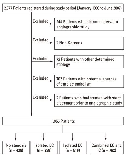
Among the 2,977 registered patients, 2,779 (91.8%) had an angiography performed [digital subtraction angiography (DSA) in 498 (17.9%), MRA in 1,699 (61.1%), and both DSA and MRA in 582 (20.9%) patients]. MRA was conducted using either a 1.5-Tesla system (Signa Horizon, GE Medical System, Milwaukee, WI or Intera or Achieva, Philips Medical System, Best, Netherlands) or a 3.0-T system (Achieva, Philips Medical System, Best, Netherlands) and almost all patients had underwent contrast enhanced MRA. Echocardiography was performed in 1,508 patients (TEE in 1,192, Transthoracic echocardiography in 182 and both in 134). For the purpose of this study, a total of 1,955 patients were finally enrolled after excluding those who had not undergone an angiographic study, non-Korean, those with potential cardiac sources of embolism (PCSE) or other determined etiologies, such as arterial dissection, Moyamoya disease, coagulation disorders, etc., and those who had been treated with stent insertion/angioplasty or carotid endarterectomy before the admission at our hospital. In patients with both DSA and MRA performed, the results of DSA were used for analysis, and in patients with MRA studies performed at different times due to re-admission or recurrent attacks, the most recent results were used for analysis. Therefore, data of only one angiographic study from each patient was used. Finally, data from 725 DSA (37.1%) and 1,230 MRA (62.9%) were analyzed for this study (Fig. 1).
Fig. 1.
Flow chart of patient selection and grouping. EC, extracranial; IC, intracranial.
Definition of atherosclerosis and degree of stenosis
Any abnormalities on angiographic studies were determined at a weekly stroke conference based on a neuroradiologist's report and the consensus of stroke specialists. Angiographic findings were defined as normal, stenotic, or occluded in each arterial segment and branch arteries of the EC and IC cerebral arteries. The degree of stenosis was measured using the North American Symptomatic Carotid Endarterectomy Trial18 or Warfarin vs. Aspirin for Symptomatic intracranial disease method.19 When multiple stenotic lesions were present in one artery, data of the most severe degree was used. The stenotic lesions were divided into EC, IC, or combined EC and IC atherosclerosis, and were also classified as mild (< 50%) or severe (≥ 50% or occluded). The number of atherosclerotic lesions was the sum of the number of stenotic lesions in the predetermined portion at each side; the common carotid artery, extracranial internal carotid artery (ICA), intracranial ICA, branch arteries of the ICA, anterior cerebral artery, middle cerebral artery, extracranial vertebral artery, intracranial vertebral artery, basilar artery, posterior inferior cerebellar artery, anterior inferior cerebellar artery, superior cerebellar artery, and posterior cerebral artery.
Statistical analysis
Statistical analysis were performed using the Windows SPSS package (version 15.0) (SPSS Inc, Chicago, IL, USA). The independent sample t-test, the Pearson chi-square test, or ANOVA test was used to compare means and proportions. The temporal changes of atherosclerosis were compared by the Mantel-Haenszel test. Multivariate logistic analysis was used to compute odds ratios of vascular risk factors. The correlation between the number of lesions and other factors was determined by Spearman's rank test. Predicted changes in the number of lesions were analyzed by a Poisson regression analysis.
RESULTS
Demographic characteristics of the study group
Enrolled for the final analysis were 1,955 patients with a mean age of 63.7 ± 10.9 years (range, 24-93), 1,230 (62.9%) of which were men. These demographic data were not different from those of the entire 2,977 patients registered during the study period in that the mean age of the 2,977 patients was 63.7 ± 11.8 years (range 6-95) and men comprised 62.1% (1,850 patients).
During the eight-year study period, there was an increasing tendency in performing angiographic studies, either MRA or DSA, from 83.4% in 1999 to 94.1% in 2007 among the entire 2,977 patients. In 1,957 patients, the proportion of angiographic study with MRA increased from 53.9% in 1999 to 66.2% in 2007.
Frequency and distribution of atherosclerosis
Among 1,955 patients, 1,517 patients (77.6%) were found to have atherosclerosis in one or more arteries of the EC or IC artery. Isolated EC atherosclerosis was found in 239 (12.2%), isolated IC atherosclerosis in 516 (26.4%), and combined EC and IC atherosclerosis in 762 (39.0%) patients.
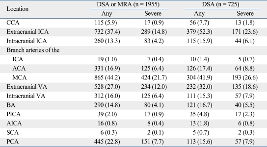
Among the arteries involved, the MCA was most common (44.2%), followed by the extracranial ICA (37.4%), and the extracranial vertebral artery (27.0%). The same pattern of distribution was noted among cases with severe atherosclerosis (Table 1). However, in a subgroup having had DSA performed, involvement of the extracranial ICA was most common (52.3%) (Table 1).
Table 1.
Distribution of Atherosclerosis
Data are given as the number (percentage) of arteries with atherosclerosis. Branch arteries of the ICA include ophthalmic artery, posterior communicating artery, and anterior choroidal artery. Abbreviations are defined as follows. ACA, Anterior cerebral artery; AICA, Anterior inferior cerebellar artery; BA, Basilar artery; CCA, Common carotid artery; DSA, Digital subtraction angiography; ECA, External carotid artery; ICA, Internal carotid artery; MCA, Middle cerebral artery; MRA, Magnetic resonance angiography; PCA, Posterior cerebral artery; PICA, Posterior inferior cerebellar artery; SCA, Superior cerebellar artery; VA, Vertebral artery.
Temporal changes in the presence and burden of atherosclerosis
After excluding patients with PCSE
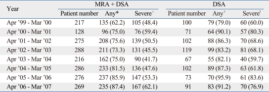
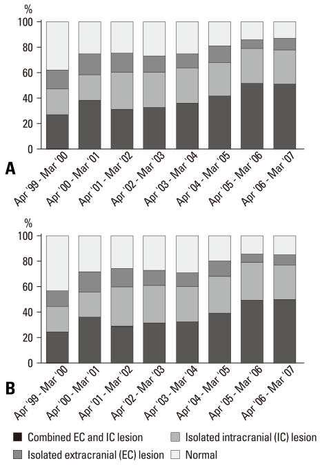
The prevalence of atherosclerosis in 1,955 ischemic stroke patients during the past eight-year period was compared annually using the Mantel-Haenszel test. There was a significant increase in atherosclerosis in this study population (p < 0.001) (Table 2). An increasing tendency of atherosclerosis was also observed in the analysis of the DSA subgroup findings (p = 0.010). These increases appear to be ascribable to the increased frequency of combined EC and IC atherosclerosis (p < 0.001) (Fig. 2A). Likewise, the proportion of severe atherosclerosis showed a similar increasing trend (p = 0.043), although it did not reach statistical significance in the analysis using the DSA findings (p = 0.075).
Table 2.
Yearly Frequency of Atherosclerosis
Data are given as the number (percentage) of patients with atherosclerosis.
*p < 0.001, †p = 0.042, ‡p = 0.010, §p = 0.077 by Mantel-Haenszel test.
Fig. 2.
Temporal changes in the frequency of atherosclerosis in the cerebral arteries in 1,955 patients excluding those with potential cardiac sources of embolism (PCSE) (A), and in 2,657 patients including those with PCSE (B). The frequencies of atherosclerosis in both study groups have been increasing during the eight-year study period. The proportion of involved sites is expressed as the percentage in each subgroup.
The relationship between the number of arterial lesions involved and the year was investigated using Spearman's rank test. There was a significant trend toward an increase in the number of arterial lesions (r = 0.244, p < 0.001). However, there was no relationship between the number of severe arterial lesions and the year (r = 0.045, p = 0.044).
After including patients with PCSE
We excluded the patients with PCSE for the analysis because occlusive lesions of the cerebral arteries caused by embolism from the heart are often indistinguishable from those by atherosclerosis. However, the exclusion of patients with PCSE may also result in bias of patient selection and data interpretation since those with PCSE may have concomitant cerebral atherosclerotic lesions. In addition, exclusion of one subtype of stroke group (cardioembolism) may prevent uncovering the features in entire stroke patients. Therefore, we further analyzed the data after including 702 patients with PCSE (total 2,657 patients), and the increasing tendency of atherosclerosis was also observed (p < 0.001) (Fig. 2B).
Burden of atherosclerosis
A total of 5,151 atherosclerotic lesions were found in 1,517 patients with atherosclerosis, 1,763 of which were severe. There were 1,868 lesions in EC portions and 3,283 lesions in IC portions. Of 1,517 patients, 374 (24.7%) patients showed a lesion in only one artery, 311 (20.5%) in two arteries, 236 (15.6%) in three arteries, 183 (12.1%) in four arteries, and the remaining 413 (27.2%) in five or more arteries.
Vascular risk factors and their association with atherosclerotic burden
A total of 1,794 patients (91.8 %) had at least one or more classic risk factors for atherosclerosis. There has been a significant increase in the presence of risk factors (p = 0.029) during the study period. Among individual risk factors, the frequencies of hypertension (p < 0.001) and diabetes mellitus (p = 0.003) have increased whereas history of previous stroke has decreased (p = 0.003). There was no difference in the mean age of the patients among the years (p = 0.162).
The patients with atherosclerosis were older than those without atherosclerosis and were more likely to have vascular risk factors (p < 0.001). The number of arteries with atherosclerosis increased as the number of risk factors increased (r = 0.223, p < 0.001). Among the patients with atherosclerosis, those with combined EC and IC atherosclerosis were older (p < 0.001) and had a higher possibility (p < 0.001) of having risk factors and increased number (p < 0.001) of risk factors identified than those with isolated EC or IC atherosclerosis.
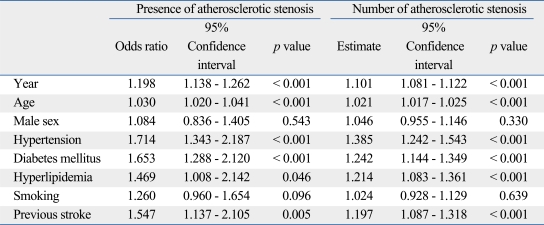
On univariate analysis, age (64.48 ± 10.49 years in a group with atherosclerosis versus 60.85 ± 11.60 years in a group without, p < 0.001), hypertension (p < 0.001), diabetes mellitus (p < 0.001), hyperlipidemia (p = 0.025), and previous stroke (p = 0.001) were associated with the presence of atherosclerosis. On multivariate logistic regression analysis, the year was also significant (Table 3). With respect to the number of arteries with atherosclerosis, the year and vascular risk factors including age, hypertension, diabetes mellitus, hypercholesterolemia, and previous cerebrovascular attacks were independently associated (Table 3).
Table 3.
Multivariate Analysis on the Presence and the Number of Atherosclerotic Stenosis
DISCUSSION
The distribution of atherosclerosis is known to differ among races in that atherosclerosis of the IC arteries is more common in non-Caucasians while that of the EC arteries is more common in Caucasians.2,3,14 However, most of the recent data from Asian patients has been based upon findings from a small number of patients or from transcranial Doppler studies, which may result in a selection bias of patients included or arteries assessed.3,14-16 It is not possible to perform conventional angiography, which is invasive, in all stroke patients. Therefore, in addition to a potential bias in patient selection, it might have been difficult to discern the pattern of EC or IC atherosclerosis in stroke patients as a whole. There was virtually no selection bias in using the angiographic studies in our study cohort by using MRA as well as DSA. Therefore, we could estimate global frequency as well as distribution of atherosclerosis. As a result, we showed that combined EC and IC atherosclerosis is most common in Korean stroke patients.
Among individual arteries, the MCA was the most commonly involved site, consistent with previous findings in Asian populations.13,16 Though in a DSA-only subgroup the involvement of the extracranial ICA was most common, this result could have been biased as patients who were found to have severe stenosis of the extracranial ICA on MRA would have undergone additional DSA before carotid endarterectomy or stenting. In a recent German study, the extracranial ICA was the most common symptomatic site of atherosclerosis, followed by the MCA.20 These findings suggest that the ICA in the extracranial portion and the MCA in the intracranial portion are the most vulnerable sites for atherosclerosis regardless of race, though a difference in frequency exists. While the exact etiology of this pattern of racial difference remains uncertain, environmental as well as genetic factors have been suggested as contributing causes.21 The difference in risk factors among races has been also suggested as a cause, as the frequency of hypertension, diabetes, and obesity is greater in black, Chinese, Japanese, and Hispanic individuals compared to Caucasians.22
Because approximately 92% of the consecutive patients enrolled for this study period had angiographic studies, we could investigate a temporally changing pattern of atherosclerosis. Of note, the frequency and burden of atherosclerosis have been rapidly increasing in our study cohort, which were coupled to an increase in vascular risk factors. Korea is a representative country in Asia that is rapidly developing and has been significantly affected by westernized lifestyles during the last decade. It has been suggested that westernized lifestyles aggravate risk factors for atherosclerosis and contribute to the progression of preclinical atherosclerosis.23 Accumulating evidence indicates that there have been rapid changes in the prevalence of vascular risk factors among the Korean population, most of which are related to lifestyle changes.7,24,25 The prevalence of hypertension in Korea has increased from 19.6% in 1990 to 27.0% in 2001.8 Moreover, in the 2001 survey, only 10.7% of hypertensive patients had their blood pressures adequately controlled. Thus, a recent and rapid increase in several vascular risk factors may in part be responsible for the increasing atherosclerotic burden. In the present study, a significant increase in the presence of risk factors was also noted. Among these risk factors, hypertension and diabetes mellitus have increased, consistent with observations in the Korean general population.8,26
With the exception of smoking, classic risk factors for stroke were associated with EC or IC atherosclerosis with multiple logistic regression analysis. In addition, the number of risk factors was associated with the burden of atherosclerosis, suggesting a cumulative impact of risk factors on EC and IC atherosclerosis. Previous studies in stroke-free patients showed that the prevalence of IC atherosclerosis escalated with an increasing number of vascular risk factors27 and that combined EC and IC atherosclerosis was associated with a greater number of risk factors, consistent with our findings. The incidence of ischemic and total cerebrovascular events was greater in the combined EC and IC group when compared to the isolated EC and IC groups.28 These findings suggest that atherosclerosis in cerebral arteries could be substantially decreased by reducing the prevalence of vascular risk factors. Given their cumulative impacts, it is essential to identify and reduce the well-known determinants of these risk factors.
Although the findings of this study were based upon a Korean population, many Asian countries may also undergo rapid lifestyle changes. Studies in Asian populations, including Japanese and Chinese, have shown that migration from traditional to westernized environments is associated with modification in diet and lifestyle, often leading to undesirable changes in blood sugar, lipid profile, and BMI and an increase in the risk of cardiovascular events.12,23,29,30 Likewise, the incidence of hypertension has been increasing in Asian counties, and the proportion of adequately controlled hypertensives remains low.31 Actually, in a study comparing the angiographic findings of Japanese stroke patients between 1963-65 and 1989-93, the proportion of patients with EC atherosclerosis was found to be higher in recent years.32 It is therefore likely that other Asian countries share the issues raised in this study, which further increases the burden of cerebral artery atherosclerosis.
Our study has several potential limitations. This is a single hospital-based study in patients with a symptomatic ischemic stroke, and the results were obtained from a certain part of Korea. Therefore, application of our data to the general population and to other countries should be cautious. While DSA is the gold standard for evaluating cerebral arteries, MRA as well as DSA was used in this study. MRA could erroneously detect false atherosclerotic lesions, particularly in intracranial arteries. However, an increasing trend of atherosclerosis was also observed in a subgroup using DSA, indicating that the findings and conclusions in our study were not impacted by the use of MRA data. While patients with PCSE were supposed to be excluded from the analysis, some patients who had PCSE that are particularly dependent on echocardiographic studies might be included because TEE was not performed in many patients. However, it is impossible to perform TEE in all patients since it requires that a patient is cooperating, and most PCSE, which are detected on echocardiographic studies in patients with sinus rhythm, are medium or low risk sources such as patent foramen ovale or spontaneous echo contrast.33 Furthermore, findings of increasing frequency and burden of atherosclerosis during the recent eight year period were not changed whether or not patients with PCSE were excluded for the analysis in this study. Finally, although the metabolic syndrome may be one of the important risk factors for atherosclerosis that can be affected by lifestyle changes, it was not included in the present analysis because the presence of the metabolic syndrome was not added to our registry until 2003.
Despite these potential limitations, the present large-scaled study of Korean stroke patients who were enrolled consecutively for the recent eight-year period demonstrated that combined EC and IC atherosclerosis is the most common form and that its presence and burden have been increasing. The increase of atherosclerotic burden was coupled to an increase in risk factors for atherosclerosis, which is presumed to be, in part, related to lifestyle changes. Urgent concerns and efforts to prevent or reduce atherosclerotic burden are necessary in Korea, and possibly in other Asian countries that may also undergo westernized lifestyle changes.
ACKNOWLEDGEMENTS
This work was supported by a grant of the Korea Healthcare Technology R & D Project, Ministry for Health, Welfare & Family Affairs, Republic of Korea (A060171, A085136) and supported by KOSEF through the National Core Research Center for Nanomedical Technology (R15-2004-024-00000-0).
Footnotes
The authors have no financial conflicts of interest.
References
- 1.Gorelick PB, Caplan LR, Hier DB, Patel D, Langenberg P, Pessin MS, et al. Racial differences in the distribution of posterior circulation occlusive disease. Stroke. 1985;16:785–790. doi: 10.1161/01.str.16.5.785. [DOI] [PubMed] [Google Scholar]
- 2.Caplan LR, Gorelick PB, Hier DB. Race, sex and occlusive cerebrovascular disease: a review. Stroke. 1986;17:648–655. doi: 10.1161/01.str.17.4.648. [DOI] [PubMed] [Google Scholar]
- 3.Wong KS, Huang YN, Gao S, Lam WW, Chan YL, Kay R. Intracranial stenosis in Chinese patients with acute stroke. Neurology. 1998;50:812–813. doi: 10.1212/wnl.50.3.812. [DOI] [PubMed] [Google Scholar]
- 4.Dahlöf B. Prevention of stroke in patients with hypertension. Am J Cardiol. 2007;100:17J–24J. doi: 10.1016/j.amjcard.2007.05.010. [DOI] [PubMed] [Google Scholar]
- 5.Enbergs A, Bürger R, Reinecke H, Borggrefe M, Breithardt G, Kerber S. Prevalence of coronary artery disease in a general population without suspicion of coronary artery disease: angiographic analysis of subjects aged 40 to 70 years referred for catheter ablation therapy. Eur Heart J. 2000;21:45–52. doi: 10.1053/euhj.1999.1763. [DOI] [PubMed] [Google Scholar]
- 6.Bang OY, Kim JW, Lee JH, Lee MA, Lee PH, Joo IS, et al. Association of the metabolic syndrome with intracranial atherosclerotic stroke. Neurology. 2005;65:296–298. doi: 10.1212/01.wnl.0000168862.09764.9f. [DOI] [PubMed] [Google Scholar]
- 7.Park HS, Kim SM, Lee JS, Lee J, Han JH, Yoon DK, et al. Prevalence and trends of metabolic syndrome in Korea: Korean National Health and Nutrition Survey 1998-2001. Diabetes Obes Metab. 2007;9:50–58. doi: 10.1111/j.1463-1326.2005.00569.x. [DOI] [PubMed] [Google Scholar]
- 8.Choi KM, Park HS, Han JH, Lee JS, Lee J, Ryu OH, et al. Prevalence of prehypertension and hypertension in a Korean population: Korean National Health and Nutrition Survey 2001. J Hypertens. 2006;24:1515–1521. doi: 10.1097/01.hjh.0000239286.02389.0f. [DOI] [PubMed] [Google Scholar]
- 9.Hachinski V. Stroke in Korean. Stroke. 2008;39:1067. doi: 10.1161/STROKEAHA.108.516708. [DOI] [PubMed] [Google Scholar]
- 10.Zhang LF, Yang J, Hong Z, Yuan GG, Zhou BF, Zhao LC, et al. Proportion of different subtypes of stroke in China. Stroke. 2003;34:2091–2096. doi: 10.1161/01.STR.0000087149.42294.8C. [DOI] [PubMed] [Google Scholar]
- 11.Korean Neurological Association. Epidemiology of cerebrovascular disease in Korea-a Collaborative Study, 1989-1990. J Korean Med Sci. 1993;8:281–289. doi: 10.3346/jkms.1993.8.4.281. [DOI] [PMC free article] [PubMed] [Google Scholar]
- 12.Sekikawa A, Kuller LH, Ueshima H, Park JE, Suh I, Jee SH, et al. Coronary heart disease mortality trends in men in the post World War II birth cohorts aged 35-44 in Japan, South Korea and Taiwan compared with the United States. Int J Epidemiol. 1999;28:1044–1049. doi: 10.1093/ije/28.6.1044. [DOI] [PubMed] [Google Scholar]
- 13.Tomita T, Mihara H. Cerebral angiographic study on C.V.D. in Japan. Angiology. 1972;23:228–239. doi: 10.1177/000331977202300408. [DOI] [PubMed] [Google Scholar]
- 14.De Silva DA, Woon FP, Lee MP, Chen CP, Chang HM, Wong MC. South Asian patients with ischemic stroke: intracranial large arteries are the predominant site of disease. Stroke. 2007;38:2592–2594. doi: 10.1161/STROKEAHA.107.484584. [DOI] [PubMed] [Google Scholar]
- 15.Tan TY, Chang KC, Liou CW, Schminke U. Prevalence of carotid artery stenosis in Taiwanese patients with one ischemic stroke. J Clin Ultrasound. 2005;33:1–4. doi: 10.1002/jcu.20081. [DOI] [PubMed] [Google Scholar]
- 16.Suh DC, Lee SH, Kim KR, Park ST, Lim SM, Kim SJ, et al. Pattern of atherosclerotic carotid stenosis in Korean patients with stroke: different involvement of intracranial versus extracranial vessels. AJNR Am J Neuroradiol. 2003;24:239–244. [PMC free article] [PubMed] [Google Scholar]
- 17.Lee BI, Nam HS, Heo JH, Kim DI Yonsei Stroke Team. Yonsei Stroke Registry. Analysis of 1,000 patients with acute cerebral infarctions. Cerebrovasc Dis. 2001;12:145–151. doi: 10.1159/000047697. [DOI] [PubMed] [Google Scholar]
- 18.North American Symptomatic Carotid Endarterectomy Trial Collaborators. Beneficial effect of carotid endarterectomy in symptomatic patients with high-grade carotid stenosis. N Engl J Med. 1991;325:445–453. doi: 10.1056/NEJM199108153250701. [DOI] [PubMed] [Google Scholar]
- 19.Warfarin-Aspirin Symptomatic Intracramial Disease (WASID) Trial Investigators. Design, progress and challenges of a double-blind trial of warfarin versus aspirin for symptomatic intracranial arterial stenosis. Neuroepidemiology. 2003;22:106–117. doi: 10.1159/000068744. [DOI] [PubMed] [Google Scholar]
- 20.Weimar C, Goertler M, Harms L, Diener HC. Distribution and outcome of symptomatic stenoses and occlusions in patients with acute cerebral ischemia. Arch Neurol. 2006;63:1287–1291. doi: 10.1001/archneur.63.9.1287. [DOI] [PubMed] [Google Scholar]
- 21.Lee SH, Kim MK, Park MS, Choi SM, Kim JT, Kim BC, et al. beta-Fibrinogen Gene -455 G/A Polymorphism in Korean Ischemic Stroke Patients. J Clin Neurol. 2008;4:17–22. doi: 10.3988/jcn.2008.4.1.17. [DOI] [PMC free article] [PubMed] [Google Scholar]
- 22.Sacco RL, Kargman DE, Gu Q, Zamanillo MC. Race-ethnicity and determinants of intracranial atherosclerotic cerebral infarction. The Northern Manhattan Stroke Study. Stroke. 1995;26:14–20. doi: 10.1161/01.str.26.1.14. [DOI] [PubMed] [Google Scholar]
- 23.Egusa G, Watanabe H, Ohshita K, Fujikawa R, Yamane K, Okubo M, et al. Influence of the extent of westernization of lifestyle on the progression of preclinical atherosclerosis in Japanese subjects. J Atheroscler Thromb. 2002;9:299–304. doi: 10.5551/jat.9.299. [DOI] [PubMed] [Google Scholar]
- 24.Park HS, Song YM, Cho SI. Obesity has a greater impact on cardiovascular mortality in younger men than in older men among non-smoking Koreans. Int J Epidemiol. 2006;35:181–187. doi: 10.1093/ije/dyi213. [DOI] [PubMed] [Google Scholar]
- 25.Kim HM, Park J, Kim HS, Kim DH, Park SH. Obesity and cardiovascular risk factors in Korean children and adolescents aged 10-18 years from the Korean National Health and Nutrition Examination Survey, 1998 and 2001. Am J Epidemiol. 2006;164:787–793. doi: 10.1093/aje/kwj251. [DOI] [PubMed] [Google Scholar]
- 26.Choi YJ, Cho YM, Park CK, Jang HC, Park KS, Kim SY, et al. Rapidly increasing diabetes-related mortality with socio-environmental changes in South Korea during the last two decades. Diabetes Res Clin Pract. 2006;74:295–300. doi: 10.1016/j.diabres.2006.03.029. [DOI] [PubMed] [Google Scholar]
- 27.Wong KS, Ng PW, Tang A, Liu R, Yeung V, Tomlinson B. Prevalence of asymptomatic intracranial atherosclerosis in high-risk patients. Neurology. 2007;68:2035–2038. doi: 10.1212/01.wnl.0000264427.09191.89. [DOI] [PubMed] [Google Scholar]
- 28.Takahashi W, Ohnuki T, Ide M, Takagi S, Shinohara Y. Stroke risk of asymptomatic intra- and extracranial large-artery disease in apparently healthy adults. Cerebrovasc Dis. 2006;22:263–270. doi: 10.1159/000094014. [DOI] [PubMed] [Google Scholar]
- 29.Nakanishi S, Okubo M, Yoneda M, Jitsuiki K, Yamane K, Kohno N. A comparison between Japanese-Americans living in Hawaii and Los Angeles and native Japanese: the impact of lifestyle westernization on diabetes mellitus. Biomed Pharmacother. 2004;58:571–577. doi: 10.1016/j.biopha.2004.10.001. [DOI] [PubMed] [Google Scholar]
- 30.Woo KS, Chook P, Raitakari OT, McQuillan B, Feng JZ, Celermajer DS. Westernization of Chinese adults and increased subclinical atherosclerosis. Arterioscler Thromb Vasc Biol. 1999;19:2487–2493. doi: 10.1161/01.atv.19.10.2487. [DOI] [PubMed] [Google Scholar]
- 31.Kearney PM, Whelton M, Reynolds K, Whelton PK, He J. Worldwide prevalence of hypertension: a systematic review. J Hypertens. 2004;22:11–19. doi: 10.1097/00004872-200401000-00003. [DOI] [PubMed] [Google Scholar]
- 32.Nagao T, Sadoshima S, Ibayashi S, Takeya Y, Fujishima M. Increase in extracranial atherosclerotic carotid lesions in patients with brain ischemia in Japan. An angiographic study. Stroke. 1994;25:766–770. doi: 10.1161/01.str.25.4.766. [DOI] [PubMed] [Google Scholar]
- 33.Han SW, Nam HS, Kim SH, Lee JY, Lee KY, Heo JH. Frequency and significance of cardiac sources of embolism in the TOAST classification. Cerebrovasc Dis. 2007;24:463–468. doi: 10.1159/000108438. [DOI] [PubMed] [Google Scholar]


