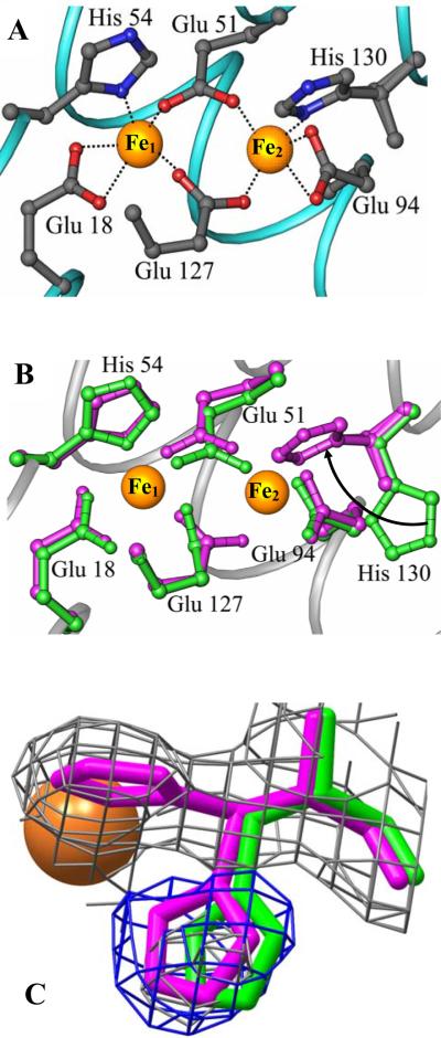
Figure 6.
Ferroxidase center of Fe soaked Pa BfrB. (A) Coordination of Fe ions at the ferroxidase center of Pa BfrB. (B) Overlay of the mineralized (green) and Fe soaked (magenta) Pa BfrB structures showing their ferroxidase centers. All residues adopt similar conformations with the exception of His130, which in the Fe-soaked structure is rotated toward the ferroxidase center, as indicated by the arrow, to facilitate binding of Fe2. (C) His130 from the Fe soaked structure (magenta) in its coordinative and non-coordinative conformations; the latter is nearly superimposed with the His130 side chain of the mineralized structure shown in green. The 2Fo-Fc map (grey mesh) is contoured at 1σ and the Fo-Fc omit map (blue mesh) at 3σ. The Fe2 atom is represented as an orange sphere.

