Abstract
Vascular dysfunction has a critical role in Alzheimer’s disease (AD). Recent data from brain imaging studies in humans and animal models suggest that cerebrovascular dysfunction may precede cognitive decline and onset of neurodegenerative changes in AD and AD models. Cerebral hypoperfusion and impaired amyloid β-peptide (Aβ) clearance across the blood–brain barrier (BBB) may contribute to the onset and progression of dementia AD type. Decreased cerebral blood flow (CBF) negatively affects the synthesis of proteins required for memory and learning, and may eventually lead to neuritic injury and neuronal death. Impaired clearance of Aβ from the brain by the cells of the neurovascular unit may lead to its accumulation on blood vessels and in brain parenchyma. The accumulation of Aβ on the cerebral blood vessels, known as cerebral amyloid angiopathy (CAA), is associated with cognitive decline and is one of the hallmarks of AD pathology. CAA can severely disrupt the integrity of the blood vessel wall resulting in micro or macro intracerebral bleedings that exacerbates neurodegenerative process and inflammatory response and may lead to hemorrhagic stroke, respectively.Here, we review the role of the neurovascular unit and molecular mechanisms in vascular cells behind AD and CAA pathogenesis. First, we discuss apparent vascular changes, including the cerebral hypoperfusion and vascular degeneration that contribute to different stages of the disease process in AD individuals. We next discuss the role of the low-density lipoprotein receptor related protein-1 (LRP), a key Aβ clearance receptor at the BBB and along the cerebrovascular system, whose expression is suppressed early in AD. We also discuss how brain-derived apolipoprotein E isoforms may influence Aβ clearance across the BBB. We then review the role of two interacting transcription factors, myocardin and serum response factor, in cerebral vascular cells in controlling CBF responses and LRP-mediated Aβ clearance. Finally, we discuss the role of microglia and perivascular macrophages in Aβ clearance from the brain. The data reviewed here support an essential role of neurovascular and BBB mechanisms in contributing to both, onset and progression of AD.
Keywords: Alzheimer’s disease, Neurovascular, Blood–brain barrier, Aβ, Clearance
Neurovascular unit and blood–brain barrier
Dynamic communication between the cells of the neurovascular unit is required for normal brain functioning [45, 105]. The neurovascular unit consists of all the major cellular components of the brain including neurons, astrocytes, brain endothelium, pericytes, vascular smooth muscle cells (VSMC), microglia and perivascular macrophages.
The vascular tree of the brain originates from large arteries at the base of the brain at the circle of Willis. These large arteries travel through the dura mater branching into the leptomeningeal or pial arteries that travel on the surface of the brain in the subarachnoid space [104]. The surface pial arteries branch next into penetrating intracerebral arteries and arterioles (20–90 µm in diameter in human brain). The cerebral arteries consist of three layers: the tunica intima (endothelium), tunica media (mainly VSMC), and tunica adventitia (collagen, fibroblasts, running nerves). VSMC can control cerebral blood flow (CBF) [15], which is essential to the maintenance of the neurovascular unit.
It is generally thought that the penetrating intracerebral vessels are separated from brain parenchyma by the surrounding perivascular spaces also known as Virchow-Robin spaces [12, 20, 33, 59]. It has been viewed for a long time that the Virchow-Robin space is an extension of the subarachnoid space and contains fluid that is close in composition to the cerebrospinal fluid [12]. However, more recent work has suggested that at the surface of the human brain there is a direct continuity of the subarachnoid space with these perivascular spaces, but the penetrating arterial perivascular spaces are separated from subpial and subarachnoid spaces by a membrane of leptomeningeal cells continuous with the pia mater [67, 101]. It has been suggested that perivascular spaces are expandable in the white matter, central gray matter and midbrain [67]. Immunocompetent perivascular cells are normally found in these perivascular spaces [54].
The penetrating arteries further branch into the arterioles and capillary microvasculature (6–10 µm in diameter) composed of endothelium and pericytes that are partially separated by the basal lamina extracellular matrix that provides support for capillaries. The total length of capillaries in human brain is about 400 miles and the capillary surface area available for molecular transport is about 20 m2 [7]. The tightly sealed monolayer of brain endothelial cells making these capillaries is the key component of the blood–brain barrier (BBB) which prevents the passive exchange of solutes between the blood and brain. Pericyte processes wrap the endothelium and communicate directly with endothelial cells through the specialized synapse-like “peg-socket” contacts [94]. Pericytes have an essential role in maintaining the stability of microvessels [94], and have also been show to modulate CBF [65]. Astrocytic end feets also contact the abluminal capillary surface providing the brain with physical support. Sporadic microglia can also be found in the surrounding pericapillary area in normal brain.
A healthy brain relies on all of the cells of the neurovascular unit to function properly and communicate with each other in order for neuronal synapses and circuitries to maintain normal cognitive functions.
Alzheimer’s disease
Alzheimer’s disease (AD) is characterized by cerebrovascular and neuronal dysfunctions leading to a progressive decline in cognitive functions [105]. Pathological hallmarks of AD include neurofibrillary tangles consisting of hyper-phosphorylated microtubule-associated protein called tau and extracellular amyloid plaques. The main component of amyloid plaques in AD brains is amyloid β (Aβ) peptide. Aβ (38–43 amino acids) is a proteolytic by-product from the amyloid precursor protein (APP) generated by the sequential β-secretase and γ-secretase cleavage. Recently, it has been shown that oligomeric Aβ species (smallest of which are dimers) isolated from AD brains are the most synaptotoxic forms [79]. In the rodent hippocampus, oligomeric Aβ species decrease neuronal long-term potentiation after high frequency stimulations and act through metabotropic glutamate receptors to enhance long-term depression and reduce neuronal dendritic spine density.
Rare, early onset AD (<1% of all AD cases) is caused by genetic mutations in the APP, presenilin 1 (PSEN1) or presenilin 2 (PSEN2) which lead to increased processing of APP [10]. Alternatively, mutations in the Aβ portion of APP, such as the Dutch (E693Q) [88], Iowa (D694N) [34], Flemish (A692G) [39], Arctic (E693G) [62] and Italian (E693K) [82], all lead to vasculotropic Aβ peptides that may cause cerebral hemorrhage without any evidence of increased APP processing. For instance, the Dutch mutant Aβ peptide preferentially accumulates on the cerebral vasculature, which is seemingly because of vascular clearance deficit of this mutant Aβ [19]. The majority of late-onset AD patients (>99%), however, do not have a mutation that cause an increase in APP processing [84]. This suggests that other pathogenic mechanisms besides increased proteolytic cleavage of APP can lead to the accumulation of Aβ and amyloid, such as impaired Aβ clearance [106].
Vascular changes in Alzheimer’s disease
It was suggested 20 years ago that vascular defects present in AD may be important in the development of the disease [77]. More recently, data from clinical imaging, epidemiological and pharmacotherapy studies have indicated that vascular changes play an important role early in AD pathogenesis [21]. Magnetic resonance imaging (MRI), transcranial doppler measurements, and single photon excitation computed tomography (SPECT) in humans have established that the resting CBF is significantly reduced in AD patients, and this may be an early event in AD pathogenesis. Arterial spin-labeling MRI has demonstrated cerebral hypoperfusion in AD patients [48]. Functional MRI (fMRI) studies using blood oxygenation level dependent (BOLD) contrast to measure increases in CBF during a task that assess episodic memory have established that there is a delay in the CBF response in patients with mild cognitive impairment (MCI), and that this delay in fMRI-BOLD signal becomes more pronounced in AD patients [70]. This suggests that CBF reductions are present in the early stages of AD pathogenesis, as MCI is considered a potential transitional state between normal aging and dementia.
Longitudinal data from a large population-based study (1,730 participants of the Rotterdam Study), showed that higher CBF velocity, measured by transcranial doppler flowmetry, was related to a lower prevalence of cognitive decline [72]. MRI scans showed less hippocampal and amygdalar atrophy in the elderly patients with greater CBF. Furthermore, this study suggested that low CBF may contribute early to the progression of dementia, prior to the cognitive decline and cerebral atrophy. Another longitudinal study examining the conversion of MCI to AD using SPECT imaging showed significant CBF reductions in the parietal lobule, angular gyrus, and precuneous of MCI patients that had a high-predictive value of conversion to AD [41].These data also suggest that regional reduction in CBF is an early event in AD.
Studies using 2-[18F] fluoro-2-deoxy-d-glucose (FDG)-PET, which measures cerebral glucose transport across the BBB, have shown reduction in cerebral glucose uptake in individuals with MCI or probable and possible AD prior to conversion to AD [26, 44]. These studies have indicated that reduced brain glucose uptake is not a result of brain atrophy, but, on the contrary, it may precede neurodegeneration [75]. A longitudinal FDG-PET study has also suggested hippocampal reductions in glucose uptake during normal aging as a predictive factor of cognitive decline [61].
Several epidemiological and pathological studies have demonstrated positive links and overlap between cerebrovascular disorder, such as atherosclerosis and AD. For example, it was found that there is a threefold increase in the risk of developing AD or vascular dementia in people with severe atherosclerosis [42]. More recently, it was found in a large population-based study (678 participants of the Rotterdam Study) that atherosclerosis, primarily in the carotid arteries, is positively associated with the risk of developing dementia [89]. Furthermore, postmortem grading of circle of Willis atherosclerotic lesions has showed that atherosclerosis was more severe in cases with AD and vascular dementia than in non-demented controls [6]. It has been suggested that the atherosclerotic changes in the arteries of the circle of Willis may account for the observed hemodynamic disturbances present in brains of AD individuals [52].
Another hypothesis of CBF reductions in AD has suggested that loss or abnormal cholinergic innervations of intracerebral blood vessels may contribute to brain hypoperfusion [29, 47]. More recently, the upregulation of two transcription factors myocardin (MYOCD) and serum response factor (SRF) in AD cerebral VSMC has been shown to lead to arterial hypercontractility potentiating reduced CBF [15], as discussed below.
Vascular anatomical defects observed in AD further support the importance of vascular disorder in AD pathogeneses. These are atrophy and irregularities of arterioles and capillaries, swelling and increased number of pinocytic vesicles in endothelial cells, increase in collagen IV, heparan sulfate proteoglycans and laminin deposition in the basement membrane, disruption of the basement membrane, reduced total microvascular density and occasional swelling of astrocytic end feets [5, 11, 29, 35, 51, 90]. Reduced staining of endothelial markers CD34 and CD31 observed in AD brains suggests that there is an extensive degeneration of the endothelium during the disease progression [50]. Recent genomic profiling of brain endothelial cells has uncovered that extremely low expression of vascular-restricted mesenchyme homeobox 2 gene in AD individuals leads to aberrant angiogenesis and premature pruning of capillary networks resulting in reductions in cerebral microcirculation [99]. Thus, it is possible that brain endothelial morphological changes seen in AD are not caused necessarily by direct ischemic vascular injury, but may reflect a state of a failing vascular remodeling in the presence of overwhelming angiogenic stimuli and unresponsive endothelium.
Reduced smooth muscle alpha actin (SMA) expression has been suggested in AD vessels based on the immunostaining studies [28]. However, more recent quantitative Western blot analysis indicated that SMA expression may in fact be increased in AD VSMC when the SMA protein expression levels in cerebral VSMC were normalized to the levels of proteins whose expression was not altered by AD process [15].
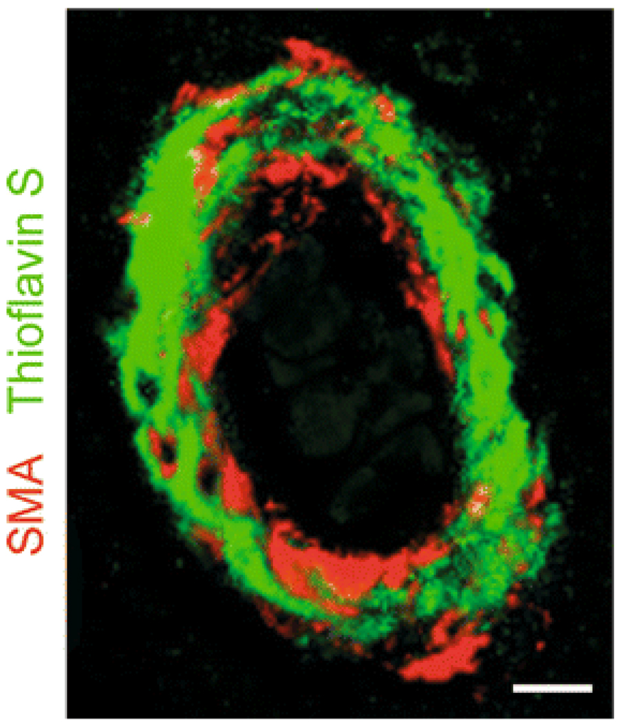
Finally, cerebral amyloid angiopathy (CAA) with Aβ deposits in the VSMC layer of small cerebral arteries (Fig. 1) is a major pathological insult to the neurovascular unit in AD [85]. Aβ plaques also accumulate onto and around cerebral capillaries [16, 92]. Decreased clearance of Aβ across the BBB and by cells of the neurovascular unit may contribute to CAA and parenchymal Aβ deposits, as discussed below. The prevalence of CAA in AD individuals and in the elderly population without AD is >80% and 10–40%, respectively [3, 36]. A strong association between CAA and cognitive impairment has also been established [4]. CAA is a significant cause of cerebral hemorrhage in elderly population [46]. Clinical topographical imaging has shown cerebral microbleedings are present in roughly 30% of AD cases [66]. On the other hand, lobar cerebral hemorrhage occurs in 7–18% of AD cases [36], which may be due to a significant loss of the VSMC layer [53] leading to vessel rupture.
Fig. 1.
Cerebral amyloid angiopathy in AD. Immunofluorescent staining of smooth muscle α actin (SMA; red) and amyloid staining (thioflavin S, green) in an AD cerebral vessel from Brodmann area 9. Staining shows significant amyloid accumulation in the vascular smooth muscle cell (VSMC) layer of this blood vessel. Amyloid accumulation may result from decreased Aβ clearance along the perivascular spaces caused by a decreased low-density lipoprotein receptor related protein-1 (LRP)-mediated Aβ clearance by VSMC, faulty Aβ clearance by perivascular macrophages and/or reduced passive Aβ drainage due to reductions in the arterial pulsatile blood flow. CAA can lead to spontaneous hemorrhage and rupture of the vessel wall due to a loss of the VSMC layer, enzymatically-induced breakdown of the vessel wall, oxidant stress and cytokine-mediated vascular injury. Scale bar 25 µm
Matrix metalloproteinases, reactive oxygen species (ROS) and CAA
Aβ treatment of human VSMC has been shown to significantly increase the activation of matrix metalloproteinase 2 (MMP2) via increasing the mRNA expression of membrane type 1 (MT1)-MMP, the primary MMP2 activator at the cell surface [49]. MMP9 specifically has been shown to be significantly present in postmortem AD tissue [2]. Activated MMP9 can degrade basement membranes, extracellular matrix proteins and tight junction proteins subsequently damaging the integrity of the BBB [31, 71, 100] and potentially leading to spontaneous cerebral hemorrhages.
In vivo microdialysis administration of Aβ40 in the rat brain has been shown to directly increase ROS, as measured by 2, 3-dihydroxybenzoic acid (reaction product of salicylate with hydroxyl radicals and/or peroxynitrite) [64]. Intracerebroventricular infusion of Aβ42 in rats was also shown to increase ROS by downregulating important endogenous antioxidant enzymes such as mitochondrial magnesium-superoxide dismutase 2 (SOD2), glutathione peroxidase and glutathione-S-transferase [55].
High levels of ROS in AD may damage proteins essentials for important neurovascular mechanisms. For example, Alzheimer’s patients have been shown to have high levels of oxidized soluble form of the low-density lipoprotein receptor related protein 1 (sLRP), which under normal conditions is the key endogenous Aβ chaperone protein in plasma [73] as discussed below. 14-3-3 ζ and γ isoforms, proteins that are involved in cell growth, survival and differentiation, were also found to be highly oxidized in brain extracts from AD and CAA patients [76]. Interestingly, it has been shown in Tg2576 mice overexpressing Swedish mutant APP695 that vascular oxidative stress precedes parenchymal oxidative stress [63], suggesting that vascular insults may be an early event in the development of AD-like pathology in this model.
Transport of Aβ via the receptor for advanced glycation endproducts (RAGE) across the BBB provides a major source of Aβ that can deposit in the brain and can directly lead to neuroinflammation via activating nuclear factor-κB mediated secretion of proinflammatory cytokines, such as tumor necrosis factor-α and interleukin 6 [23], that may reduce the BBB patency. The breakdown of the BBB may in turn disrupt the normal transport of nutrients, vitamins and electrolytes across the BBB, which are essential for proper neuronal functioning. Therefore, therapies that reduce ROS, MMP2, and MMP9, as for example with some forms of non-anticoagulant-activated protein C (APC) analogs [14, 37], or that block RAGE-Aβ interaction may be potentially useful strategies to correct BBB dysfunction in AD.
Aβ clearance hypothesis
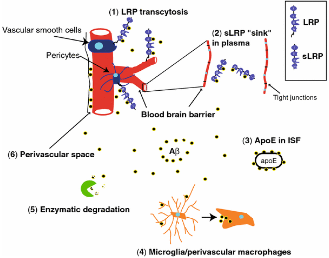
There is a significant body of evidence suggesting that Aβ accumulates in the wall of cerebral vessels and in the brains of AD individuals because of imbalances between its production and clearance from the brain [106]. Figure 2 illustrates some of the major Aβ clearance routes with emphasis on vascular clearance.
Fig. 2.
Essential Aβ clearance vascular and other routes. Aβ clearance can occur via several routes: 1 LRP-mediated transcytosis (purple, receptor) across the blood–brain barrier (red, capillaries) removes Aβ from brain interstitial fluid to blood and LRP-mediated degradation of Aβ on vascular smooth muscle cells and pericytes lowers Aβ levels in perivascular spaces (blue, cells), 2 soluble LRP, sLRP-mediated (purple, soluble receptor) endogenous Aβ “sink” action in plasma increases peripheral Aβ clearance and lowers the levels of free Aβ in the circulation which in turn promotes the cell surface LRP-mediated clearance of brain-derived Aβ across the blood–brain barrier, 3 Aβ chaperones in brain interstitial fluid such as ApoE isoforms may reduce clearance of brain-derived Aβ in an isoform-specific manner, i.e., apoE4 > apoE3 or apoE2, 4 clearance of Aβ by microglia and perivascular brain macrophages (orange, cells) from brain parenchyma and perivascular spaces, respectively, 5 direct enzymatic degradation of Aβ in the brain (green, enzymes), and 6 elimination of Aβ along the perivascular spaces by passive drainage that is influenced by the arterial pulstatile flow. The illustrated pathways by all means do not cover in detail all possible routes that control Aβ levels in the brain
Aβ that is produced both in the brain and periphery by a number of different cell types is transported across the BBB via receptor-mediated transcytosis. RAGE is the key receptor that transports Aβ from the blood into the brain [23], which occurs at a rate that is five- to sixfold lower than the rate determined for the transport of large neutral amino acids [78]. LRP is the major cell surface Aβ clearance receptor that transports Aβ out of the brain across the BBB [9, 25, 80] and promotes Aβ clearance on VSMC [87] (Fig. 2). Aβ is not only cleared from the brain interstitial fluid (ISF) [9] as a soluble peptide, but can also be transported by its chaperone proteins in the ISF, such as apolipoprotein E (apoE), apolipoprotein J, and α2-macroglobulin [103].
Aβ clearance directly into the blood seems to be the most prominent pathway, but it has also been suggested that there exists a perivascular route for the clearance of Aβ in the human brain [67] (Fig. 2). The pulsation force of cerebral blood vessels has been proposed to drive a Aβ drainage route along the perivascular spaces [97]. In this context, vessel constriction [15] and/or stiffening [97] may reduce Aβ clearance along the perivascular spaces by reducing the pulsatile flow which in turn would increase Aβ deposition in the arterial wall of AD patients.
Aβ can also be directly degraded by cells of the neurovascular unit. A vast body of evidence also suggests an important role of Aβ degrading enzymes such as insulin degrading enzyme and neprilysin in the clearance of Aβ (Fig. 2), which is not discussed in detail here, but is reviewed elsewhere [74].
If any of the clearance pathways are disrupted, soluble Aβ will accumulate and promote the formation of toxic Aβ oligomeric and aggregated species, which have devastating effects on the neurovascular unit. Furthermore, vasculotropic Aβ mutations, such as the Dutch mutation, decrease the clearance rate of the peptide by approximately 6.8-fold when compared with the wild-type Aβ peptide clearance rates [60].
sLRP “sink” mechanism for Aβ clearance
Soluble LRP (sLRP) in plasma has recently been identified as a major endogenous peripheral “sink” agent of Aβ [73] (Fig. 2). The N-terminus of LRP can be cleaved at the cell surface by β-secretase [93] and this extracellular domain of LRP exists in plasma as sLRP [68]. Aβ can bind directly to sLRP in vitro and µ70% of Aβ in human plasma is associated with sLRP [73]. Furthermore, sLRP in AD patient’s plasma is significantly oxidized and does not efficiently bind to Aβ. These data have suggested that sLRP in human plasma acts as peripheral sink pulling Aβ from the brain into the blood, and carrying it to the liver for degradation. Cell surface LRP also mediates systemic Aβ clearance by the liver [83]. Understanding the specific Aβ–LRP interactions will greatly advance our understanding of this Aβ clearance mechanism. It has been recently reported that Aβ binds to clusters II and IV of LRP via its C-terminal end [22].
ApoE and Aβ clearance across the BBB
ApoE is a reactive apolipoprotein that has a major function in the transport of lipids and cholesterol in our body. ApoE exists in 3 isoforms (apoE2, apoE3, and apoE4) in humans. Roughly, 40–65% of individuals that develop AD carry at least one apoE4 allele, and apoE4 homozygosity increases ones chance of developing AD from 20 to 90% [18]. On the other hand, carrying an apoE2 allele seems to reduce the risk of individuals to develop AD [17]. The exact relationship how apoE genotype influences AD pathogenesis and Aβ metabolism is still a subject of intense research.
With respect to vascular clearance, it has been shown that apoE is an Aβ chaperone protein [98] (Fig. 2). ApoE has been associated with impaired transport of Aβ across the BBB [9, 24, 58]. Particularly, apoE2-Aβ and apoE3-Aβ complexes are cleared at the BBB at a substantially faster rate than apoE4-Aβ complexes, and lipidated apoE-Aβ complexes are cleared across the BBB significantly slower than their respective lipid-poor apoE-Aβ complexes [24]. It seems that while free Aβ can be rapidly cleared from the brain mainly via LRP, Aβ-ApoE complexes are cleared by the very low-density lipoprotein receptor at a much slower rate, causing Aβ retention in the brain.
Interestingly, pericytes with apoE2 or apoE3 genotype compared to pericytes with an apoE4 allele were more resistant to toxic effects of Aβ40 carrying an E22Q mutation, as seen in hereditary cerebral hemorrhage with amyloidosis of the Dutch type [91]. The distribution of cerebrovascular amyloid in AD varies with apoE genotype and specifically the increasing dose of apoE4 alleles has been associated with increased CAA [1, 86]. Future research understanding cellular and molecular mechanism by which apoE genotype influences the disease process in AD individuals may be useful in developing both the diagnostic tools and new therapeutic opportunities for AD.
Myocardin and serum response factor transcriptional control of Aβ clearance
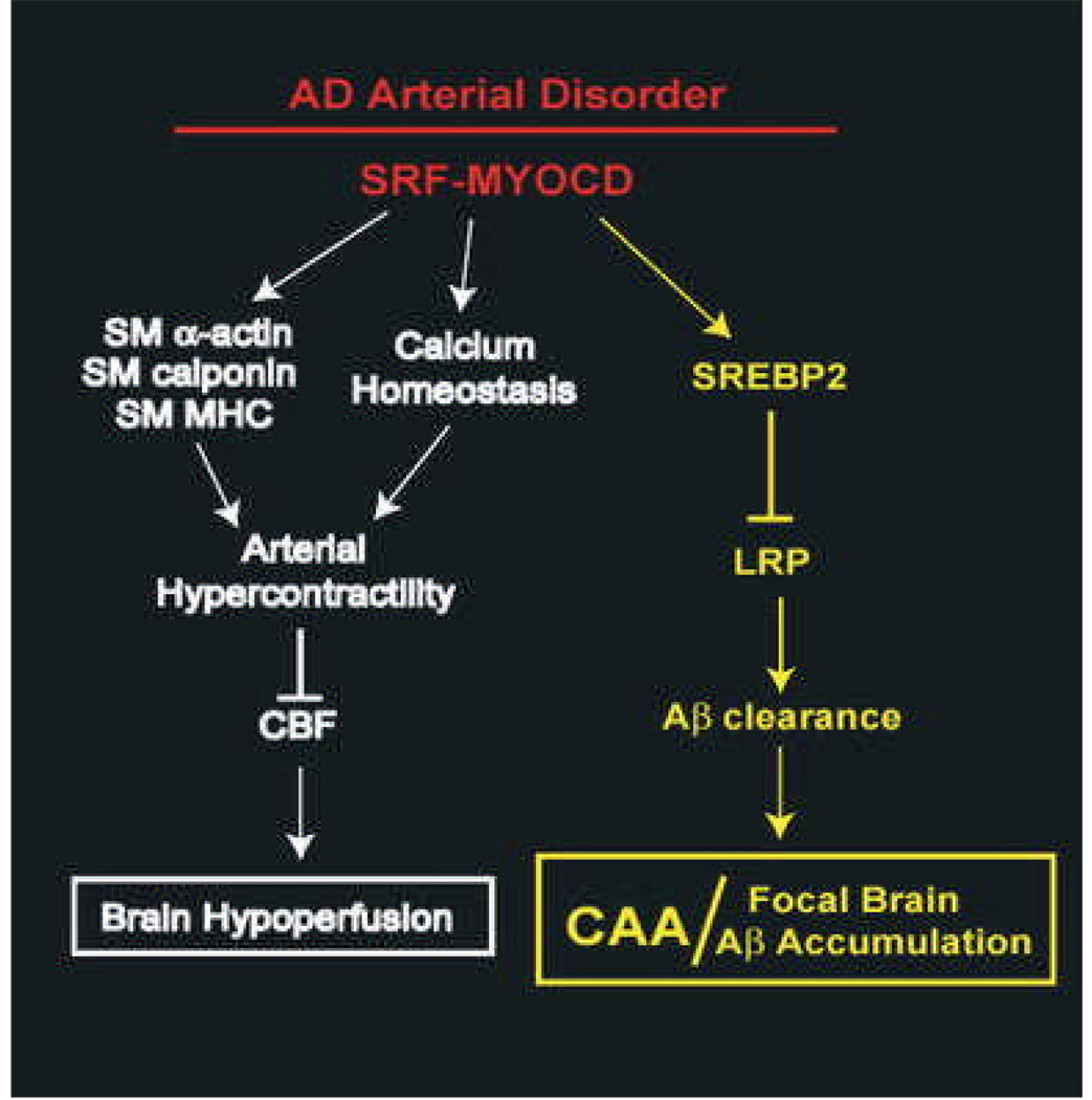
MYOCD, a SAF-A/B, Acinus and PIAS domain family nuclear protein, is a smooth muscle specific transcriptional co-activator that binds SRF to induce gene expression [95]. SRF is a ubiquitously expressed transcription factor that binds to a 10 base pair cis element called a CArG box, which is located in the regulatory region of numerous target genes [81]. MYOCD and SRF constitute a molecular switch for the VSMC differentiation program [13, 56]. We have shown that increased MYOCD and SRF expressed in AD VSMC contribute to arterial hypercontractility and cerebral hypoperfusion [15]. Specifically, MYOCD and SRF increase the levels of genes encoding contractile proteins and regulating calcium homeostasis causing arterial hypercontractility and reduced CBF (Fig. 3 white pathway). This chronic arterial hypercontractility may reduce the arterial pulsations because the vessel no longer properly relaxes, and therefore the contribution of Aβ clearance along the perivascular spaces may also be reduced.
Fig. 3.
MYOCD and SRF hypothesis of AD arterial pathology. High levels of SRF-MYOCD in AD VSMC contribute to brain hypoperfusion (white pathway) by increasing the expression of contractile proteins, such as smooth muscle (SM) α-actin, calponin, and myosin heavy chain (MHC) and by increasing the expression of genes that regulate calcium homeostasis. This leads to arterial hypercontractility, reduced resting cerebral blood flow (CBF) and attenuated CBF responses to brain activation, which ultimately creates a chronic hypoperfusion state. Furthermore, SRF-MYOCD potentiate CAA and focal brain Aβ accumulation (yellow pathway) via CArG-box dependent activation of SREBP2, which acts as transcriptional suppressor of LRP, a key Aβ clearance receptor
Recently, CAA-positive cerebral blood vessels isolated from AD patients have also been shown to have a significant increase in MYOCD and SRF levels [8] confirming the above findings. However, neither Aβ nor ROS effect the expression of MYOCD or SRF [15], therefore it seems that the increase in MYOCD and SRF is not due to oxidant stress or Aβ-related VSMC injury. Therefore, the high expression of MYOCD and SRF in CAA-positive AD vessels may be just by coincidence or alternatively, high levels of MYOCD and SRF may negatively affect the clearance of Aβ from the brain. Recent data, suggesting the latter, show that increased levels of MYOCD and SRF in AD VSMC may suppress Aβ clearance and thus exacerbate CAA. We have shown that high levels of MYOCD and SRF in vitro and in vivo directly lead to increased expression of sterol response element binding protein 2 (SREBP2), which mediates LRP transcriptional repression, causing CAA and focal brain Aβ accumulations due to reduced LRP-dependent clearance (Fig. 3 yellow pathway).
MYOCD and SRF activate SREBP2 transcription in a CArG dependent mechanism. SREBP2 represses LRP transcription via binding to the sterol response element in the LRP 5′ untranslated region [57]. Low levels of LRP in VSMC reduce the clearance of Aβ and therefore potentiate CAA and focal Aβ accumulation. It is of note that small pial and intracerebral arteries in AD do not typically develop atherosclerotic changes and accumulation of aggregated low-density lipoproteins and cholesterol esters [43]. According to the present data this could be due to MYOCD-SRF-mediated SREBP-2 stabilization and SREBP-2 dependent LRP downregulation. However, a potential side effect of such an “anti-atherosclerotic” VSMC phenotype in small resistant brain arteries, may be the loss of the LRP-dependent Aβ clearing function and development of CAA with focal brain amyloidosis.
Hypoxia has been shown to increase MYOCD levels in VSMC [8, 15, 69]. The molecular mechanism by which hypoxia increases MYOCD is not yet completely understood. It has been suggested that midkine upregulation under hypoxic conditions is responsible for MYOCD overexpression [69]. However, we have recently identified a putative conserved hypoxia response element in the MYOCD promoter, suggesting possible direct activation of MYOCD by hypoxia inducible factor 1α. More experimental data will be required to fully elucidate how hypoxia induces MYOCD.
Aβ clearance by microglia and perivascular macrophages
Microglial and perivascular macrophages may play role in clearing Aβ from the brain (Fig. 2) and in enhancing AD-related inflammation. Microglia have been shown to be significantly activated in AD brains and localized at sites of amyloid deposition [96]. Early activation of microglia in early AD pathogenesis has been show to be beneficial in scavenging and clearing toxic Aβ from the brain. Specifically, decreased microglia accumulation in CC-chemokine receptor deficient Tg-2576 AD transgenic mice increased mortality and Aβ accumulation particularly around blood vessels [27]. However, as the disease progresses the microglial activation may lead to inflammation, reduced Aβ clearance and severe neurodegeneration [40].
Perivascular macrophages are CD163 (hemoglobin-haptoglobin scavenger receptor) and CD206 (mannose receptor) positive immune cells located in outer aspects of blood vessels within the brain. Perivascular macrophages are antigen-presenting phagocytotic cells, and have been shown to respond to CNS inflammation. Interestingly, blood-derived macrophages from AD patients were shown to be less effective at phagocytosing Aβ compared with cells derived from non-demented control patients [30]. It has also been shown that Aβ induces migration of monocytes across the human BBB via RAGE and platelet endothelial cell adhesion molecule 1 [32]. Clodronate-mediated depletion of perivascular macrophages in vivo significantly increased CAA in TgCRND8 AD transgenic mice, whereas chitin stimulated perivascular macrophages turnover and promoted Aβ clearance [38]. These data suggest an important role of microglia and perivascular macrophages in Aβ clearance, and provide additional evidence that Aβ is cleared along perivascular spaces. Whether the role of these cells in AD is more beneficial or harmful still remains controversial. Further experimental data are needed to resolve this issue.
Conclusions
The elaborate interactions between the different cell types of the neurovascular unit provide the brain with a healthy environment that is required for proper cognitive functioning. The role of the neurovasculature and BBB in the progression of neurodegenerative diseases, such as AD reviewed here, are now becoming more fully appreciated. Recently, breakdown of the blood spinal cord barrier and reductions in microcirculation have been implicated in the early disease onset prior to motor neuron degeneration in mutant SOD1 expressing transgenic mice, a model of familial amyotrophic lateral sclerosis (ALS) [102]. It will be important to continue evaluating the degree of neurovascular and BBB involvement in other neurodegenerative diseases, such as ALS, multiple sclerosis, and Parkinson’s disease. Ultimately understanding the different molecular mechanisms of the neurovascular uncoupling and BBB breakdown present in neurodegenerative diseases will allow for the development of more effective diagnostic procedures and treatment strategies for different forms of neurodegeneration.
Acknowledgments
This work was supported by the United States National Institutes of Health grants R37AG023084 and R37NS34467 to B.V.Z.
References
- 1.Alonzo NC, Hyman BT, Rebeck GW, Greenberg SM. Progression of cerebral amyloid angiopathy: accumulation of amyloid-beta40 in affected vessels. J Neuropathol Exp Neurol. 1998;57:353–359. doi: 10.1097/00005072-199804000-00008. doi:10.1097/00005072-199804000-00008. [DOI] [PubMed] [Google Scholar]
- 2.Asahina M, Yoshiyama Y, Hattori T. Expression of matrix metalloproteinase-9 and urinary-type plasminogen activator in Alzheimer’s disease brain. Clin Neuropathol. 2001;20:60–63. [PubMed] [Google Scholar]
- 3.Attems J, Jellinger KA, Lintner F. Alzheimer’s disease pathology influences severity and topographical distribution of cerebral amyloid angiopathy. Acta Neuropathol. 2005;110:222–231. doi: 10.1007/s00401-005-1064-y. doi:10.1007/s00401-005-1064-y. [DOI] [PubMed] [Google Scholar]
- 4.Attems J, Quass M, Jellinger KA, Lintner F. Topographical distribution of cerebral amyloid angiopathy and its effect on cognitive decline are influenced by Alzheimer disease pathology. J Neurol Sci. 2007;257:49–55. doi: 10.1016/j.jns.2007.01.013. doi:10.1016/j.jns.2007.01.013. [DOI] [PubMed] [Google Scholar]
- 5.Bailey TL, Rivara CB, Rocher AB, Hof PR. The nature and effects of cortical microvascular pathology in aging and Alzheimer’s disease. Neurol Res. 2004;26:573–578. doi: 10.1179/016164104225016272. doi:10.1179/016164104225016272. [DOI] [PubMed] [Google Scholar]
- 6.Beach TG, Wilson JR, Sue LI, et al. Circle of Willis atherosclerosis: association with Alzheimer’s disease, neuritic plaques and neurofibrillary tangles. Acta Neuropathol. 2007;113:13–21. doi: 10.1007/s00401-006-0136-y. doi:10.1007/s00401-006-0136-y. [DOI] [PubMed] [Google Scholar]
- 7.Begley DJ, Brightman MW. Structural and functional aspects of the blood-brain barrier. Prog Drug Res. 2003;61:39–78. doi: 10.1007/978-3-0348-8049-7_2. [DOI] [PubMed] [Google Scholar]
- 8.Bell RD, Deane R, Chow N, et al. SRF and myocardin regulate LRP-mediated amyloid-beta clearance in brain vascular cells. Nat Cell Biol. 2009;11:143–153. doi: 10.1038/ncb1819. doi:10.1038/ncb1819. [DOI] [PMC free article] [PubMed] [Google Scholar]
- 9.Bell RD, Sagare AP, Friedman AE, et al. Transport pathways for clearance of human Alzheimer’s amyloid beta-peptide and apolipoproteins E and J in the mouse central nervous system. J Cereb Blood Flow Metab. 2007;27:909–918. doi: 10.1038/sj.jcbfm.9600419. [DOI] [PMC free article] [PubMed] [Google Scholar]
- 10.Bertram L, Tanzi RE. Thirty years of Alzheimer’s disease genetics: the implications of systematic meta-analyses. Nat Rev Neurosci. 2008;9:768–778. doi: 10.1038/nrn2494. doi:10.1038/nrn2494. [DOI] [PubMed] [Google Scholar]
- 11.Berzin TM, Zipser BD, Rafii MS, et al. Agrin and microvascular damage in Alzheimer’s disease. Neurobiol Aging. 2000;21:349–355. doi: 10.1016/s0197-4580(00)00121-4. doi:10.1016/S0197-4580(00)00121-4. [DOI] [PubMed] [Google Scholar]
- 12.Bradbury M. Cerebral arterial supply. In: Bradbury M, editor. The concept of a blood–brain barrier. New York: Wiley; 1979. pp. 18–19. [Google Scholar]
- 13.Chen J, Kitchen CM, Streb JW, Miano JM. Myocardin: a component of a molecular switch for smooth muscle differentiation. J Mol Cell Cardiol. 2002;34:1345–1356. doi: 10.1006/jmcc.2002.2086. doi:10.1006/jmcc.2002.2086. [DOI] [PubMed] [Google Scholar]
- 14.Cheng T, Petraglia AL, Li Z, et al. Activated protein C inhibits tissue plasminogen activator-induced brain hemorrhage. Nat Med. 2006;12:1278–1285. doi: 10.1038/nm1498. doi:10.1038/nm1498. [DOI] [PubMed] [Google Scholar]
- 15.Chow N, Bell RD, Deane R, et al. Serum response factor and myocardin mediate arterial hypercontractility and cerebral blood flow dysregulation in Alzheimer’s phenotype. Proc Natl Acad Sci USA. 2007;104:823–828. doi: 10.1073/pnas.0608251104. doi:10.1073/pnas.0608251104. [DOI] [PMC free article] [PubMed] [Google Scholar]
- 16.Clifford PM, Zarrabi S, Siu G, et al. Abeta peptides can enter the brain through a defective blood-brain barrier and bind selectively to neurons. Brain Res. 2007;1142:223–236. doi: 10.1016/j.brainres.2007.01.070. doi:10.1016/j.brainres.2007.01.070. [DOI] [PubMed] [Google Scholar]
- 17.Corder EH, Saunders AM, Risch NJ, et al. Protective effect of apolipoprotein E type 2 allele for late onset Alzheimer disease. Nat Genet. 1994;7:180–184. doi: 10.1038/ng0694-180. doi:10.1038/ng0694-180. [DOI] [PubMed] [Google Scholar]
- 18.Corder EH, Saunders AM, Strittmatter WJ, et al. Gene dose of apolipoprotein E type 4 allele and the risk of Alzheimer’s disease in late onset families. Science. 1993;261:921–923. doi: 10.1126/science.8346443. doi:10.1126/science.8346443. [DOI] [PubMed] [Google Scholar]
- 19.Davis J, Xu F, Deane R, et al. Early-onset and robust cerebral microvascular accumulation of amyloid beta-protein in transgenic mice expressing low levels of a vasculotropic Dutch/Iowa mutant form of amyloid beta-protein precursor. J Biol Chem. 2004;279:20296–20306. doi: 10.1074/jbc.M312946200. doi:10.1074/jbc.M312946200. [DOI] [PubMed] [Google Scholar]
- 20.Davson H, Welch K, Segal MB. Morphological aspects of the barriers: perivascular spaces. In: Davson H, editor. The physiology and pathophysiology of the cerebrospinal fluid. New York: Churchill Livingstone; 1987. pp. 135–137. [Google Scholar]
- 21.de la Torre JC. Is Alzheimer’s disease a neurodegenerative or a vascular disorder? Data, dogma, and dialectics. Lancet Neurol. 2004;3:184–190. doi: 10.1016/S1474-4422(04)00683-0. doi:10.1016/S1474-4422(04)00683-0. [DOI] [PubMed] [Google Scholar]
- 22.Deane R, Bell RD, Sagare A, Zlokovic BZ. Clearance of amyloid-beta peptide across the blood–brain barrier: implications for therapies in Alzhiemer’s disease. CNS Neurol Disord Drug Targets. 2009;8 doi: 10.2174/187152709787601867. doi:10.2174/187152709787601867 (in press) [DOI] [PMC free article] [PubMed] [Google Scholar]
- 23.Deane R, Du Yan S, Submamaryan RK, et al. RAGE mediates amyloid-beta peptide transport across the blood-brain barrier and accumulation in brain. Nat Med. 2003;9:907–913. doi: 10.1038/nm890. doi:10.1038/nm890. [DOI] [PubMed] [Google Scholar]
- 24.Deane R, Sagare A, Hamm K, et al. apoE isoform-specific disruption of amyloid beta peptide clearance from mouse brain. J Clin Invest. 2008;118:4002–4013. doi: 10.1172/JCI36663. doi:10.1172/JCI36663. [DOI] [PMC free article] [PubMed] [Google Scholar]
- 25.Deane R, Wu Z, Sagare A, et al. LRP/amyloid beta-peptide interaction mediates differential brain efflux of Abeta isoforms. Neuron. 2004;43:333–344. doi: 10.1016/j.neuron.2004.07.017. doi:10.1016/j.neuron.2004.07.017. [DOI] [PubMed] [Google Scholar]
- 26.Drzezga A, Lautenschlager N, Siebner H, et al. Cerebral metabolic changes accompanying conversion of mild cognitive impairment into Alzheimer’s disease: a PET follow-up study. Eur J Nucl Med Mol Imaging. 2003;30:1104–1113. doi: 10.1007/s00259-003-1194-1. doi:10.1007/s00259-003-1194-1. [DOI] [PubMed] [Google Scholar]
- 27.El Khoury J, Toft M, Hickman SE, et al. Ccr2 deficiency impairs microglial accumulation and accelerates progression of Alzheimer-like disease. Nat Med. 2007;13:432–438. doi: 10.1038/nm1555. doi:10.1038/nm1555. [DOI] [PubMed] [Google Scholar]
- 28.Ervin JF, Pannell C, Szymanski M, Welsh-Bohmer K, Schmechel DE, Hulette CM. Vascular smooth muscle actin is reduced in Alzheimer disease brain: a quantitative analysis. J Neuropathol Exp Neurol. 2004;63:735–741. doi: 10.1093/jnen/63.7.735. [DOI] [PubMed] [Google Scholar]
- 29.Farkas E, Luiten PG. Cerebral microvascular pathology in aging and Alzheimer’s disease. Prog Neurobiol. 2001;64:575–611. doi: 10.1016/s0301-0082(00)00068-x. doi:10.1016/S0301-0082(00)00068-X. [DOI] [PubMed] [Google Scholar]
- 30.Fiala M, Lin J, Ringman J, et al. Ineffective phagocytosis of amyloid-beta by macrophages of Alzheimer’s disease patients. J Alzheimers Dis. 2005;7:221–232. doi: 10.3233/jad-2005-7304. discussion 55–62. [DOI] [PubMed] [Google Scholar]
- 31.Fukuda S, Fini CA, Mabuchi T, Koziol JA, Eggleston LL, Jr, del Zoppo GJ. Focal cerebral ischemia induces active proteases that degrade microvascular matrix. Stroke. 2004;35:998–1004. doi: 10.1161/01.STR.0000119383.76447.05. doi:10.1161/01.STR.0000119383.76447.05. [DOI] [PMC free article] [PubMed] [Google Scholar]
- 32.Giri R, Shen Y, Stins M, et al. Beta-amyloid-induced migration of monocytes across human brain endothelial cells involves RAGE and PECAM-1. Am J Physiol Cell Physiol. 2000;279:C1772–C1781. doi: 10.1152/ajpcell.2000.279.6.C1772. [DOI] [PubMed] [Google Scholar]
- 33.Girouard H, Iadecola C. Neurovascular coupling in the normal brain and in hypertension, stroke, and Alzheimer disease. J Appl Physiol. 2006;100:328–335. doi: 10.1152/japplphysiol.00966.2005. doi:10.1152/japplphysiol.00966.2005. [DOI] [PubMed] [Google Scholar]
- 34.Grabowski TJ, Cho HS, Vonsattel JP, Rebeck GW, Greenberg SM. Novel amyloid precursor protein mutation in an Iowa family with dementia and severe cerebral amyloid angiopathy. Ann Neurol. 2001;49:697–705. doi: 10.1002/ana.1009. doi:10.1002/ana.1009. [DOI] [PubMed] [Google Scholar]
- 35.Grammas P, Yamada M, Zlokovic B. The cerebromicrovasculature: a key player in the pathogenesis of Alzheimer’s disease. J Alzheimers Dis. 2002;4:217–223. doi: 10.3233/jad-2002-4311. [DOI] [PubMed] [Google Scholar]
- 36.Greenberg SM, Gurol ME, Rosand J, Smith EE. Amyloid angiopathy-related vascular cognitive impairment. Stroke. 2004;35:2616–2619. doi: 10.1161/01.STR.0000143224.36527.44. doi:10.1161/01.STR.0000143224.36527.44. [DOI] [PubMed] [Google Scholar]
- 37.Griffin JH, Zlokovic B, Fernandez JA. Activated protein C: potential therapy for severe sepsis, thrombosis, and stroke. Semin Hematol. 2002;39:197–205. doi: 10.1053/shem.2002.34093. doi:10.1053/shem.2002.34093. [DOI] [PubMed] [Google Scholar]
- 38.Hawkes CA, McLaurin J. Selective targeting of perivascular macrophages for clearance of beta-amyloid in cerebral amyloid angiopathy. Proc Natl Acad Sci USA. 2009;106:1261–1266. doi: 10.1073/pnas.0805453106. doi:10.1073/pnas.0805453106. [DOI] [PMC free article] [PubMed] [Google Scholar]
- 39.Hendriks L, van Duijn CM, Cras P, et al. Presenile dementia and cerebral haemorrhage linked to a mutation at codon 692 of the beta-amyloid precursor protein gene. Nat Genet. 1992;1:218–221. doi: 10.1038/ng0692-218. doi:10.1038/ng0692-218. [DOI] [PubMed] [Google Scholar]
- 40.Hickman SE, Allison EK, El Khoury J. Microglial dysfunction and defective beta-amyloid clearance pathways in aging Alzheimer’s disease mice. J Neurosci. 2008;28:8354–8360. doi: 10.1523/JNEUROSCI.0616-08.2008. doi:10.1523/JNEUROSCI.0616-08.2008. [DOI] [PMC free article] [PubMed] [Google Scholar]
- 41.Hirao K, Ohnishi T, Hirata Y, et al. The prediction of rapid conversion to Alzheimer’s disease in mild cognitive impairment using regional cerebral blood flow SPECT. Neuroimage. 2005;28:1014–1021. doi: 10.1016/j.neuroimage.2005.06.066. doi:10.1016/j.neuroimage.2005.06.066. [DOI] [PubMed] [Google Scholar]
- 42.Hofman A, Ott A, Breteler MM, et al. Atherosclerosis, apolipoprotein E, and prevalence of dementia and Alzheimer’s disease in the Rotterdam Study. Lancet. 1997;349:151–154. doi: 10.1016/S0140-6736(96)09328-2. doi:10.1016/S0140-6736(96)09328-2. [DOI] [PubMed] [Google Scholar]
- 43.Honig LS, Kukull W, Mayeux R. Atherosclerosis and AD: analysis of data from the US National Alzheimer’s Coordinating Center. Neurology. 2005;64:494–500. doi: 10.1212/01.WNL.0000150886.50187.30. [DOI] [PubMed] [Google Scholar]
- 44.Hunt A, Schonknecht P, Henze M, Seidl U, Haberkorn U, Schroder J. Reduced cerebral glucose metabolism in patients at risk for Alzheimer’s disease. Psychiatry Res. 2007;155:147–154. doi: 10.1016/j.pscychresns.2006.12.003. doi:10.1016/j.pscychresns.2006.12.003. [DOI] [PubMed] [Google Scholar]
- 45.Iadecola C. Neurovascular regulation in the normal brain and in Alzheimer’s disease. Nat Rev Neurosci. 2004;5:347–360. doi: 10.1038/nrn1387. doi:10.1038/nrn1387. [DOI] [PubMed] [Google Scholar]
- 46.Itoh Y, Yamada M, Hayakawa M, Otomo E, Miyatake T. Cerebral amyloid angiopathy: a significant cause of cerebellar as well as lobar cerebral hemorrhage in the elderly. J Neurol Sci. 1993;116:135–141. doi: 10.1016/0022-510x(93)90317-r. doi:10.1016/0022-510X(93)90317-R. [DOI] [PubMed] [Google Scholar]
- 47.Jellinger KA. Alzheimer disease and cerebrovascular pathology: an update. J Neural Transm. 2002;109:813–836. doi: 10.1007/s007020200068. doi:10.1007/s007020200068. [DOI] [PubMed] [Google Scholar]
- 48.Johnson NA, Jahng GH, Weiner MW, et al. Pattern of cerebral hypoperfusion in Alzheimer disease and mild cognitive impairment measured with arterial spin-labeling MR imaging: initial experience. Radiology. 2005;234:851–859. doi: 10.1148/radiol.2343040197. doi:10.1148/radiol.2343040197. [DOI] [PMC free article] [PubMed] [Google Scholar]
- 49.Jung SS, Zhang W, Van Nostrand WE. Pathogenic A beta induces the expression and activation of matrix metalloproteinase-2 in human cerebrovascular smooth muscle cells. J Neurochem. 2003;85:1208–1215. doi: 10.1046/j.1471-4159.2003.01745.x. doi:10.1046/j.1471-4159.2003.01745.x. [DOI] [PubMed] [Google Scholar]
- 50.Kalaria RN, Hedera P. Differential degeneration of the cerebral microvasculature in Alzheimer’s disease. Neuroreport. 1995;6:477–480. doi: 10.1097/00001756-199502000-00018. doi:10.1097/00001756-199502000-00018. [DOI] [PubMed] [Google Scholar]
- 51.Kalaria RN, Pax AB. Increased collagen content of cerebral microvessels in Alzheimer’s disease. Brain Res. 1995;705:349–352. doi: 10.1016/0006-8993(95)01250-8. doi:10.1016/0006-8993(95)01250-8. [DOI] [PubMed] [Google Scholar]
- 52.Kalback W, Esh C, Castano EM, et al. Atherosclerosis, vascular amyloidosis and brain hypoperfusion in the pathogenesis of sporadic Alzheimer’s disease. Neurol Res. 2004;26:525–539. doi: 10.1179/016164104225017668. doi:10.1179/016164104225017668. [DOI] [PubMed] [Google Scholar]
- 53.Kawai M, Kalaria RN, Cras P, et al. Degeneration of vascular muscle cells in cerebral amyloid angiopathy of Alzheimer disease. Brain Res. 1993;623:142–146. doi: 10.1016/0006-8993(93)90021-e. doi:10.1016/0006-8993(93)90021-E. [DOI] [PubMed] [Google Scholar]
- 54.Kida S, Steart PV, Zhang ET, Weller RO. Perivascular cells act as scavengers in the cerebral perivascular spaces and remain distinct from pericytes, microglia and macrophages. Acta Neuropathol. 1993;85:646–652. doi: 10.1007/BF00334675. doi:10.1007/BF00334675. [DOI] [PubMed] [Google Scholar]
- 55.Kim HC, Yamada K, Nitta A, et al. Immunocytochemical evidence that amyloid beta (1–42) impairs endogenous antioxidant systems in vivo. Neuroscience. 2003;119:399–419. doi: 10.1016/s0306-4522(02)00993-4. doi:10.1016/S0306-4522(02)00993-4. [DOI] [PubMed] [Google Scholar]
- 56.Li S, Wang DZ, Wang Z, Richardson JA, Olson EN. The serum response factor coactivator myocardin is required for vascular smooth muscle development. Proc Natl Acad Sci USA. 2003;100:9366–9370. doi: 10.1073/pnas.1233635100. doi:10.1073/pnas.1233635100. [DOI] [PMC free article] [PubMed] [Google Scholar]
- 57.Llorente-Cortes V, Costales P, Bernues J, Camino-Lopez S, Badimon L. Sterol regulatory element-binding protein-2 negatively regulates low density lipoprotein receptor-related protein transcription. J Mol Biol. 2006;359:950–960. doi: 10.1016/j.jmb.2006.04.008. doi:10.1016/j.jmb.2006.04.008. [DOI] [PubMed] [Google Scholar]
- 58.Martel CL, Mackic JB, Matsubara E, et al. Isoform-specific effects of apolipoproteins E2, E3, and E4 on cerebral capillary sequestration and blood-brain barrier transport of circulating Alzheimer’s amyloid beta. J Neurochem. 1997;69:1995–2004. doi: 10.1046/j.1471-4159.1997.69051995.x. [DOI] [PubMed] [Google Scholar]
- 59.McComb JG, Zlokovic BV. Cerebrospinal fluid and the blood–brain interface. In: Cheek WR, editor. Pediatric neurosurgery: surgery of the developing nervous system. 3rd edn. Philladelphia: Saunders; 1994. pp. 180–198. [Google Scholar]
- 60.Monro OR, Mackic JB, Yamada S, et al. Substitution at codon 22 reduces clearance of Alzheimer’s amyloid-beta peptide from the cerebrospinal fluid and prevents its transport from the central nervous system into blood. Neurobiol Aging. 2002;23:405–412. doi: 10.1016/s0197-4580(01)00317-7. doi:10.1016/S0197-4580(01)00317-7. [DOI] [PubMed] [Google Scholar]
- 61.Mosconi L, Sorbi S, de Leon MJ, et al. Hypometabolism exceeds atrophy in presymptomatic early-onset familial Alzheimer’s disease. J Nucl Med. 2006;47:1778–1786. [PubMed] [Google Scholar]
- 62.Nilsberth C, Westlind-Danielsson A, Eckman CB, et al. The ‘Arctic’ APP mutation (E693G) causes Alzheimer’s disease by enhanced Abeta protofibril formation. Nat Neurosci. 2001;4:887–893. doi: 10.1038/nn0901-887. doi:10.1038/nn0901-887. [DOI] [PubMed] [Google Scholar]
- 63.Park L, Anrather J, Forster C, Kazama K, Carlson GA, Iadecola C. Abeta-induced vascular oxidative stress and attenuation of functional hyperemia in mouse somatosensory cortex. J Cereb Blood Flow Metab. 2004;24:334–342. doi: 10.1097/01.WCB.0000105800.49957.1E. doi:10.1097/01.WCB.0000105800.49957.1E. [DOI] [PubMed] [Google Scholar]
- 64.Parks JK, Smith TS, Trimmer PA, Bennett JP, Jr, Parker WD., Jr Neurotoxic Abeta peptides increase oxidative stress in vivo through NMDA-receptor and nitric-oxide-synthase mechanisms, and inhibit complex IV activity and induce a mitochondrial permeability transition in vitro. J Neurochem. 2001;76:1050–1056. doi: 10.1046/j.1471-4159.2001.00112.x. doi:10.1046/j.1471-4159.2001.00112.x. [DOI] [PubMed] [Google Scholar]
- 65.Peppiatt CM, Howarth C, Mobbs P, Attwell D. Bidirectional control of CNS capillary diameter by pericytes. Nature. 2006;443:700–704. doi: 10.1038/nature05193. doi:10.1038/nature05193. [DOI] [PMC free article] [PubMed] [Google Scholar]
- 66.Pettersen JA, Sathiyamoorthy G, Gao FQ, et al. Microbleed topography, leukoaraiosis, and cognition in probable Alzheimer disease from the Sunnybrook dementia study. Arch Neurol. 2008;65:790–795. doi: 10.1001/archneur.65.6.790. doi:10.1001/archneur.65.6.790. [DOI] [PubMed] [Google Scholar]
- 67.Preston SD, Steart PV, Wilkinson A, Nicoll JA, Weller RO. Capillary and arterial cerebral amyloid angiopathy in Alzheimer’s disease: defining the perivascular route for the elimination of amyloid beta from the human brain. Neuropathol Appl Neurobiol. 2003;29:106–117. doi: 10.1046/j.1365-2990.2003.00424.x. doi:10.1046/j.1365-2990.2003.00424.x. [DOI] [PubMed] [Google Scholar]
- 68.Quinn KA, Grimsley PG, Dai YP, Tapner M, Chesterman CN, Owensby DA. Soluble low density lipoprotein receptor-related protein (LRP) circulates in human plasma. J Biol Chem. 1997;272:23946–23951. doi: 10.1074/jbc.272.38.23946. doi:10.1074/jbc.272.38.23946. [DOI] [PubMed] [Google Scholar]
- 69.Reynolds PR, Mucenski ML, Le Cras TD, Nichols WC, Whitsett JA. Midkine is regulated by hypoxia and causes pulmonary vascular remodeling. J Biol Chem. 2004;279:37124–37132. doi: 10.1074/jbc.M405254200. doi:10.1074/jbc.M405254200. [DOI] [PubMed] [Google Scholar]
- 70.Rombouts SA, Goekoop R, Stam CJ, Barkhof F, Scheltens P. Delayed rather than decreased BOLD response as a marker for early Alzheimer’s disease. Neuroimage. 2005;26:1078–1085. doi: 10.1016/j.neuroimage.2005.03.022. doi:10.1016/j.neuroimage.2005.03.022. [DOI] [PubMed] [Google Scholar]
- 71.Rosenberg GA. Matrix metalloproteinases and their multiple roles in neurodegenerative diseases. Lancet Neurol. 2009;8:205–216. doi: 10.1016/S1474-4422(09)70016-X. doi:10.1016/S1474-4422(09)70016-X. [DOI] [PubMed] [Google Scholar]
- 72.Ruitenberg A, den Heijer T, Bakker SL, et al. Cerebral hypoperfusion and clinical onset of dementia: the Rotterdam Study. Ann Neurol. 2005;57:789–794. doi: 10.1002/ana.20493. doi:10.1002/ana.20493. [DOI] [PubMed] [Google Scholar]
- 73.Sagare A, Deane R, Bell RD, et al. Clearance of amyloid-beta by circulating lipoprotein receptors. Nat Med. 2007;13:1029–1031. doi: 10.1038/nm1635. doi:10.1038/nm1635. [DOI] [PMC free article] [PubMed] [Google Scholar]
- 74.Saido TC, Iwata N. Metabolism of amyloid beta peptide and pathogenesis of Alzheimer’s disease. Towards presymptomatic diagnosis, prevention and therapy. Neurosci Res. 2006;54:235–253. doi: 10.1016/j.neures.2005.12.015. doi:10.1016/j.neures.2005.12.015. [DOI] [PubMed] [Google Scholar]
- 75.Samuraki M, Matsunari I, Chen WP, et al. Partial volume effect-corrected FDG PET and grey matter volume loss in patients with mild Alzheimer’s disease. Eur J Nucl Med Mol Imaging. 2007;34:1658–1669. doi: 10.1007/s00259-007-0454-x. doi:10.1007/s00259-007-0454-x. [DOI] [PubMed] [Google Scholar]
- 76.Santpere G, Puig B, Ferrer I. Oxidative damage of 14-3-3 zeta and gamma isoforms in Alzheimer’s disease and cerebral amyloid angiopathy. Neuroscience. 2007;146:1640–1651. doi: 10.1016/j.neuroscience.2007.03.013. doi:10.1016/j.neuroscience.2007.03.013. [DOI] [PubMed] [Google Scholar]
- 77.Scheibel AB, Duong TH, Jacobs R. Alzheimer’s disease as a capillary dementia. Ann Med. 1989;21:103–107. doi: 10.3109/07853898909149194. doi:10.3109/07853898909149194. [DOI] [PubMed] [Google Scholar]
- 78.Segal MB, Preston JE, Collis CS, Zlokovic BV. Kinetics and Na independence of amino acid uptake by blood side of perfused sheep choroid plexus. Am J Physiol. 1990;258:F1288–F1294. doi: 10.1152/ajprenal.1990.258.5.F1288. [DOI] [PubMed] [Google Scholar]
- 79.Shankar GM, Li S, Mehta TH, et al. Amyloid-beta protein dimers isolated directly from Alzheimer’s brains impair synaptic plasticity and memory. Nat Med. 2008;14:837–842. doi: 10.1038/nm1782. doi:10.1038/nm1782. [DOI] [PMC free article] [PubMed] [Google Scholar]
- 80.Shibata M, Yamada S, Kumar SR, et al. Clearance of Alzheimer’s amyloid-beta, (1–40) peptide from brain by LDL receptor-related protein-1 at the blood-brain barrier. J Clin Invest. 2000;106:1489–1499. doi: 10.1172/JCI10498. doi:10.1172/JCI10498. [DOI] [PMC free article] [PubMed] [Google Scholar]
- 81.Sun Q, Chen G, Streb JW, et al. Defining the mammalian CArGome. Genome Res. 2006;16:197–207. doi: 10.1101/gr.4108706. doi:10.1101/gr.4108706. [DOI] [PMC free article] [PubMed] [Google Scholar]
- 82.Tagliavini FRG, Padovani A, Magoni M, Andora G, Sgarzi M, Bizzi ASM, Carella F, Morbin M, Giaccone G, Bugiani O. A new beta APP mutation related to hereditary cerebral haemorrhage. Alzheimers Rep. 1999;2:S28. [Google Scholar]
- 83.Tamaki C, Ohtsuki S, Iwatsubo T, et al. Major involvement of low-density lipoprotein receptor-related protein 1 in the clearance of plasma free amyloid beta-peptide by the liver. Pharm Res. 2006;23:1407–1416. doi: 10.1007/s11095-006-0208-7. doi:10.1007/s11095-006-0208-7. [DOI] [PubMed] [Google Scholar]
- 84.Tanzi RE, Bertram L. Twenty years of the Alzheimer’s disease amyloid hypothesis: a genetic perspective. Cell. 2005;120:545–555. doi: 10.1016/j.cell.2005.02.008. doi:10.1016/j.cell.2005.02.008. [DOI] [PubMed] [Google Scholar]
- 85.Thal DR, Griffin WS, de Vos RA, Ghebremedhin E. Cerebral amyloid angiopathy and its relationship to Alzheimer’s disease. Acta Neuropathol. 2008;115:599–609. doi: 10.1007/s00401-008-0366-2. doi:10.1007/s00401-008-0366-2. [DOI] [PubMed] [Google Scholar]
- 86.Trembath D, Ervin JF, Broom L, et al. The distribution of cerebrovascular amyloid in Alzheimer’s disease varies with ApoE genotype. Acta Neuropathol. 2007;113:23–31. doi: 10.1007/s00401-006-0162-9. doi:10.1007/s00401-006-0162-9. [DOI] [PubMed] [Google Scholar]
- 87.Urmoneit B, Prikulis I, Wihl G, et al. Cerebrovascular smooth muscle cells internalize Alzheimer amyloid beta protein via a lipoprotein pathway: implications for cerebral amyloid angiopathy. Lab Invest. 1997;77:157–166. [PubMed] [Google Scholar]
- 88.Van Broeckhoven C, Haan J, Bakker E, et al. Amyloid beta protein precursor gene and hereditary cerebral hemorrhage with amyloidosis (Dutch) Science. 1990;248:1120–1122. doi: 10.1126/science.1971458. doi:10.1126/science.1971458. [DOI] [PubMed] [Google Scholar]
- 89.van Oijen M, de Jong FJ, Witteman JC, Hofman A, Koudstaal PJ, Breteler MM. Atherosclerosis and risk for dementia. Ann Neurol. 2007;61:403–410. doi: 10.1002/ana.21073. doi:10.1002/ana.21073. [DOI] [PubMed] [Google Scholar]
- 90.Verbeek MM, Otte-Holler I, van den Born J, et al. Agrin is a major heparan sulfate proteoglycan accumulating in Alzheimer’s disease brain. Am J Pathol. 1999;155:2115–2125. doi: 10.1016/S0002-9440(10)65529-0. [DOI] [PMC free article] [PubMed] [Google Scholar]
- 91.Verbeek MM, Van Nostrand WE, Otte-Holler I, Wesseling P, De Waal RM. Amyloid-beta-induced degeneration of human brain pericytes is dependent on the apolipoprotein E genotype. Ann N Y Acad Sci. 2000;903:187–199. doi: 10.1111/j.1749-6632.2000.tb06368.x. doi:10.1111/j.1749-6632.2000.tb06368.x. [DOI] [PubMed] [Google Scholar]
- 92.Vinters HV, Secor DL, Read SL, et al. Microvasculature in brain biopsy specimens from patients with Alzheimer’s disease: an immunohistochemical and ultrastructural study. Ultrastruct Pathol. 1994;18:333–348. doi: 10.3109/01913129409023202. doi:10.3109/01913129409023202. [DOI] [PubMed] [Google Scholar]
- 93.von Arnim CA, Kinoshita A, Peltan ID, et al. The low density lipoprotein receptor-related protein (LRP) is a novel beta-secretase (BACE1) substrate. J Biol Chem. 2005;280:17777–17785. doi: 10.1074/jbc.M414248200. doi:10.1074/jbc.M414248200. [DOI] [PubMed] [Google Scholar]
- 94.von Tell D, Armulik A, Betsholtz C. Pericytes and vascular stability. Exp Cell Res. 2006;312:623–629. doi: 10.1016/j.yexcr.2005.10.019. doi:10.1016/j.yexcr.2005.10.019. [DOI] [PubMed] [Google Scholar]
- 95.Wang D, Chang PS, Wang Z, et al. Activation of cardiac gene expression by myocardin, a transcriptional cofactor for serum response factor. Cell. 2001;105:851–862. doi: 10.1016/s0092-8674(01)00404-4. doi:10.1016/S0092-8674(01)00404-4. [DOI] [PubMed] [Google Scholar]
- 96.Wegiel J, Wisniewski HM. The complex of microglial cells and amyloid star in three-dimensional reconstruction. Acta Neuropathol. 1990;81:116–124. doi: 10.1007/BF00334499. doi:10.1007/BF00334499. [DOI] [PubMed] [Google Scholar]
- 97.Weller RO, Djuanda E, Yow HY, Carare RO. Lymphatic drainage of the brain and the pathophysiology of neurological disease. Acta Neuropathol. 2009;117:1–14. doi: 10.1007/s00401-008-0457-0. doi:10.1007/s00401-008-0457-0. [DOI] [PubMed] [Google Scholar]
- 98.Wisniewski T, Frangione B. Apolipoprotein E: a pathological chaperone protein in patients with cerebral and systemic amyloid. Neurosci Lett. 1992;135:235–238. doi: 10.1016/0304-3940(92)90444-c. doi:10.1016/0304-3940(92)90444-C. [DOI] [PubMed] [Google Scholar]
- 99.Wu Z, Guo H, Chow N, et al. Role of the MEOX2 homeobox gene in neurovascular dysfunction in Alzheimer disease. Nat Med. 2005;11:959–965. doi: 10.1038/nm1287. [DOI] [PubMed] [Google Scholar]
- 100.Yang Y, Estrada EY, Thompson JF, Liu W, Rosenberg GA. Matrix metalloproteinase-mediated disruption of tight junction proteins in cerebral vessels is reversed by synthetic matrix metalloproteinase inhibitor in focal ischemia in rat. J Cereb Blood Flow Metab. 2007;27:697–709. doi: 10.1038/sj.jcbfm.9600375. doi:10.1038/sj.jcbfm.9600440. [DOI] [PubMed] [Google Scholar]
- 101.Zhang ET, Inman CB, Weller RO. Interrelationships of the pia mater and the perivascular (Virchow-Robin) spaces in the human cerebrum. J Anat. 1990;170:111–123. [PMC free article] [PubMed] [Google Scholar]
- 102.Zhong Z, Deane R, Ali Z, et al. ALS-causing SOD1 mutants generate vascular changes prior to motor neuron degeneration. Nat Neurosci. 2008;11:420–422. doi: 10.1038/nn2073. doi:10.1038/nn2073. [DOI] [PMC free article] [PubMed] [Google Scholar]
- 103.Zlokovic BV. Cerebrovascular transport of Alzheimer’s amyloid beta and apolipoproteins J and E: possible anti-amyloidogenic role of the blood-brain barrier. Life Sci. 1996;59:1483–1497. doi: 10.1016/0024-3205(96)00310-4. doi:10.1016/0024-3205(96)00310-4. [DOI] [PubMed] [Google Scholar]
- 104.Zlokovic BV. Neurovascular mechanisms of Alzheimer’s neurodegeneration. Trends Neurosci. 2005;28:202–208. doi: 10.1016/j.tins.2005.02.001. doi:10.1016/j.tins.2005.02.001. [DOI] [PubMed] [Google Scholar]
- 105.Zlokovic BV. The blood-brain barrier in health and chronic neurodegenerative disorders. Neuron. 2008;57:178–201. doi: 10.1016/j.neuron.2008.01.003. doi:10.1016/j.neuron.2008.01.003. [DOI] [PubMed] [Google Scholar]
- 106.Zlokovic BV, Yamada S, Holtzman D, Ghiso J, Frangione B. Clearance of amyloid beta-peptide from brain: transport or metabolism? Nat Med. 2000;6:718. doi: 10.1038/77397. doi:10.1038/773977. [DOI] [PubMed] [Google Scholar]


