ABSTRACT
Chordomas are rare malignant tumors arising from embryonic remnants of the primitive notochord, around which the skull base and vertebral column develop. They are locally aggressive but metastasize rarely. To our knowledge, this is the first reported case of synchronous intraosseous chordomas. A 32-year-old man presented with intermittent double vision secondary to a right-side abducent nerve palsy. Imaging revealed a clivus chordoma and an asymptomatic synchronous second primary chordoma in the fifth lumbar vertebra. Both chordomas were surgically excised: the clivus using the endonasal, endoscopic route and the L5 vertebra by total vertebral excision and replacement with a titanium prosthesis. The patient made an uneventful and complete recovery. We have modified our departmental practice as we believe that all patients diagnosed with chordoma should have magnetic resonance imaging of their entire spinal tract to exclude a second primary chordoma.
Keywords: Clivus chordoma, synchronous, multiple, imaging, endonasal excision, abducent nerve palsy
Chordomas are rare malignant tumors arising from embryonic remnants of the primitive notochord, around which the skull base and vertebral column develop. They are locally aggressive but metastasize rarely.1 Chordomas account for 1% of intracranial tumors and 4% of all primary bone tumors.1,2 They can arise from any site along the embryonic notochord (spheno-occiput to sacrococcygeal) but approximately one-third arise around the clivus.1,3 Computed tomography (CT) and magnetic resonance imaging (MRI) are the methods of choice to assess the degree of bone destruction and tumor relation to critical local soft tissue structures. Management is usually surgical excision followed by radiotherapy. We report, according to our knowledge, the first case in the world literature of two synchronous intraosseous primary chordomas.
CASE REPORT
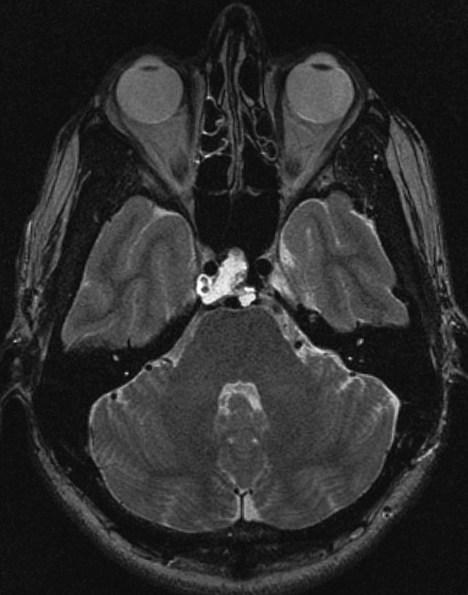
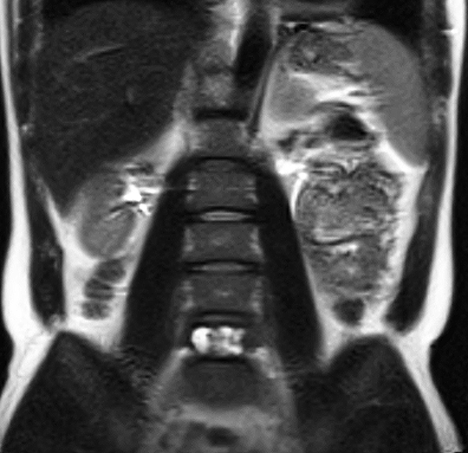
A 32-year-old management consultant was referred by a local otorhinolaryngologist to our institution with a 4-month history of intermittent double vision secondary to a right-side abducent nerve palsy. CT and MRI scans performed by the referring physician were first incorrectly interpreted as showing an inflammatory mass in the clivus (Fig. 1). This was in fact a chordoma that was subsequently excised endonasally using the endoscopic technique. Histology confirmed “classical chordoma with no atypical features.” The patient's VI nerve palsy resolved postoperatively, and at his 2-month follow up appointment, after researching the subject himself on the Internet, the patient asked if there was any chance of a second tumor in the remainder of the spinal tract. Despite being completely asymptomatic and having a normal neurological examination, he underwent an MRI of his entire spine. To our surprise, this showed a second primary chordoma in his fifth lumbar vertebra (Fig. 2). He endured total L5 vertebral excision and replacement with a titanium prosthesis at another institution. Histology again confirmed “classical chordoma.” There were no complications, and he made an uneventful and complete recovery. He was referred to our radio-oncologist for radiotherapy assessment. Because both tumors were completely excised macroscopically and on follow-up imaging, and with favorable histology, radiotherapy was not indicated for the time being and the patient remains under follow up.
Figure 1.
Axial T2-weighted magnetic resonance imaging scan showing clivus chordoma wrapping around the right internal carotid artery.
Figure 2.
Coronal T2-weighted magnetic resonance imaging scan showing synchronous L5 vertebra chordoma.
DISCUSSION
Spread of chordomas in the form of intradural seeding, surgical extradural seeding, and local metastasis has been previously described.4,5 Miller et al6 reported 19 cases of cutaneous involvement of chordoma and described the condition as “chordoma cutis.” These patients had metastatic chordomas and not multiple synchronous primary tumors. In contrast, the case we report had two synchronous distant intraosseous chordomas, one of which was completely asymptomatic. Badwal et al7 reported on a patient with multiple “synchronous” spinal extraosseous intradural chordomas. However, the authors reported on the result of autopsy findings and could not differentiate between multiple tumors arising at the same time or tumor spread as intradural seeding. There are also many reports of chordomas associated with different primary malignant tumors.8,9,10 Ji et al11 studied 2546 primary bone tumor patients in Sweden and quantified the risk of a second primary malignancy in patients with a chordoma as 1.99 (standardized incidence ratio11), but the risk was higher in patients under the age of 20 years. It is impossible to state with 100% certainty that the second tumor in our patient was a second primary chordoma; however, there are many reasons that make the alternative (a metastasis) highly unlikely, including the smaller size of the symptomatic primary, the radiological characteristics of the two tumors, the histological findings with no evidence of atypia, the short time frame (2 months) between the two being diagnosed, and the distance between the two tumors with absence of any other tumor in 12 months of follow-up. Our case is the first description of two distant synchronous primary intraosseous chordomas.
CONCLUSION
To our knowledge, we report the first case in the literature of synchronous primary intraosseous chordomas. The first chordoma was in the clivus and resulted in an abducent cranial nerve palsy, but the second in L5 vertebra was completely asymptomatic. We have modified our departmental practice as we believe that all patients diagnosed with chordoma should have MRI scanning of their entire spinal tract to exclude a second primary chordoma.
REFERENCES
- McMaster M L, Goldstein A M, Bromley C M, Ishibe N, Parry D M. Chordoma: incidence and survival patterns in the United States, 1973–1995. Cancer Causes Control. 2001;12:1–11. doi: 10.1023/a:1008947301735. [DOI] [PubMed] [Google Scholar]
- Dahlin D C, MacCarty C S. Chordoma. Cancer. 1952;5:1170–1178. doi: 10.1002/1097-0142(195211)5:6<1170::aid-cncr2820050613>3.0.co;2-c. [DOI] [PubMed] [Google Scholar]
- Krol G, Sundaresan N, Deck M. Computed tomography of axial chordomas. J Comput Assist Tomogr. 1983;7:286–289. doi: 10.1097/00004728-198304000-00015. [DOI] [PubMed] [Google Scholar]
- Asano S, Kawahara N, Kirino T. Intradural spinal seeding of a clival chordoma. Acta Neurochir (Wien) 2003;145:599–603. discussion 603. doi: 10.1007/s00701-003-0057-7. [DOI] [PubMed] [Google Scholar]
- Chambers P W, Schwinn C P. Chordoma. A clinicopathologic study of metastasis. Am J Clin Pathol. 1979;72:765–776. doi: 10.1093/ajcp/72.5.765. [DOI] [PubMed] [Google Scholar]
- Miller S D, Vinson R P, McCollough M L, Keeling J H., III Multiple smooth skin nodules. Chordoma cutis. Arch Dermatol. 1997;133:1579–1580, 1582–1583. doi: 10.1001/archderm.1997.03890480101016. [DOI] [PubMed] [Google Scholar]
- Badwal S, Pal L, Basu A, Saxena S. Multiple synchronous spinal extra-osseous intradural chordomas: is it a distinct entity? Br J Neurosurg. 2006;20:99–103. doi: 10.1080/02688690600682614. [DOI] [PubMed] [Google Scholar]
- Yamamoto Y, Moriwaki S, Takashima S, Yumoto Y. [Autopsy case of simultaneous triple malignancies—ovarian, chordoma and thyroid cancers] Gan No Rinsho. 1983;29:1375–1378. [PubMed] [Google Scholar]
- Harrington K J, Kelly L F, Pandha H S, McKenzie C G. Sacral chordoma, gas gangrene and bilateral renal cell carcinomas. Clin Oncol (R Coll Radiol) 1996;8:123–124. doi: 10.1016/s0936-6555(96)80121-x. [DOI] [PubMed] [Google Scholar]
- Shishkina V L, Kasumova SIu, Snigireva RIa, et al. Craniopharyngioma associated with pituitary adenoma and chordoma of Blumenbach's clivus. Zh Vopr Neirokhir Im N N Burdenko. 1981;6:52–54. [PubMed] [Google Scholar]
- Ji J, Hemminki K. Incidence of multiple primary malignancies among patients with bone cancers in Sweden. J Cancer Res Clin Oncol. 2006;132:529–535. doi: 10.1007/s00432-006-0100-1. [DOI] [PMC free article] [PubMed] [Google Scholar]


