Abstract
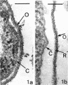
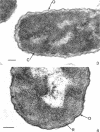
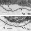
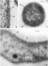
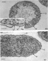
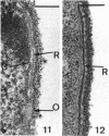
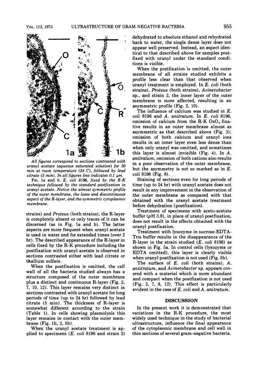
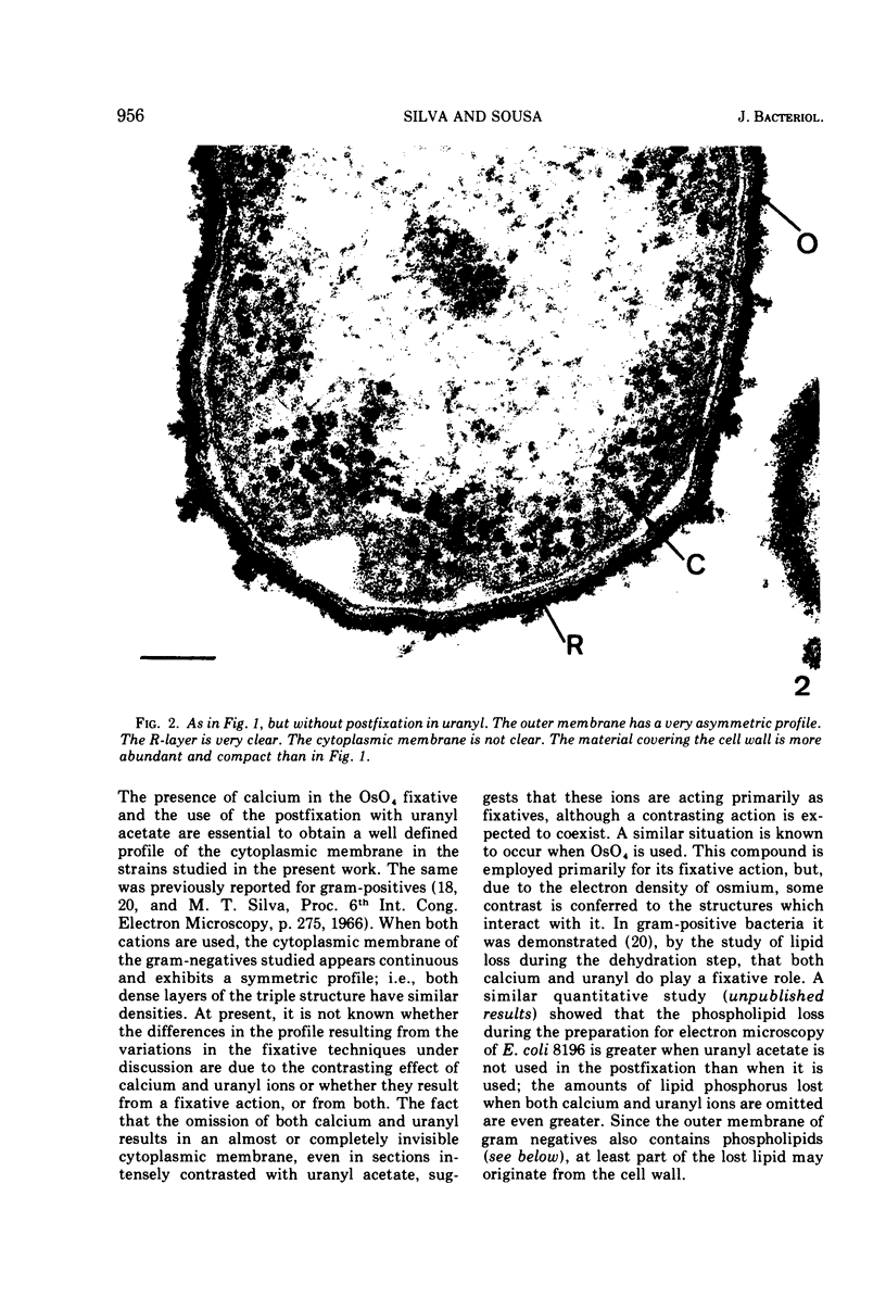
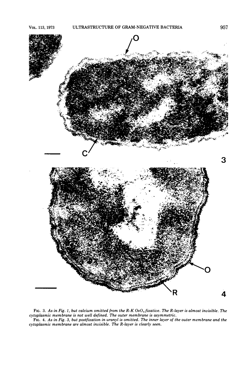
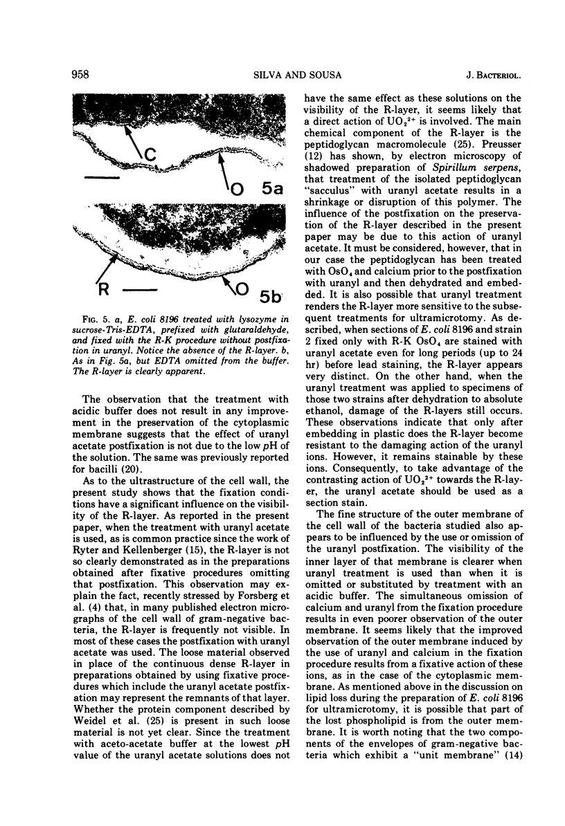
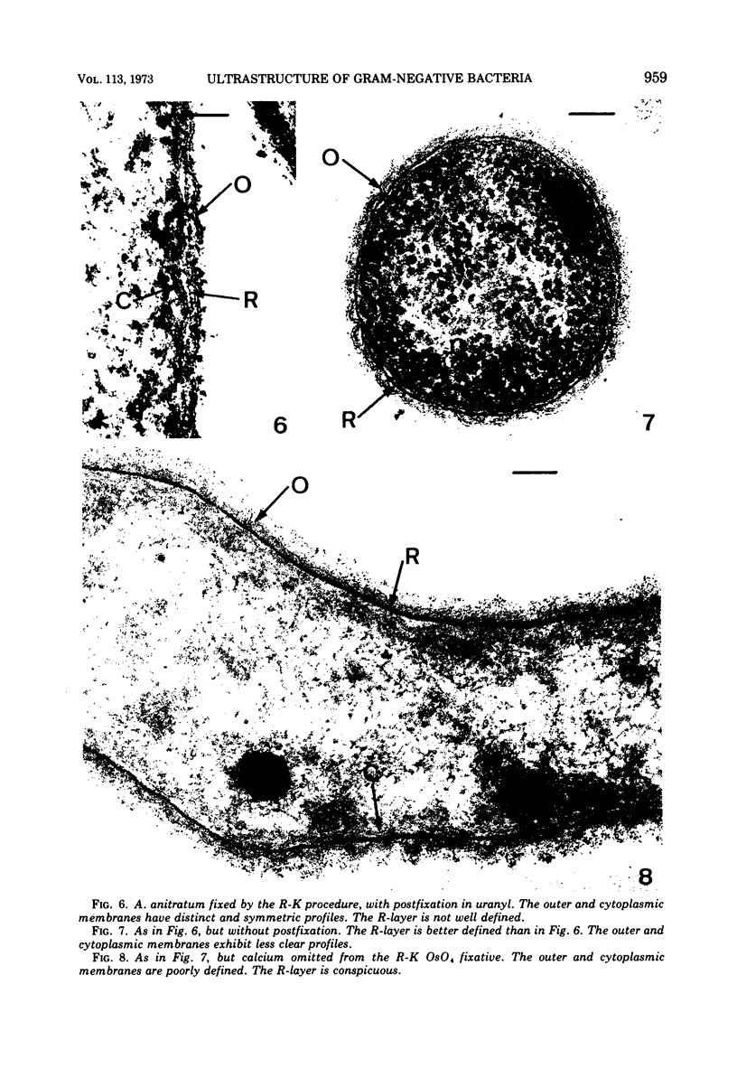
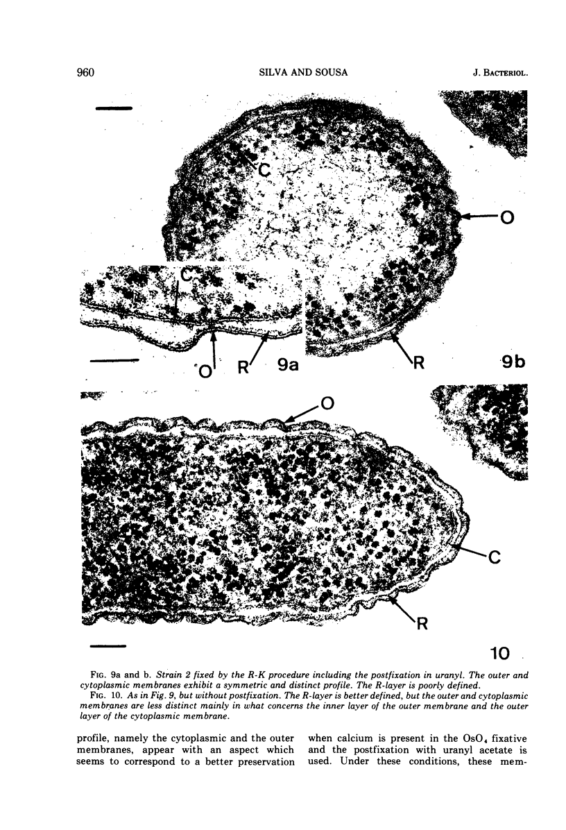
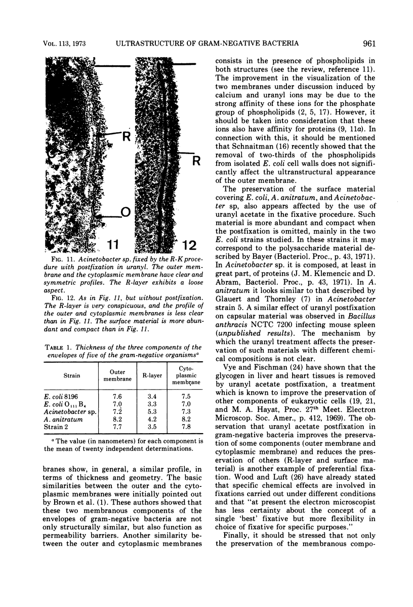
The ultrastructure of the cytoplasmic membrane and cell wall of two strains of Escherichia coli, Proteus morganii, P. vulgaris, Acinetobacter anitratum, Moraxella lacunata, Erwinia amylovora, Acinetobacter sp., and of a plant pathogen, unclassified gram-negative, fixed by the Ryter-Kellenberger procedure, was found to be significantly affected by the use or omission of the uranyl postfixation included in that procedure, and by the presence or absence of calcium in the OsO4 fixative. The omission of the uranyl treatment results in a less clear profile of both the outer membrane of the cell wall and of the cytoplasmic membrane. The observation of these two membranes is further limited when both uranyl and calcium are omitted. The R-layer and the material covering the surface of the cell wall appear more distinct when the uranyl postfixation is not used. Evidence is given suggesting that the influence of uranyl and calcium ions on the appearance of the outer and cytoplasmic membranes would be primarily due to their action as fixatives, whereas the influence of uranyl on the appearance of the R-layer would be due to a direct action on the peptidoglycan component of this layer. When uranyl acetate is used as a section stain after the embedding in plastic, it improves the observation of the R-layer. In this case, a well contrasted R-layer is consistently observed in all strains studied, provided that the postfixation has been omitted. The frequent difficulty in clearly observing the R-layer in many published micrographs probably results from the common use of uranyl postfixation.
Full text
PDF
Images in this article
Selected References
These references are in PubMed. This may not be the complete list of references from this article.
- Burdett I. D., Rogers H. J. Modification of the appearance of mesosomes in sections of Bacillus licheniformis according to the fixation procedures. J Ultrastruct Res. 1970 Feb;30(3):354–367. doi: 10.1016/s0022-5320(70)80068-5. [DOI] [PubMed] [Google Scholar]
- Forsberg C. W., Costerton J. W., Macleod R. A. Separation and localization of cell wall layers of a gram-negative bacterium. J Bacteriol. 1970 Dec;104(3):1338–1353. doi: 10.1128/jb.104.3.1338-1353.1970. [DOI] [PMC free article] [PubMed] [Google Scholar]
- Forstner J., Manery J. F. Calcium binding by human erythrocyte membranes. Biochem J. 1971 Sep;124(3):563–571. doi: 10.1042/bj1240563. [DOI] [PMC free article] [PubMed] [Google Scholar]
- Glauert A. M., Thornley M. J. Fine structure and radiation resistance in Acinetobacter: a comparison of a range of strains. J Cell Sci. 1971 Jan;8(1):19–41. doi: 10.1242/jcs.8.1.19. [DOI] [PubMed] [Google Scholar]
- Glauert A. M., Thornley M. J. The topography of the bacterial cell wall. Annu Rev Microbiol. 1969;23:159–198. doi: 10.1146/annurev.mi.23.100169.001111. [DOI] [PubMed] [Google Scholar]
- LUFT J. H. Improvements in epoxy resin embedding methods. J Biophys Biochem Cytol. 1961 Feb;9:409–414. doi: 10.1083/jcb.9.2.409. [DOI] [PMC free article] [PubMed] [Google Scholar]
- MURRAY R. G., STEED P., ELSON H. E. THE LOCATION OF THE MUCOPEPTIDE IN SECTIONS OF THE CELL WALL OF ESCHERICHIA COLI AND OTHER GRAM-NEGATIVE BACTERIA. Can J Microbiol. 1965 Jun;11:547–560. doi: 10.1139/m65-072. [DOI] [PubMed] [Google Scholar]
- Manery J. F. Effects of Ca ions on membranes. Fed Proc. 1966 Nov-Dec;25(6):1804–1810. [PubMed] [Google Scholar]
- PASSOW H., ROTHSTEIN A., CLARKSON T. W. The general pharmacology of the heavy metals. Pharmacol Rev. 1961 Jun;13:185–224. [PubMed] [Google Scholar]
- Preusser H. J. Strukturveränderungen am "Murein-Sacculus" von Spirillum serpens nach Einwirkung von Uranylacetat. Arch Mikrobiol. 1969 Oct;68(2):150–164. [PubMed] [Google Scholar]
- REPASKE R. Lysis of gram-negative bacteria by lysozyme. Biochim Biophys Acta. 1956 Oct;22(1):189–191. doi: 10.1016/0006-3002(56)90240-2. [DOI] [PubMed] [Google Scholar]
- RYTER A., KELLENBERGER E., BIRCHANDERSEN A., MAALOE O. Etude au microscope électronique de plasmas contenant de l'acide désoxyribonucliéique. I. Les nucléoides des bactéries en croissance active. Z Naturforsch B. 1958 Sep;13B(9):597–605. [PubMed] [Google Scholar]
- Schnaitman C. A. Effect of ethylenediaminetetraacetic acid, Triton X-100, and lysozyme on the morphology and chemical composition of isolate cell walls of Escherichia coli. J Bacteriol. 1971 Oct;108(1):553–563. doi: 10.1128/jb.108.1.553-563.1971. [DOI] [PMC free article] [PubMed] [Google Scholar]
- Shah D. O. Interaction of uranyl ions with phospholipid and cholesterol monolayers. J Colloid Interface Sci. 1969 Feb;29(2):210–215. doi: 10.1016/0021-9797(69)90188-x. [DOI] [PubMed] [Google Scholar]
- Silva M. T. Changes induced in the ultrastructure of the cytoplasmic and intracytoplasmic membranes of several Gram-positive bacteria by variations in OsO 4 fixation. J Microsc. 1971 Jun;93(3):227–232. doi: 10.1111/j.1365-2818.1971.tb02285.x. [DOI] [PubMed] [Google Scholar]
- Silva M. T., Guerra F. C., Magalhães M. M. The fixative action of uranyl acetate in electron microscopy. Experientia. 1968 Oct 15;24(10):1074–1074. doi: 10.1007/BF02138757. [DOI] [PubMed] [Google Scholar]
- Silva M. T., Santos Mota J. M., Melo J. V., Guerra F. C. Uranyl salts as fixatives for electron microscopy. Study of the membrane ultrastructure and phospholipid loss in bacilli. Biochim Biophys Acta. 1971 Jun 1;233(3):513–520. doi: 10.1016/0005-2736(71)90151-9. [DOI] [PubMed] [Google Scholar]
- Terzakis J. A. Uranyl acetate, a stain and a fixative. J Ultrastruct Res. 1968 Jan;22(1):168–184. doi: 10.1016/s0022-5320(68)90055-5. [DOI] [PubMed] [Google Scholar]
- VENABLE J. H., COGGESHALL R. A SIMPLIFIED LEAD CITRATE STAIN FOR USE IN ELECTRON MICROSCOPY. J Cell Biol. 1965 May;25:407–408. doi: 10.1083/jcb.25.2.407. [DOI] [PMC free article] [PubMed] [Google Scholar]
- Vye M. V., Fischman D. A. The morphological alteration of particulate glycogen by en bloc staining with uranyl acetate. J Ultrastruct Res. 1970 Nov;33(3):278–291. doi: 10.1016/s0022-5320(70)90022-5. [DOI] [PubMed] [Google Scholar]
- Vörös J., Goodman R. N. Filamentous forms of Erwinia amylovora. Phytopathology. 1965 Aug;55(8):876–879. [PubMed] [Google Scholar]
- WEIDEL W., PELZER H. BAGSHAPED MACROMOLECULES--A NEW OUTLOOK ON BACTERIAL CELL WALLS. Adv Enzymol Relat Areas Mol Biol. 1964;26:193–232. doi: 10.1002/9780470122716.ch5. [DOI] [PubMed] [Google Scholar]
- WOOD R. L., LUFT J. H. THE INFLUENCE OF BUFFER SYSTEMS ON FIXATION WITH OSMIUM TETROXIDE. J Ultrastruct Res. 1965 Feb;12:22–45. doi: 10.1016/s0022-5320(65)80004-1. [DOI] [PubMed] [Google Scholar]


