Abstract
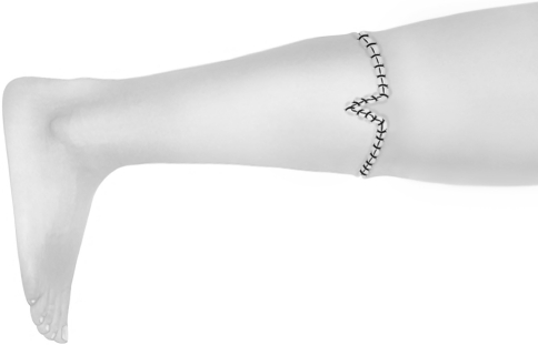
Rotationplasty provides stable and durable biologic reconstruction after tumor resection around the knee and renders reliable results, in young patients. However, after resection of the tumor, there is often a mismatch between the circumference of the proximal (femoral) and the distal (tibial) parts. Because rotationplasty includes an intercalary amputation where the ends are readapted, there is always a mismatch of the proximal and distal circumferences of the soft tissue envelope. To facilitate skin closure without tension and to avoid impaired wound healing and subsequent infections, the type of incision is critical and must be carefully planned. We present a new incision technique for rotationplasty about the knee. Half of the difference of the incision length of the proximal and distal circumferences represents the base of the triangle proximally, medially and laterally of the thigh. After adapting both ends, the peak of this flat triangle is distally adapted via a vertical incision which allows it to match unequal circumferences. This technique was used in eight patients, in all of whom the wounds healed uneventfully.
Introduction
Rotationplasty was first described in 1930 by Borggreve for treatment of a shortened limb with ankylosis of the knee after tuberculosis [1]. In 1948, Van Nes described its use for treatment of congenital defects of the femur [17]. In the 1980s, Kotz and Salzer [11] and Salzer et al. [16] reported treating patients with malignant tumors around the knee by rotationplasty as an alternative to above-knee amputation. Today, rotationplasty is one option for surgical management of musculoskeletal tumors, particularly in children [2, 3, 8, 18] or as a salvage option after failed reconstruction [4, 5, 13]. Overall function reportedly is excellent [4, 10, 15]. There have been no problems with wound healing. Patients are able to bear their full weight, actively control the knee in coordinated gait patterns, and walk without assistive devices.
Different incisions have been described. Originally, two elliptical or oval circumferential incisions were described to produce a rhomboidal area of skin to be resected with the tumor mass [1]. However, this incision technique causes difficulties with later skin closure because of the mismatch of the amount of skin of the proximal and distal parts. Consequently, a fish-mouth–shaped incision of the proximal, ie, the femoral part, was proposed by Gebhart et al. in 1987 [6]. The calf is viewed as a cylinder with the circumference at the level of the intended distal resection. Then, the circumference of the intended proximal thigh resection is projected onto the cylinder and angled until the shadows of both circumferences meet. A height is defined by the angle of obliquity and diameter of the thigh and calf circumferences. This height represents the depth of the incision medially and laterally on the leg, which is used to produce the fish-mouth–shaped distal incision. However, there is often the problem of proximal nonfitting.
To overcome the main disadvantages of these incision techniques, we describe a new incision technique for the Borggreve/Van Nes rotationplasty and briefly describe the eight patients in whom this incision was used.
Surgical Technique
With the patient supine, the levels of the skin incision are determined based on the exact dimension of the tumor as assessed by preoperative diagnostic imaging and planning to ensure safe margins. At the end of growth, the center of rotation of the rotated ankle should be at the same level as the center of rotation of the contralateral knee accepting that the rotated limb initially is longer than the contralateral, uninvolved limb to compensate for the remaining growth discrepancy. We acknowledge that some surgeons favor a technique which adjusts the limb length at surgery even in young children.
A sterile cord is used to measure the circumference of the planned distal incision. It is laid around the leg, thereby representing the entire length of the distal incision (Fig. 1). This cord subsequently is used to match the circumference of the distal and proximal parts. For this purpose, it is cut in half and laid on the anterior and posterior circumferences of the planned proximal incision, leaving two equally sized gaps medially and laterally (Fig. 1). The resulting distance between the anterior and posterior parts is connected by a symmetric freehand triangular-shaped incision of equal size at both sides (Fig. 2). In turn, at the distal site, a straight lateral and medial incision representing the height of the proximal triangle is made. As such, the proximal soft tissue spindle adapts into the distal enlarged or spread incision to perfectly accommodate for the difference at the circumferences. These can be enlarged later according to the needs as given by the proximal triangles to match both parts (Fig. 3).
Fig. 1.
The incision is planned by measuring the circumference of the distal incision by a sterile cord. This then is used to map to the proximal incision by cutting the cord into two equally sized parts, which are placed in a symmetric fashion at the place where the proximal incision will be made (The proximal incision is shown close to the knee only for illustration purposes).
Fig. 2.
The distance between the two semicircular incisions is bridged by two symmetric triangular-shaped incisions. Distally, two incisions with the length representing the height of the proximal incision are made.
Fig. 3.
The legs of the triangles can be matched exactly, without difficulty, to the needed size.
The surgical technique after skin incision and preparation of the subcutaneous tissue has been presented in detail [5]. It is important to fix the plate not too far laterally or ventrally to prevent the limb from rotating too much laterally and furthermore, to assure the heel is in the neutral position of the foot (90° flexion) that corresponds to the level of the contralateral knee. It is important to reattach the gastrocnemius muscles as proximally as possible to the anterior fascia of the femur in the neutral position of the foot. Finally, the skin then is adapted under slight pressure thereby avoiding leaving an over length, which later would lead to an unfavorable appearance and difficulties with the prosthesis.
Case Reports
We have used this incision in eight patients (five females, three males). The average age of the patients at surgery was 25 years (range, 5–70 years). Two patients had a high-grade osteosarcoma, one had a proximal femoral focal deficiency (PFFD), and one had a longitudinal femoral defect. One patient had a diaphyseal femoral defect attributable to osteomyelitis, one had a myxoid liposarcoma (and secondary malignant fibrous histiocytoma subsequent to radiotherapy), one had a high-grade liposarcoma, and one had a leiomyosarcoma, respectively (Table 1). The mean followup was 34.5 months (range, 8–55 months). All wounds healed without skin necrosis at the triangles of the interface of the upper and lower limb. At last followup none of the patients had any sign of infection. One patient died of disease. All others were unrestricted or only slightly restricted in activities of daily living. Two patients participated in exercise and cycling (Table 1). The plate fixation material was left in place in all patients.
Table 1.
Overview of patients
| Patient number | Date of surgery | Followup (months) | Age at surgery (years) | Gender | Disease | Side | Primary or secondary procedure | Oncologic outcome | Range of motion (°) | Weight-bearing | Painless walking | Radiographic signs of plate loosening |
|---|---|---|---|---|---|---|---|---|---|---|---|---|
| 1 | 8/28/01 | 31 | 10 | F | High-grade osteosarcoma | R | Primary | DOD | 90 | Full | Yes | None |
| 2 | 2/10/03 | 55 | 13 | F | High-grade osteosarcoma | R | Primary | ANED | 50 | Full | Yes | None |
| 3 | 1/29/04 | 53 | 5 | M | Longitudinal femoral defect | L | Secondary | 90 | Full | Yes | None | |
| 4 | 2/22/07 | 29 | 20 | M | Myxoid fibrosarcoma/malignant fibrous histiocytoma | R | Primary | ANED | 50 | Full | Yes | None |
| 5 | 1/4/06 | 37 | 70 | F | Liposarcoma | R | Primary | AWD | 90 | Full with crutches | Yes | None |
| 6 | 5/2/05 | 29 | 5 | F | Proximal focal femoral defect | R | Primary | 50 | Full | Yes | None | |
| 7 | 5/16/06 | 34 | 19 | F | Diaphyseal femoral defect after osteomyelitis | R | Primary | 90 | Full with crutches | Yes | None | |
| 8 | 10/29/07 | 8 | 61 | M | Leiomyosarcoma | R | Primary | ANED | 90 | Full | Yes | None |
DOD = died of disease; ANED = alive with no evidence of disease; AWD = alive with disease.
A myxoid liposarcoma at the medial vastus muscle in a 16-year-old boy was treated by resection and radiotherapy. Four years later, he had a malignant fibrous histiocytoma of bone develop in the previous radiation field. Resection of the distal femur was performed and reconstructed using a tumor endoprosthesis. Because of aseptic loosening, this was replaced by a silver-coated prosthesis. The patient had an infection develop and we removed all hardware. Rotationplasty was chosen as salvage surgery (Fig. 4). The wound healed uneventfully without infection (Fig. 5). The patient was free of infection at 29 months of followup (Fig. 6).
Fig. 4A–B .
After having readapted and secured the proximal and distal ends, the proximal soft tissue spindle is fitted properly into the distal part without tension on the wound. (A) The intraoperative situation before wound readaptation, and (B) after wound closure are shown.
Fig. 5.

Two weeks after surgery, the wound had healed uneventfully with the staples still in place.
Fig. 6.
The patient was free of infection at 29 months followup.
Discussion
We present a new incision technique to facilitate matching the proximal and distal incisions of the leg after rotationplasty. Complications after rotationplasty include wound infection, vascular compromise, nerve palsy, and nonunion with infection being the most severe complication of rotationplasty [12, 14, 19]. Our new incision technique may reduce the risk of soft tissue complications because the soft tissues can be sutured without tension as a result of better fitting of the proximal and distal incisions after resection. Using the rhomboid-shaped incision as originally described, it is often impossible to exactly match the incisions. Specific complications of this incision include harm to soft tissues, wound healing complications, and unfavorable cosmetic results [4, 5, 7, 9, 12]. In addition, this technique requires much experience to perform the correct incisions to perfectly match the circumferences. However, the advantage of the classic incision is that it requires less preoperative and intraoperative planning and is relatively easy to perform. Other conventional incisions described such as the rhombus-shaped skin incision around the thigh and calf as proposed [6, 12] aimed to at least partially eliminate the problem of major discrepancies of the circumferences of the proximal and distal skin borders. However, that incision technique causes difficulties with later skin closure because of the mismatch of the amount of skin of the proximal and distal parts, which is avoided with the technique we present.
Acknowledgments
We thank Carol de Simio for drawing and preparation of the figures.
Footnotes
Each author certifies that he or she has no commercial associations (eg, consultancies, stock ownership, equity interest, patent/licensing arrangements, etc) that might pose a conflict of interest in connection with the submitted article.
Each author certifies that his or her institution has approved or waived approval for the reporting of this case and that all investigations were conducted in conformity with ethical principles of research.
References
- 1.Borggreve J. [Replacement of the knee joint by the rotated ankle] [in German] Arch Orthop Unfall-Chir. 1930;28:175–178. doi: 10.1007/BF02581614. [DOI] [Google Scholar]
- 2.Cammisa FP, Jr, Glasser DB, Otis JC, Kroll MA, Lane JM, Healey JH. The Van Nes tibial rotationplasty: a functionally viable reconstructive procedure in children who have a tumor of the distal end of the femur. J Bone Joint Surg Am. 1990;72:1541–1547. [PubMed] [Google Scholar]
- 3.Bari A, Krajbich JI, Langer F, Hamilton EL, Hubbard S. Modified Van Nes rotationplasty for osteosarcoma of the proximal tibia in children. J Bone Joint Surg Br. 1990;72:1065–1069. doi: 10.1302/0301-620X.72B6.2246290. [DOI] [PubMed] [Google Scholar]
- 4.Fuchs B, Kotajarvi BR, Kaufman KR, Sim FH. Functional outcome of patients with rotationplasty about the knee. Clin Orthop Relat Res. 2003;415:52–58. doi: 10.1097/01.blo.0000093896.12372.c1. [DOI] [PubMed] [Google Scholar]
- 5.Fuchs B, Sim FH. Rotationplasty about the knee: surgical technique and anatomical considerations. Clin Anat. 2004;17:345–353. doi: 10.1002/ca.10211. [DOI] [PubMed] [Google Scholar]
- 6.Gebhart MJ, McCormack RR, Jr, Healey JH, Otis JC, Lane JM. Modification of the skin incision for the Van Nes limb rotationplasty. Clin Orthop Relat Res. 1987;216:179–182. [PubMed] [Google Scholar]
- 7.Hardes J, Gebert C, Schwappach A, Ahrens H, Streitburger A, Winkelmann W, Gosheger G. Characteristics and outcome of infections associated with tumor endoprostheses. Arch Orthop Trauma Surg. 2006;126:289–296. doi: 10.1007/s00402-005-0009-1. [DOI] [PubMed] [Google Scholar]
- 8.Hardes J, Gosheger G, Vachtsevanos L, Hoffmann C, Ahrens H, Winkelmann W. Rotationplasty type BI versus type BIIIa in children under the age of ten years: should the knee be preserved? J Bone Joint Surg Br. 2005;87:395–400. doi: 10.1302/0301-620x.87b3.14793. [DOI] [PubMed] [Google Scholar]
- 9.Hillmann A, Hoffmann C, Gosheger G, Krakau H, Winkelmann W. Malignant tumor of the distal part of the femur or the proximal part of the tibia: endoprosthetic replacement or rotationplasty. Functional outcome and quality-of-life measurements. J Bone Joint Surg Am. 1999;81:462–468. doi: 10.1302/0301-620X.81B3.9134. [DOI] [PubMed] [Google Scholar]
- 10.Hopyan S, Tan JW, Graham HK, Torode IP. Function and upright time following limb salvage, amputation, and rotationplasty for pediatric sarcoma of bone. J Pediatr Orthop. 2006;26:405–408. doi: 10.1097/01.bpo.0000203016.96647.43. [DOI] [PubMed] [Google Scholar]
- 11.Kotz R, Salzer M. Rotation-plasty for childhood osteosarcoma of the distal part of the femur. J Bone Joint Surg Am. 1982;64:959–969. [PubMed] [Google Scholar]
- 12.Lindner NJ, Ramm O, Hillmann A, Roedl R, Gosheger G, Brinkschmidt C, Juergens H, Winkelmann W. Limb salvage and outcome of osteosarcoma: the University of Muenster experience. Clin Orthop Relat Res. 1999;358:83–89. doi: 10.1097/00003086-199901000-00011. [DOI] [PubMed] [Google Scholar]
- 13.Ramseier LE, Dumont CE, Exner GU. Rotationplasty (Borggreve/Van Nes and modifications) as an alternative to amputation in failed reconstructions after resection of tumours around the knee joint. Scand J Plast Reconstr Surg Hand Surg. 2008;42:199–201. doi: 10.1080/02844310802069434. [DOI] [PubMed] [Google Scholar]
- 14.Renard AJ, Veth RP, Schreuder HW, Loon CJ, Koops HS, Horn JR. Function and complications after ablative and limb-salvage therapy in lower extremity sarcoma of bone. J Surg Oncol. 2000;73:198–205. doi: 10.1002/(SICI)1096-9098(200004)73:4<198::AID-JSO3>3.0.CO;2-X. [DOI] [PubMed] [Google Scholar]
- 15.Rodl RW, Pohlmann U, Gosheger G, Lindner NJ, Winkelmann W. Rotationplasty: quality of life after 10 years in 22 patients. Acta Orthop Scand. 2002;73:85–88. doi: 10.1080/000164702317281468. [DOI] [PubMed] [Google Scholar]
- 16.Salzer M, Knahr K, Kotz R, Kristen H. Treatment of osteosarcomata of the distal femur by rotationplasty. Arch Orthop Trauma Surg. 1981;99:131–136. doi: 10.1007/BF00389748. [DOI] [PubMed] [Google Scholar]
- 17.Nes CP. Transplantation of the tibia and fibula to replace the femur following resection: turn-up-plasty of the leg. J Bone Joint Surg Am. 1948;30:854–858. [PubMed] [Google Scholar]
- 18.Winkelmann W. Rotation grafts] [in German. Orthopade. 1993;22:152–159. [PubMed] [Google Scholar]
- 19.Winkelmann WW. Rotationplasty. Orthop Clin North Am. 1996;27:503–523. [PubMed] [Google Scholar]


