Abstract
Stevens-Johnson syndrome and its more severe variant, toxic epidermal necrolysis, have relatively low overall incidence; however, this disease presents with high morbidity and mortality. The majority of patients develop ocular inflammation and ulceration at the acute stage. Due to the hidden nature of these ocular lesions and the concentration of effort toward life-threatening issues, current acute management has not devised a strategy to preclude blinding cicatricial complications. This review summarizes recent literature data, showing how sight-threatening corneal complications can progressively develop from cicatricial pathologies of lid margin, tarsus, and fornix at the chronic stage. It illustrates how such pathologies can be prevented with the early intervention of cryopreserved amniotic membrane transplantation to suppress inflammation and promote epithelial healing at the acute stage. Significant dry eye problems and photophobia can also be avoided with this intervention. This new therapeutic strategy can avert the catastrophic ophthalmic sequelae of this rare but devastating disease.
Keywords: amniotic membrane transplantation, blindness, cicatricial pathology, corneal complication, scarring, Stevens-Johnson syndrome, toxic epidermal necrolysis
In 1922, Stevens and Johnson69 reported the cases of two boys with eruptive fever, stomatitis, and opthalmalia, later named Stevens-Johnson syndrome (SJS). Lyell40 described toxic epidermal necrolysis (TEN), a condition characterized by extensive epidermal scalding. Although initially described separately, both diseases are now considered a continuum of epidermal bullous diseases in which epidermal cell death results in subepidermal separation that can be elicited by stroking the skin (i.e., positive Nikolsky sign). A prodrome of fever, malaise, and upper respiratory infection symptoms is usually followed by a vesicular eruption of the mucous membranes. Although there is no universally accepted definition, both diseases are characterized by eruptive lesion involving at least two mucosal surfaces, and can be separated by the percentage of detachment of the body surface area (BSA).57 SJS involves less than 10% total BSA, overlapping SJS/TEN involves 10–30% BSA, and TEN involves more than 30% BSA.
Although SJS and TEN are rare, they are important because of high morbidity and mortality; the mortality rate is 3% for SJS and 25–40% for TEN.42,49,82 Acute ocular complications develop in more than half (i.e., 43–81%) of hospitalized patients for both SJS and TEN, and among them, 25% exhibit severe involvement.53 Chronic ocular sequelae occur in up to 35% of patients.5 Corneal damage leading to blindness is the most severe long-term complication for survivors of SJS/TEN.5,53,83 Although ocular morbidity and visual loss can be caused by acute corneal complications, progressive conjunctival scarring is significantly associated with subsequent loss of vision.16 This review describes the ocular manifestations of SJS/TEN, summarizes literature evidence illustrating how ocular cicatricial complications may emerge even if the vision is not immediately threatened, and how these sight-threatening complications can be prevented by a novel therapy based on amniotic membrane transplantation (AMT) delivered during the acute stage of SJS/TEN. For more information on other aspects of SJS/TEN, see recent reviews10,37 and references cited therein.
Acute Ocular Manifestation
The estimated annual incidence of SJS/TEN ranges from 0.6 to 10 cases per million person-year.8,12,45,62 TEN may occur more commonly in older people and people with AIDS.7 The most common cause of the disease is an idiopathic reaction to medications such as antibiotics (penicillin and sulfa), anticonvulsants (phenytoin, carbamazepine, and barbiturate), and nonsteroidal anti-inflammatory drugs.37,56 Other causes included infection (herpes simplex, mycoplasma pneumoniae, measles, mycobacterium, group A streptococci, Epstein-Barr, yersinia, enterovirus, smallpox vaccination)17,47,72 and some remained undetermined.86 Although it is known that SJS/TEN causes epidermal cell apoptosis, the pathogenesis of the disease is unclear. Some studies have suggested the involvement of Fas-Fas ligand interaction,80,81 cytotoxic T-cells,46,52 tumor necrosis factor-alpha,48,51 and nitric oxide synthase36 (for a review of pathogenesis of SJS/TEN, see Khalili and Bahna27).
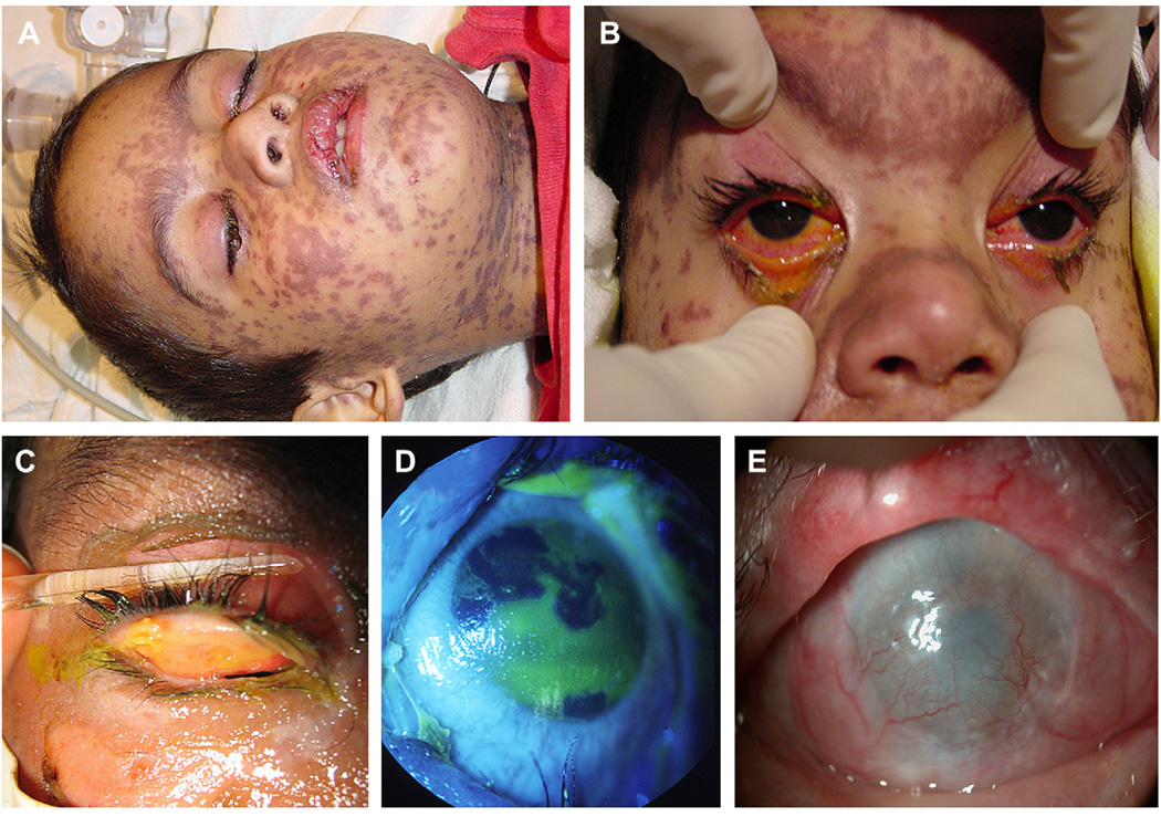
In the acute stage of SJS/TEN, defined as the first two weeks after onset of symptoms, the aforementioned immune dysregulation can attack mucous membranes of the whole body. As stated previously, an overwhelming majority of hospitalized patients develop ocular lesions during the acute stage,53 with 15–75% of them manifesting bilateral conjunctivitis.6,25,50 In the eye, although lid skin vesicles may erupt into ulcers like the rest of the body (Fig. 1A), external examination at the bedside frequently only reveals conjunctivitis with redness and mucous discharge (Fig. 1B). However, it should be stressed that epithelial defects or ulcers involve tarsi and fornices in nearly all cases with ocular involvement. These hidden lesions are impossible to ascertain without everting the eyelids (Fig. 1C), a maneuver sometimes hard to practice as acute care often includes respiratory support. As reported, approximately 25% of hospitalized patients develop more severe ocular involvement,53 manifesting corneal epithelial defects and inflammation (Fig. 1D).
Fig. 1.
Acute ocular manifestation of SJS/TEN. A: External appearance of a SJS patient shows diffuse skin rashes, oral mucosal ulceration, and closure of both eyes with crust. B: External examination at the bedside reveals only ulcers involving the lid margin skin and conjunctival redness. (Reproduced from Di Pascuale et al16 with permission of Ophthalmology.) C: Eversion of the eyelids reveals diffuse ulcers involving the tarsus and fornix. D: Acute ocular involvement manifesting a geographical corneal epithelial defect. (C and D are reproduced from Kobayashi et al31 with permission of Ophthalmology.) E: Limbal stem cell deficiency can occur acutely, resulting in conjunctivalization and formation of an extensive fibrovascular scar, causing blindness.
If ocular surface inflammation and ulceration is not quickly managed, the ensuing wound healing usually results in scarring. It remains a mystery why the inflammatory process tends to be relentless and prolonged in patients with SJS/TEN even after they are released from acute care or discharged from the hospital. In a proportion of patients, persistent inflammation and ulceration of the conjunctiva or cornea may extend to destroy corneal epithelial stem cells at the limbus, leading to a pathological state termed limbal stem cell deficiency (Fig. 1E) (for reviews see the literature20,33,74). Without limbal epithelial stem cells, the cornea will be covered by the surrounding conjunctival epithelium as well as an extensive fibrovascular scar, causing an immediate loss of vision. Limbal stem cell deficiency cannot be treated by conventional corneal transplantation, and because limbal stem cell deficiency is usually bilateral in this disease, visual rehabilitation efforts are limited to transplantation of allogeneic limbal stem cells (for reviews see the literature18,21,75) or keratoprosthesis.38,59,84 Due to continuous ocular surface inflammation and other cicatricial complications detailed subsequently, eyes with total limbal stem cell deficiency caused by SJS/ TEN have the worst prognosis even if subjected to transplantation of allogeneic limbal stem cells.65 Furthermore, there are also risks derived from taking prolonged, if not indefinite, systemic immunosuppressive agents, while potential complications of keratoprosthesis use include glaucoma, retroprosthetic membrane, tissue necrosis, retinal detatchment, and endopthalmitis.84 For all these reasons, it is highly desirable to adopt a new strategy to suppress ocular surface inflammation, promote epithelial healing, retain remaining limbal epithelial stem cells, and encourage their expansion at the acute stage.
Chronic Ocular Complications
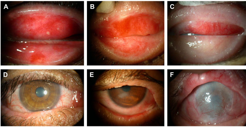
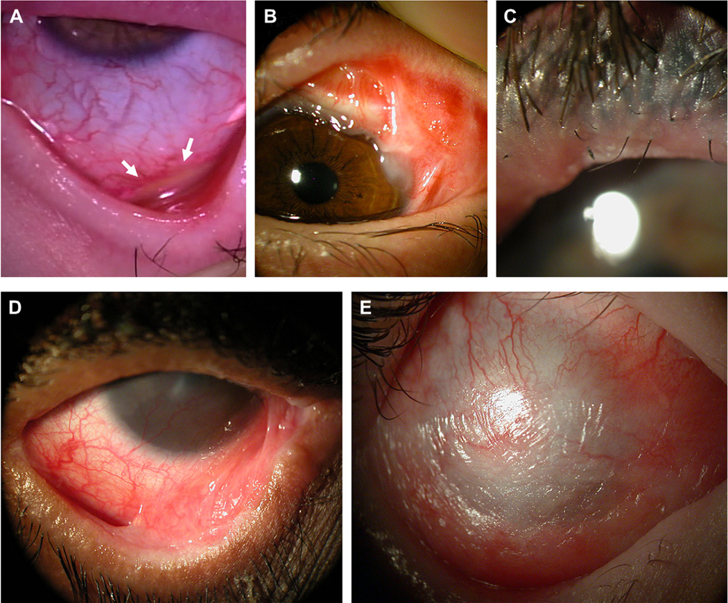
Chronic ocular sequelae occur in up to 35% of patients.5 It has been recognized that sight-threatening corneal scarring is the most severe long term complication for survivors of SJS/TEN.43,53 Even if the cornea is not affected during the acute stage or after discharge from the hospital, patients with SJS/ TEN may suffer from severe loss of vision due to chronic corneal complications. A potential causative relationship between cicatricial complications of the lid margin and the tarsus and sight-threatening corneal scarring was revealed by a recent study of 38 SJS/TEN patients (Fig. 2).16 These cicatricial complications are derived in part from the persistent and prolonged conjunctival inflammation and ulceration (Fig.3A). Thus conjunctival scarring commonly occurs as a chronic sequel.86 This scarring frequently leads to symblepharon and foreshortening of the fornix (Fig. 3B).28,76 Depending on the location and severity of symblepharon, a number of pathogenic elements ensue. For example, scarring at the lid margin or the tarsus can deform the eyelid, causing entropion or ectropion and allowing lashes and scar tissue to elicit friction-related microtrauma to the cornea during blinking (Fig. 3C). If symblepharon obliterates the tear meniscus, it will interfere with aqueous tear flow and spread to the ocular surface (Fig. 3D). If the superior or inferior fornix is involved, untoward exposure arises to render more dryness both day and night because of inadequate blinking, closure, or limitation of Bell’s phenomenon. In the superotemporal fornix, scarring will obstruct secretory lacrimal ductules (Fig. 3B). If scarring involves the extraocular muscle or the upper lid, diplopia and ptosis occur. When scarring affects the lid margin, it frequently causes keratinization and seals meibomian gland orifices resulting in lipid layer anomaly of the tear film (see Fig. 2). SJS/TEN can also cause squamous metaplasia with extensive loss of conjunctival goblet cells (Fig. 3E).5,55 Collectively, these cicatricial complications exacerbate blink-related microtrauma and produce severe dry eye. Cumulative insults of the aforementioned pathogenic elements will not only lead to severe loss of vision but also cause multiple deficiencies in the ocular surface defense, rendering subsequent reconstructive efforts difficult, if not impossible. Because of these complicating factors, it is no wonder that SJS/TEN patients find it difficult to escape photophobia, constant irritation, and the devastating outcome of ocular surface failure. For additional reviews and studies about the chronic complications of ocular SJS/TEN see the literature.5,17,83 Although AMT and other surgical strategies have been used for treating cicatricial complications and limbal stem cell deficiency at the chronic stage,32,66,76 they are outside the scope of this review and have been discussed by others.11,58
Fig. 2.
Significant correlation between lid margin or tarsal keratinization and scarring with corneal scarring and vascularization. The severity of grade 1 (A), grade 2 (B), and grade 3 (C) of lid margin and tarsal scarring and keratinization of representative cases is correlated well with their corneas, which showed clear (D), mild scarring (E), and severe scarring and vascularization (F). (Reproduced from Di Pascuale et al16 with permission of Ophthalmology.)
Fig. 3.
Chronic cicatricial complications of SJS/TEN. A: Progressive conjunctival inflammation and non-healing fornix ulcer (marked by arrows) in the chronic stage, seen when the eyelid was everted by a muscle hook. B: Persistent conjunctival inflammation and scarring involves the superotemporal fornix, obstructing the lacrimal ductules. C: Lid margin keratinization and scarring leads to distichiasis and meibomian gland obstruction. D: Inferior symblepharon obliterates the tear meniscus, interfering with aqueous tear flow and spread to the ocular surface E: Diffuse squamous metaplasia due to severe dry eye and exposure.
Amniotic Membrane Transplantation as a New Strategy for Minimizing Ocular Sequels in Acute SJS/TEN
Because SJS/TEN, especially TEN, is potentially life-threatening, physicians must deploy life-saving measures at the acute stage. Because patients are frequently put on a respirator, their eyes are often closed and they cannot voice any discomfort caused by inflammation or ulceration, giving the false impression that the eyes are not involved. Even if an ophthalmologist is consulted because skin ulceration involves the lid margin or the conjunctiva showed redness and discharge, the eye examination at the bedside cannot uncover deep-seated ulcers unless the eyelids are fully everted (see Fig. 1). Conventional ocular management may consist of bedside examination, application of lubricating ointment, antibiotics to prevent infection, steroids to control inflammation, cyclosporine, and periodic lysis of symblepharons with a glass rod or insertion of a symblepharon ring.
Systemic corticosteroids, cyclosporine, and intravenous immunoglobulin (IVIG) have shown some potential as treatments for SJS/TEN, although their use remains controversial. Early intervention with high doses of steroids during the acute stage may inhibit inflammation, but may increase the risk of infection and mortality.14 In addition, a large retrospective review of patients treated with systemic corticosteroids (n = 366) showed no significant reduction in ocular sequelae.53 On the other hand, a few recent case reports and small uncontrolled series proposed beneficial effects achieved by systemic cyclosporine,2,3,14 and IVIG administered early after lesion onset is thought to hold promise for improvement in survival and a reduction in long-term morbidity.22,27,37 However, a retrospective review of data from patients in France and Germany who were enrolled in Euro-SCAR, a case-control study of mortality risk factors in SJS/TEN patients, found that neither IVIG nor corticosteroids showed any significant effect on mortality as compared to supportive care alone.61 Topical corticosteroids and cyclosporine have been suggested as a possible means of decreasing the intensity of ocular surface inflammation in SJS/ TEN.60 At the present time, no prospective, randomized controlled studies for any of these systemic or topical treatments currently exist, and it remains unclear whether any of these treatments is effective enough to abort inflammation and ulceration. For recent reviews of these treatments for the acute management of SJS/TEN, see the literature.13,24,61
Emerging literature data indicate that transplantation of cryopreserved amniotic membrane (AM) in a procedure termed AMT is a new strategy to suppress inflammation, prevent ulcer formation, and promote healing during the acute stage of SJS/ TEN, thus preventing sight-threatening cicatricial complications. Anatomically, AM is the innermost layer of the placental membrane, and consists of a thick basement membrane and an avascular stroma. In ophthalmology, AMT using cryopreserved AM as a permanent graft has been shown to be effective in suppressing inflammation and scarring and promoting healing in patients suffering from a variety of ocular surface diseases (for reviews see the literature9,19,64,73). The potential action mechanisms for AMT to exert anti-inflammatory and anti-scarring actions77 as well as to promote limbal epithelial stem cell expansion23 have recently been reviewed.
AM’s anti-inflammatory actions may be mediated in part by its secretion of anti-inflammatory cytokines interleukin-10, inhibin, activin, and interleukin-1 receptor antagonist as well as anti-inflammatory protease inhibitors such as a1 anti-trypsin inhibitor and inter-a-trypsin inhibitor (for a review see Tseng et al77). AM has been shown to suppress innate immunity by trapping both mono-nuclear and polymorphonuclear granulocytes within its stromal matrix and inducing them to undergo rapid apoptosis.63 AM may also modulate acquired immunity by suppressing alloreactive responses and down regulating production of Th1 and Th2 cytokines.79 AM’s secretion of anti-inflammatory cytokines also contributes to its anti-scarring action. In addition, AM stromal matrix exerts a powerful direct anti-scarring action on ocular surface fibroblasts by suppressing TGF-β signaling at the transcriptional level, leading to downregulation of several downstream genes that are responsible for scar formation.34,78
Intriguingly, these therapeutic actions can also be delivered by cryopreserved AM used as a temporary biological bandage. For example, this mode of AMT also helps reduce inflammation and facilitate wound healing in persistent corneal epithelial defects caused by a number of ocular surface diseases.35,67 Extending from this earlier observation, AM has also successfully been used for treating chemical and thermal burns at the acute stage.29,30,41,54,68,71
Various methods have been used to preserve AM, including fresh (hypothermic storage), dry (freezedried), and preserved (cryopreservation). Fresh amniotic membrane, like cryopreserved AM, has been successful in treating several ocular surface diseases: recurrent pterygium,39,87 stenosis of the conjunctival sac,85 acute chemical burn,4 recurrent Mooren ulcer,15,39,88 viral keratitis,39 and symblepharon.15,39 There are currently no studies on the use of fresh or dry AM for the treatment of acute SJS/TEN. However, there have been two published case series comparing the use of fresh versus preserved AMT for conjunctival reconstruction after symblepharon lysis in chronic SJS/TEN patients. Both reports found that AMT resulted in successful conjunctival reconstruction, with no significant statistical difference in surgical outcome between fresh and preserved AMT.89,90 Adds et al advocated the use of frozen rather than fresh AM because of safety, logistical, and cost concerns.1 In the United States, the FDA has ruled that dry AM requires pre-market clinical trials before it can be approved as a surgical graft, and that fresh AM cannot be used as a tissue as it contains live allogeneic cells (for more information, see: www.fda.gov/BiologicBloodVaccines/TissueTissueProducts/RegulationofTissues/ucm152857.htm).
PROCEDURE
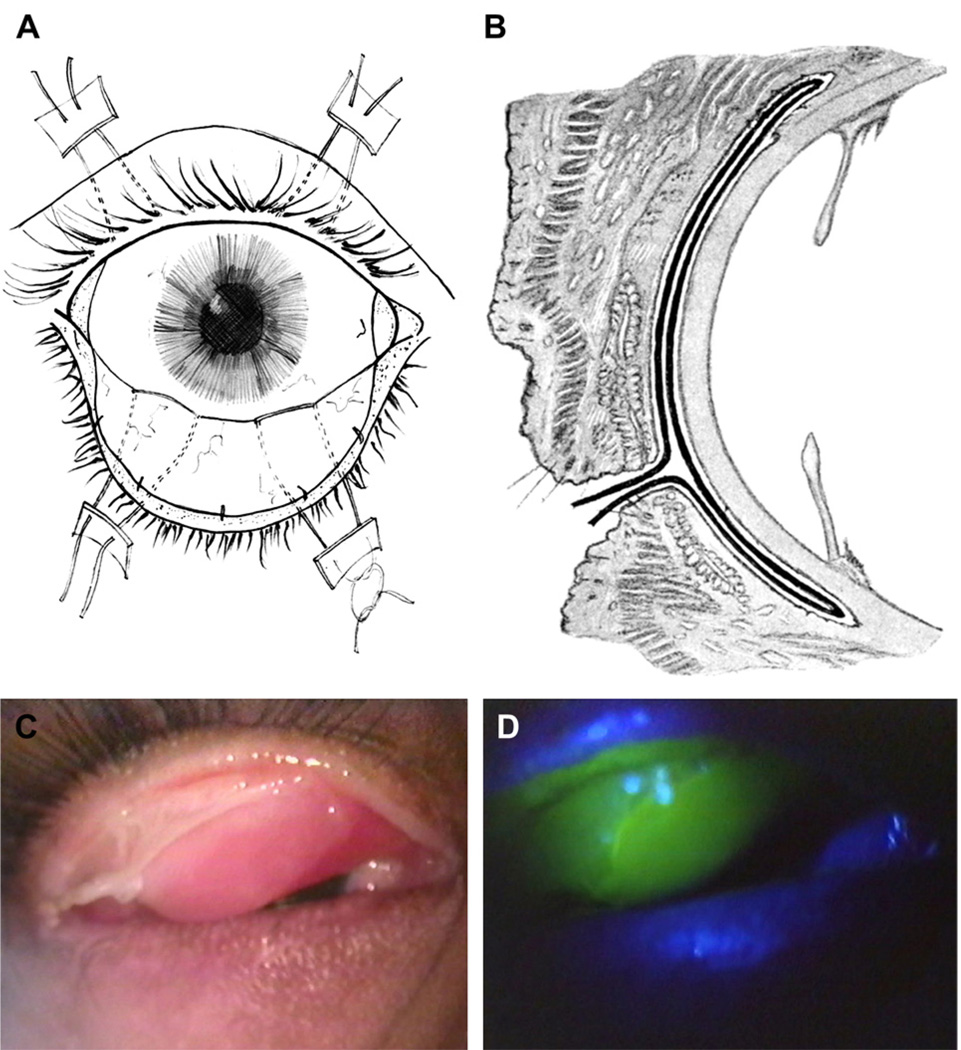
For acute SJS/TEN, cryopreserved AM is sutured to cover the entire ocular surface from lid margin to lid margin as a temporary biological bandage (shown schematically in Fig. 4A and 4B).If the lid margin skin is involved, the lashes are trimmed so that the membrane can be extended to cover the ulcer. If not, the membrane is attached to the gray line by interrupted or continuously running 10-0 or 8-0 nylon/vicryl sutures. From the upper lid margin, AM is then spread to cover the tarsal conjunctiva and fornix before being reflected to cover the bulbar conjunctiva by a muscle hook and anchored to the skin through a double-armed 4-0 black silk or 6-0 prolene suture in a mattress fashion. The same procedure is then performed on the lower lid. Both AMs are fixed to the episclera with a continuous 10-0 nylon suture in a purse-string fashion near the limbus. Alternatively, they can be secured by a symblepharon ring. For surgical videos, visit http://www.osref.org/acute-treat ment-of-severe-ocular-inflammation.aspx from the Ocular Surface Research and Education Foundation, or see a video report by Muqit et al44 at http://bjo.bmj.com/content/vol91/issue11/images/data/1536/DC1/muqitfinalfast.mov.
Fig. 4.
Schematic depiction of the sutured method of AMT for SJS/TEN. AM covers the entire ocular surface, secured by four 4-0 silk double-armed sutures from the fornix to the skin (A: front view; B: side view.) (A and B are reproduced from Meller et al41 with permission of Ophthalmology.) C: AM is attached to the ulcerated tarsus while dissolving in other areas. D: Fluorescein staining confirms that AM has covered the tarsus. This membrane facilitates conjunctival epithelialization while preventing epidermal migration from the lid margin. (C and D are reproduced from Di Pascuale et al16 with permission of Ophthalmology.)
SURGICAL OUTCOME
AM as a temporary biological bandage has been successfully used to treat six patients with acute SJS/TEN during the period from 2002 to 2007.16,26,31,44,70 Table 1 summarizes clinical characteristics and outcomes of 12 eyes of these six pediatric patients (ranging from 4–12 years old), three boys and three girls, who developed SJS (n = 2) or TEN (n = 4) precipitated by medication (n = 4) or infection (n = 2). At the acute stage, five patients presented with external eruptive lesions extending to the lid margin. In the eye, all showed conjunctival inflammation and four patients showed tarsal ulceration, leading to lid adhesion to the globe in five patients. Three patients presented with corneal epithelial defects that were total in three eyes and geographical in three eyes. One eye showed a limbal epithelial defect. AMTwas performed within the first 2 weeks from the onset of the symptoms, based on the sutured method described herein in six patients, using cryopreserved AM obtained from Bio-Tissue (Miami, FL) in four patients. In one eye, AM was stretched by a conformer before suturing.26 In three eyes, one with a corneal epithelial defect, AM did not cover the cornea.26,70 In a median follow-up of 9 months (ranging from 4–36 months) after AMT, none of these 12 eyes showed any persistent conjunctival inflammation. As a result, the entire ocular surface remained stable without limbal stem cell deficiency, and their uncorrected visual acuities ranged from 20/16 to 20/40. It is noteworthy that tarsal ulcerations were covered by AM while the rest dissolved during the acute stage (Fig. 4C and 4D). This explained why scarring was absent in five eyes, but was mild and focal in the tarsal conjunctiva of the remaining seven eyes. In these seven eyes, additional scarring involved the lid margin in four eyes and the corneal periphery in three eyes. Of these three eyes, two had undergone AMT in which AM did not cover the cornea.70 Only two of 12 eyes showed focal symblepharon, which was located in the inferior fornix. Two of the six eyes with corneal involvement healed with fine peripheral vascularization.
TABLE 1.
Literature Summary of AMT for Acute SJS/TEN
| Case Number | 1 | 2 | 3 | 4 | 5 | 6 |
|---|---|---|---|---|---|---|
| Age | 6 | 8 | 4 | 6 | 12 | 10 |
| Sex | M | F | M | M | F | F |
| Offending Agent | SMZ | Mycoplasma | Ibuprofen | Phenobarbital | Infection | Drug |
| Diagnosis | TEN | TEN | SJS | TEN | TEN | SJS |
| Initial Presentation | ||||||
| Lid | ||||||
| Margin ulceration | Yes (OU) | Yes (OU) | Yes (OU) | Yes (OU) | Yes (OU) | No |
| Adhesion to globe | Yes (OU) | Yes (OU) | Noa | Yes (OU) | Yes (OU) | Yes (OU) |
| Conjunctiva | ||||||
| Inflammation | Yes (OU) | Yes (OU) | Yes (OU) | Yes (OU) | Yes (OU) | Yes (OU) |
| Tarsal/fornix ulceration |
NA | NA | Yes (OU) | Yes (OU) | Yes (OU) | Yes (OU) |
| Corneal involvement | ||||||
| Inflammation | No | Yes (OU) | No | Yes (OU) | No | No |
| Epithelial defect | No | Total (OU) | No | Total (OD), Geographical (OS) |
No | Geographical (OU) |
| Limbal involvement | No | No | No | Yes (OS) | No | No |
| Timing of AMT (days)b | <14 | <14 | 7 | 5 | 3 | 3 |
|
Follow-up (months post-operative) |
36 | 34 | 12 | 4 | 3 | 6 |
| Outcome | ||||||
| Persistent conjunctival inflammation |
No | No | No | No | No | No |
| Cicatricial | ||||||
| complications | ||||||
| Lid margin | No | No | Mild (OU) | No | Mild (OU) | No |
| Tarsal conjunctiva | No | Mild (OU) | Mild (OU) | Mild (OD) | Mild (OU) | No |
| Symblepharon | No | No | No | Focal (OD) | Focal (OD) | No |
| Corneal complications | ||||||
| Epithelial defect | No | No | No | No | PEE (OU) | No |
| Opacity | No | No | No | Peripheral (OD) | No | Slight central haze (OS) |
| Vascularization | No | Peripheral (OS) | No | Peripheral (OD) | No | No |
| Limbal stem cell deficiency |
No | No | No | No | No | No |
| Other morbidity | No | Madarosis (OU) | No | No | Trichiasis (OU) | Trichiasis (OU) |
| VA | 20/20 | 20/30 (OD), 20/40 (OS) |
20/20a | 20/16 | 20/20 | 20/16 |
| Literature source | (26) | (26) | (16) | (31) | (70) | (44) |
AMT = amniotic membrane transplantation; NA = not available; OD = right eye; OS = left eye; OU = both eyes; PEE = punctuate epithelial erosions; SJS = Stevens-Johnson syndrome; SMZ = sulfamethoxazole; TEN = toxic epidermal necrolysis; VA = uncorrected visual acuity.
This data do not appear in original paper, but are instead obtained from personal communication with the author.
After onset of eye symptoms.
Collectively, these results demonstrate that AMT, when performed within two weeks after the onset of the disease, effectively aborts inflammation and facilitates rapid healing in AM-covered areas, thus preventing pathogenic cicatricial complications at the chronic stage. When performed at a later stage, AMT might still recover the corneal surface, but cannot prevent progressive conjunctival scarring. Further studies may be necessary to determine the optimal timing of AMT.
Conclusion
Persistent inflammation and ulceration of the conjunctiva or cornea invariably results in cicatricial complications in many types of chronic cicatricial keratoconjunctivitis. Herein literature evidence strongly demonstrates that lesions for SJS/TEN tend to be hidden in the fornix and tarsus in the acute stage and frequently evolve into sight-threatening corneal complications in the chronic stage. For the first time, the literature data also reveal an encouraging trend: AMT performed in the first two weeks after the onset of ocular involvement facilitates rapid epithelial healing and reduces inflammation and scarring of the ocular surface. As a result, this novel therapy can potentially avert the otherwise doomed visual outcome at the chronic stage of this disease. Therefore, although SJS/TEN is relatively rare, the high rate and typically severe ocular morbidity requires prompt diagnosis and early intervention with AMT at the acute stage. Further investigation may elucidate how AMT exerts such anti-inflammatory and anti-scarring actions and help unravel new therapeutic modalities that will promote regeneration rather than repair in SJS/ TEN as well as other mucous membrane inflammatory and ulcerative diseases.
Method of Literature Search
Literature search was performed in Medline using the following keywords: epidemiology, pathogenesis, Stevens-Johnson syndrome, toxic epidermal necrolysis, as well as relevant references cited in those articles. For foreign language publications, no translation was obtained, however, abstracts of relevant non-English articles were used. For the first section focusing on a short review of SJS and TEN and its ocular morbidity, we chose key review articles or studies with large series dealing with these topics back to 1860. For the second and third sections describing the clinical significance of ocular SJS/TEN including epidemiology, pathogenesis, and natural history, we focus on the literature connecting cicatricial complications and blindness. For the medical treatments of acute SJS detailed in the fourth section, studies of the treatments as well as recent reviews were chosen from 1990. Regarding action mechanism and ocular applications of AMT, we only cite recent key reviews dated after AMT was introduced for ocular surface reconstruction in 1995. For the statements that are frequently mentioned by others but have not changed from time to time, we chose the earliest publication and other important articles. For surgical outcome, we conducted a Medline search with the keywords acute, Stevens-Johnson syndrome, and amniotic membrane. All articles dealing with AMT in the acute management of SJS and TEN were reviewed and succinctly summarized as a Table for meta-analysis.
Acknowledgments
Dr Tseng has obtained a patent for the method of preparation and clinical uses of amniotic membrane and has licensed the right to Bio-Tissue, which procures, processes, and distributes preserved amniotic membrane for clinical and research uses. The other authors reported no proprietary or commercial interest in any product mentioned or concept discussed in this article. Dr. Ahmad Kheirkhah is a recipient of Joseph Swiger and Eye Foundation of America Fellowship from Ocular Surface Research and Education Foundation, Miami, FL. Dr. Lingyi Liang received the Exchange Scholarship Grant for PhD candidates from the Scholarship Council of China.
References
- 1.Adds PJ, Hunt CJ, Dart JK. Amniotic membrane grafts, “fresh” or frozen? A clinical and in vitro comparison. Br J Ophthalmol. 2001;85:905–907. doi: 10.1136/bjo.85.8.905. [DOI] [PMC free article] [PubMed] [Google Scholar]
- 2.Aihara Y, Ito R, Ito S, et al. Toxic epidermal necrolysis in a child successfully treated with cyclosporin A and methyl-prednisolone. Pediatr Int. 2007 Oct;49(5):659–662. doi: 10.1111/j.1442-200X.2007.02439.x. [DOI] [PubMed] [Google Scholar]
- 3.Arevalo JM, Lorente JA, Gonzalez-Herrada C, Jimenez-Reyes J. Treatment of toxic epidermal necrolysis with cyclosporin A. J Trauma. 2000;48(3):473–478. doi: 10.1097/00005373-200003000-00017. [DOI] [PubMed] [Google Scholar]
- 4.Arora R, Mehta D, Jain V. Amniotic membrane transplantation in acute chemical burns. Eye. 2005 Mar;19(3):273–278. doi: 10.1038/sj.eye.6701490. [DOI] [PubMed] [Google Scholar]
- 5.Arstikaitis MJ. Ocular aftermath of Stevens-Johnson syndrome. Arch Ophthalmol. 1973 Nov;90(5):376–379. doi: 10.1001/archopht.1973.01000050378008. [DOI] [PubMed] [Google Scholar]
- 6.Ashby DW, Lazar T. Erythema multiforme exudativum major (Stevens-Johnson syndrome) Lancet. 1951;1(20):1091–1095. doi: 10.1016/s0140-6736(51)92611-6. [DOI] [PubMed] [Google Scholar]
- 7.Belfort R, Jr, de SM, Whitcup SM, et al. Ocular complications of Stevens-Johnson syndrome and toxic epidermal necrolysis in patients with AIDS. Cornea. 1991;10(6):536–538. doi: 10.1097/00003226-199111000-00013. [DOI] [PubMed] [Google Scholar]
- 8.Bottinger LE, Strandberg I, Westerholm B. Drug-induced febrile mucocutaneous syndrome: with a review of literature. Acta Med Scand. 1975;198:229–233. [PubMed] [Google Scholar]
- 9.Bouchard CS, John T. Amniotic membrane transplantation in the management of severe ocular surface disease: indications and outcomes. Ocular Surface. 2004;2(3):201–211. doi: 10.1016/s1542-0124(12)70062-9. [DOI] [PubMed] [Google Scholar]
- 10.Brilakis HS, Palmon FE, Webster GF, Holland EJ. Erythema multiforme, Stevens-Johnson syndrome, and toxic epidermal necrolysis. In: Krachmer JH, Mannis MJ, Holland EJ, editors. Cornea. Philadelphia, Mosby. ed. 2. 2005. pp. 691–702. [Google Scholar]
- 11.Burman S, Tejwani S, Vemuganti GK, et al. Ophthalmic applications of preserved human amniotic membrane: a review of current indications. Cell Tissue Bank. 2004;5(3):161–175. doi: 10.1023/B:CATB.0000046067.25057.0a. [DOI] [PubMed] [Google Scholar]
- 12.Chan HL, Stern RS, Arndt KA, et al. The incidence of erythema multiforme, Stevens-Johnson syndrome, and toxic epidermal necrolysis. A population-based study with particular reference to reactions caused by drugs among outpatients. Arch Dermatol. 1990;126(1):43–47. [PubMed] [Google Scholar]
- 13.Chang YS, Huang FC, Tseng SH, et al. Erythema multiforme, Stevens-Johnson syndrome, and toxic epidermal necrolysis: acute ocular manifestations, causes, and management. Cornea. 2007;26(2):123–129. doi: 10.1097/ICO.0b013e31802eb264. [DOI] [PubMed] [Google Scholar]
- 14.Chave TA, Mortimer NJ, Sladden MJ, et al. Toxic epidermal necrolysis: current evidence, practical management and future directions. Br J Dermatol. 2005;153(2):241–253. doi: 10.1111/j.1365-2133.2005.06721.x. [DOI] [PubMed] [Google Scholar]
- 15.Chen J, Zhou S, Huang T, et al. [A clinical study on fresh amniotic membrane transplantation for treatment of severe ocular surface disorders at acute inflammatory and cicatricial stage] Zhonghua Yan Ke Za Zhi. 2000;36(1):13–17. [PubMed] [Google Scholar]
- 16.Di Pascuale MA, Espana EM, Liu DT, et al. Correlation of corneal complications with eyelid cicatricial pathologies in patients with Stevens-Johnson syndrome and toxic epidermal necrolysis syndrome. Ophthalmology. 2005;112(5):904–912. doi: 10.1016/j.ophtha.2004.11.035. [DOI] [PubMed] [Google Scholar]
- 17.Dohlman CH, Doughman DJ. The Stevens-Johnson Syndrome. Trans New Orleans Acad Ophthalmol. 1972;24:236–252. [Google Scholar]
- 18.Dua HS, Azuara-Blanco A. Allo-limbal transplantation in patients with limbal stem cell deficiency. Br J Ophthalmol. 1999;83:414–419. doi: 10.1136/bjo.83.4.414. [DOI] [PMC free article] [PubMed] [Google Scholar]
- 19.Dua HS, Gomes JA, King AJ, Maharajan VS. The amniotic membrane in ophthalmology. Surv Ophthalmol. 2004 Jan;49(1):51–77. doi: 10.1016/j.survophthal.2003.10.004. [DOI] [PubMed] [Google Scholar]
- 20.Dua HS, Jagjit SS, Azuara-Blanco A, Gupta P. Limbal stem cell deficiency: concept, aetiology, clinical presentation, diagnosis and management. Indian J Ophthalmol. 2000;48:83–92. [PubMed] [Google Scholar]
- 21.Espana EM, Di Pascuale M, Grueterich M, et al. Keratolimbal allograft in corneal reconstruction. Eye. 2004;18(4):406–417. doi: 10.1038/sj.eye.6700670. [DOI] [PubMed] [Google Scholar]
- 22.French LE. Toxic epidermal necrolysis and Stevens-Johnson syndrome: our current understanding. Allergol Int. 2006;55(1):9–16. doi: 10.2332/allergolint.55.9. [DOI] [PubMed] [Google Scholar]
- 23.Grueterich M, Espana EM, Tseng SCG. Ex vivo expansion of limbal epithelial stem cells: amniotic membrane serving as a stem cell niche. Surv Ophthalmol. 2003;48:631–646. doi: 10.1016/j.survophthal.2003.08.003. [DOI] [PubMed] [Google Scholar]
- 24.Hazin R, Ibrahimi OA, Hazin MI, Kimyai-Asadi A. Stevens-Johnson syndrome: pathogenesis, diagnosis, and management. Ann Med. 2008;40(2):129–138. doi: 10.1080/07853890701753664. [DOI] [PubMed] [Google Scholar]
- 25.Howard GM. The Stevens-Johnson syndrome Ocular prognosis and treatment. Am J Ophthalmol. 1963;55:893–900. [PubMed] [Google Scholar]
- 26.John T, Foulks GN, John ME, et al. Amniotic membrane in the surgical management of acute toxic epidermal necrolysis. Ophthalmology. 2002;109(2):351–360. doi: 10.1016/s0161-6420(01)00900-9. [DOI] [PubMed] [Google Scholar]
- 27.Khalili B, Bahna SL. Pathogenesis and recent therapeutic trends in Stevens-Johnson syndrome and toxic epidermal necrolysis. Ann Allergy Asthma Immunol. 2006;97(3):272–280. doi: 10.1016/S1081-1206(10)60789-2. [DOI] [PubMed] [Google Scholar]
- 28.Kheirkhah A, Johnson DA, Paranjpe DR, et al. Temporary sutureless amniotic membrane patch for acute alkaline burns. Arch Ophthalmol. 2008;126(8):1059–1066. doi: 10.1001/archopht.126.8.1059. [DOI] [PMC free article] [PubMed] [Google Scholar]
- 29.Kim JS, Kim JC, Na BK, et al. Amniotic membrane patching promotes healing and inhibits protease activity on wound healing following acute corneal alkali burns. Exp Eye Res. 2000;70:329–337. doi: 10.1006/exer.1999.0794. [DOI] [PubMed] [Google Scholar]
- 30.Kobayashi A, Shirao Y, Yoshita T, et al. Temporary amniotic membrane patching for acute chemical burns. Eye. 2003;17:149–158. doi: 10.1038/sj.eye.6700316. [DOI] [PubMed] [Google Scholar]
- 31.Kobayashi A, Yoshita T, Sugiyama K, et al. Amniotic membrane transplantation in acute phase of toxic epidermal necrolysis with severe corneal involvement. Ophthalmology. 2006;113(1):126–132. doi: 10.1016/j.ophtha.2005.09.001. [DOI] [PubMed] [Google Scholar]
- 32.Koizumi N, Inatomi T, Suzuki T, et al. Cultivated corneal epithelial stem cell transplantation in ocular surface disorders. Ophthalmology. 2001;108:1569–1574. doi: 10.1016/s0161-6420(01)00694-7. [DOI] [PubMed] [Google Scholar]
- 33.Lavker RM, Tseng SC, Sun TT. Corneal epithelial stem cells at the limbus: looking at some old problems from a new angle. Exp Eye Res. 2004;78(3):433–446. doi: 10.1016/j.exer.2003.09.008. [DOI] [PubMed] [Google Scholar]
- 34.Lee NJ, Wang SJ, Durairaj KK, et al. Increased expression of transforming growth factor-beta1, acidic fibroblast growth factor, and basic fibroblast growth factor in fetal versus adult fibroblast cell lines. Laryngoscope. 2000;110(4):616–619. doi: 10.1097/00005537-200004000-00015. [DOI] [PubMed] [Google Scholar]
- 35.Lee SH, Tseng SCG. Amniotic membrane transplantation for persistent epithelial defects with ulceration. Am J Ophthalmol. 1997;123:303–312. doi: 10.1016/s0002-9394(14)70125-4. [DOI] [PubMed] [Google Scholar]
- 36.Lerner LH, Qureshi AA, Reddy BV, Lerner EA. Nitric oxide synthase in toxic epidermal necrolysis and Stevens-Johnson syndrome. J Invest Dermatol. 2000;114(1):196–199. doi: 10.1046/j.1523-1747.2000.00816.x. [DOI] [PubMed] [Google Scholar]
- 37.Letko E, Papaliodis DN, Papaliodis GN, et al. Stevens-Johnson syndrome and toxic epidermal necrolysis: a review of the literature. Ann Allergy Asthma Immunol. 2005;94(4):419–436. doi: 10.1016/S1081-1206(10)61112-X. [DOI] [PubMed] [Google Scholar]
- 38.Liu C, Paul B, Tandon R, et al. The osteo-odonto-keratoprosthesis (OOKP) Semin Ophthalmol. 2005;20(2):113–128. doi: 10.1080/08820530590931386. [DOI] [PubMed] [Google Scholar]
- 39.Luo HY, Peng SM, Wang YJ. [Fresh amniotic membrane transplantation for ocular surface diseases] Di Yi Jun Yi Da Xue Xue Bao. 2003;23(5):488–489. [PubMed] [Google Scholar]
- 40.Lyell A. Toxic epidermal necrolysis: an eruption resembling scalding of the skin. Br J Dermatol. 1956;68(11):355–361. doi: 10.1111/j.1365-2133.1956.tb12766.x. [DOI] [PubMed] [Google Scholar]
- 41.Meller D, Pires RT, Mack RJ, et al. Amniotic membrane transplantation for acute chemical or thermal burns. Ophthalmology. 2000;107(5):980–989. doi: 10.1016/s0161-6420(00)00024-5. [DOI] [PubMed] [Google Scholar]
- 42.Mockenhaupt M, Schopf E. Epidemiology of drug-induced severe skin reactions. Semin Cutan Med Surg. 1996;15(4):236–243. doi: 10.1016/s1085-5629(96)80036-8. [DOI] [PubMed] [Google Scholar]
- 43.Mondino BJ. Cicatricial pemphigoid and erythema multiforme. Ophthalmology. 1990;97(7):939–952. doi: 10.1016/s0161-6420(90)32479-x. [DOI] [PubMed] [Google Scholar]
- 44.Muqit MM, Ellingham RB, Daniel C. Technique of amniotic membrane transplant dressing in the management of acute Stevens-Johnson syndrome. Br J Ophthalmol. 2007;91(11):1536. doi: 10.1136/bjo.2007.131102. [DOI] [PMC free article] [PubMed] [Google Scholar]
- 45.Naldi L, Locati F, Marchesi L, Cainelli T. Incidence of toxic epidermal necrolysis in Italy. Arch Dermatol. 1990;126(8):1103–1104. doi: 10.1001/archderm.1990.01670320127028. [DOI] [PubMed] [Google Scholar]
- 46.Nassif A, Bensussan A, Dorothee G, et al. Drug specific cytotoxic T-cells in the skin lesions of a patient with toxic epidermal necrolysis. J Invest Dermatol. 2002;118(4):728–733. doi: 10.1046/j.1523-1747.2002.01622.x. [DOI] [PubMed] [Google Scholar]
- 47.Ostler HB, Conant MA, Groundwater J. Lyell’s disease, the Stevens-Johnson syndrome, and exfoliative dermatitis. Trans Am Acad Ophthalmol. Otolaryngol. 1970;74(6):1254–1265. [PubMed] [Google Scholar]
- 48.Paquet P, Nikkels A, Arrese JE, et al. Macrophages and tumor necrosis factor alpha in toxic epidermal necrolysis. Arch Dermatol. 1994;130(5):605–608. [PubMed] [Google Scholar]
- 49.Paquet P, Pierard GE, Quatresooz P. Novel treatments for drug-induced toxic epidermal necrolysis (Lyell’s syndrome) Int Arch Allergy Immunol. 2005;136(3):205–216. doi: 10.1159/000083947. [DOI] [PubMed] [Google Scholar]
- 50.Patz A. Ocular involvement in erythema multiforme. Arch Ophthalmol. 1950;43(2):244–256. doi: 10.1001/archopht.1950.00910010251007. [DOI] [PubMed] [Google Scholar]
- 51.Paul C, Wolkenstein P, Adle H, et al. Apoptosis as a mechanism of keratinocyte death in toxic epidermal necrolysis. Br J Dermatol. 1996;134(4):710–714. doi: 10.1111/j.1365-2133.1996.tb06976.x. [DOI] [PubMed] [Google Scholar]
- 52.Posadas SJ, Padial A, Torres MJ, et al. Delayed reactions to drugs show levels of perforin, granzyme B, and Fas-L to be related to disease severity. J Allergy Clin Immunol. 2002;109(1):155–161. doi: 10.1067/mai.2002.120563. [DOI] [PubMed] [Google Scholar]
- 53.Power WJ, Ghoraishi M, Merayo-Lloves J, et al. Analysis of the acute ophthalmic manifestations of the erythema multiforme/Stevens-Johnson syndrome/toxic epidermal necrolysis disease spectrum. Ophthalmology. 1995;102:1669–1676. doi: 10.1016/s0161-6420(95)30811-1. [DOI] [PubMed] [Google Scholar]
- 54.Prabhasawat P, Kosrirukvongs P, Booranapong W, Vajaradul Y. Amniotic membrane transplantation for ocular surface reconstruction. J Med Assoc Thai. 2001;84(5):705–718. [PubMed] [Google Scholar]
- 55.Ralph RA. Conjunctival goblet cell density in normal subjects and in dry eye syndromes. Invest Ophthalmol. 1975;14(4):299–302. [PubMed] [Google Scholar]
- 56.Raviglione MC, Pablos-Mendez A, Battan R. Clinical features and management of severe dermatological reactions to drugs. Drug Saf. 1990;5(1):39–64. doi: 10.2165/00002018-199005010-00005. [DOI] [PubMed] [Google Scholar]
- 57.Roujeau JC. The spectrum of Stevens-Johnson syndrome and toxic epidermal necrolysis: a clinical classification. J Invest Dermatol. 1994;102(6):28S–30S. doi: 10.1111/1523-1747.ep12388434. [DOI] [PubMed] [Google Scholar]
- 58.Sangwan VS, Burman S, Tejwani S, et al. Amniotic membrane transplantation: a review of current indications in the management of ophthalmic disorders. Indian J Ophthalmol. 2007;55(4):251–260. doi: 10.4103/0301-4738.33036. [DOI] [PubMed] [Google Scholar]
- 59.Sayegh RR, Ang LP, Foster CS, Dohlman CH. The Boston keratoprosthesis in Stevens-Johnson syndrome. Am J Ophthalmol. 2008;145(3):438–444. doi: 10.1016/j.ajo.2007.11.002. [DOI] [PubMed] [Google Scholar]
- 60.Scheinfeld N. A review of deferasirox, bortezomib, dasatinib, and cyclosporine eye drops: possible uses and known side effects in cutaneous medicine. J Drugs Dermatol. 2007;6(3):352–355. [PubMed] [Google Scholar]
- 61.Schneck J, Fagot JP, Sekula P, et al. Effects of treatments on the mortality of Stevens-Johnson syndrome and toxic epidermal necrolysis: a retrospective study on patients included in the prospective EuroSCAR Study. J Am Acad Dermatol. 2008;58(1):33–40. doi: 10.1016/j.jaad.2007.08.039. [DOI] [PubMed] [Google Scholar]
- 62.Schopf E, Stuhmer A, Rzany B, Toxic epidermal necrolysis, Stevens-Johnson syndrome, et al. An epidemiologic study from West Germany. Arch Dermatol. 1991;127(6):839–842. doi: 10.1001/archderm.1991.01680050083008. [DOI] [PubMed] [Google Scholar]
- 63.Shimmura S, Shimazaki J, Ohashi Y, Tsubota K. Anti-inflammatory effects of amniotic membrane transplantation in ocular surface disorders. Cornea. 2001;20(4):408–413. doi: 10.1097/00003226-200105000-00015. [DOI] [PubMed] [Google Scholar]
- 64.Sippel KC, Ma JJK, Foster CS. Amniotic membrane surgery. Curr Opin Ophthalmol. 2001;12:269–281. doi: 10.1097/00055735-200108000-00006. [DOI] [PubMed] [Google Scholar]
- 65.Solomon A, Ellies P, Anderson DF, et al. Long-term outcome of keratolimbal allograft with or without penetrating keratoplasty for total limbal stem cell deficiency. Ophthalmology. 2002;109(6):1159–1166. doi: 10.1016/s0161-6420(02)00960-0. [DOI] [PubMed] [Google Scholar]
- 66.Solomon A, Espana EM, Tseng SCG. Amniotic membrane transplantation for reconstruction of the conjunctival fornices. Ophthalmology. 2003;110:93–100. doi: 10.1016/s0161-6420(02)01441-0. [DOI] [PubMed] [Google Scholar]
- 67.Solomon A, Meller D, Prabhasawat P, et al. Amniotic membrane grafts for nontraumatic corneal perforations, descemetoceles, and deep ulcers. Ophthalmology. 2002;109(4):694–703. doi: 10.1016/s0161-6420(01)01032-6. [DOI] [PubMed] [Google Scholar]
- 68.Sridhar MS, Bansal AK, Sangwan VS, Rao GN. Amniotic membrane transplantation in acute chemical and thermal injury. Am J Ophthalmol. 2000;130:134–137. doi: 10.1016/s0002-9394(00)00500-6. [DOI] [PubMed] [Google Scholar]
- 69.Stevens AM, Johnson FC. A new eruptive fever associated with stomatitis and opthalmitis. Report of two cases in children. Am J Dis Child. 1922;24(6):526–533. [Google Scholar]
- 70.Tandon A, Cackett P, Mulvihill A, Fleck B. Amniotic membrane grafting for conjunctival and lid surface disease in the acute phase of toxic epidermal necrolysis. J AAPOS. 2007;11(6):612–613. doi: 10.1016/j.jaapos.2007.04.020. [DOI] [PubMed] [Google Scholar]
- 71.Tejwani S, Kolari RS, Sangwan VS, Rao GN. Role of amniotic membrane graft for ocular chemical and thermal injuries. Cornea. 2007;26(1):21–26. doi: 10.1097/ICO.0b013e31802b4201. [DOI] [PubMed] [Google Scholar]
- 72.Tonnesen MG, Soter NA. Erythema multiforme. J Am Acad Dermatol. 1979;1(4):357–364. doi: 10.1016/s0190-9622(79)70030-2. [DOI] [PubMed] [Google Scholar]
- 73.Tseng SC. Amniotic membrane transplantation for ocular surface reconstruction. Biosci Rep. 2001;21(4):481–489. doi: 10.1023/a:1017995810755. [DOI] [PubMed] [Google Scholar]
- 74.Tseng SCG. Regulation and clinical implications of corneal epithelial stem cells. Mol Biol Rep. 1996;23:47–58. doi: 10.1007/BF00357072. [DOI] [PubMed] [Google Scholar]
- 75.Tseng SCG, Chen JJY, Huang AJW, et al. Classification of conjunctival surgeries for corneal disease based on stem cell concept. Clin North Am Ophthalmol. 1990;3:595–610. [Google Scholar]
- 76.Tseng SCG, Di Pascuale MA, Liu DZ, et al. Intraoperative mitomycin C and amniotic membrane transplantation for fornix reconstruction in severe cicatricial ocular surface diseases. Ophthalmology. 2005;112:896–903. doi: 10.1016/j.ophtha.2004.11.041. [DOI] [PubMed] [Google Scholar]
- 77.Tseng SCG, Espana EM, Kawakita T, et al. How does amniotic membrane work? Ocular Surface. 2004;2(3):177–187. doi: 10.1016/s1542-0124(12)70059-9. [DOI] [PubMed] [Google Scholar]
- 78.Tseng SCG, Li DQ, Ma X. Suppression of transforming growth factor isoforms, TGF--b receptor II, and myofibro-blast differentiation in cultured human corneal and limbal fibroblasts by amniotic membrane matrix. J Cell Physiol. 1999;179:325–335. doi: 10.1002/(SICI)1097-4652(199906)179:3<325::AID-JCP10>3.0.CO;2-X. [DOI] [PubMed] [Google Scholar]
- 79.Ueta M, Kweon MN, Sano Y, et al. Immunosuppressive properties of human amniotic membrane for mixed lymphocyte reaction. Clin Exp Immunol. 2002;129(3):464–470. doi: 10.1046/j.1365-2249.2002.01945.x. [DOI] [PMC free article] [PubMed] [Google Scholar]
- 80.Viard I, Wehrli P, Bullani R, et al. Inhibition of toxic epidermal necrolysis by blockade of CD95 with human intravenous immunoglobulin. Science. 1998;282(5388):490–493. doi: 10.1126/science.282.5388.490. [DOI] [PubMed] [Google Scholar]
- 81.Viard-Leveugle I, Bullani RR, Meda P, et al. Intracellular localization of keratinocyte Fas ligand explains lack of cytolytic activity under physiological conditions. J Biol Chem. 2003;278(18):16183–16188. doi: 10.1074/jbc.M212188200. [DOI] [PubMed] [Google Scholar]
- 82.Westly ED, Wechsler HL. Toxic epidermal necrolysis Granulocytic leukopenia as a prognostic indicator. Arch Dermatol. 1984;120(6):721–726. doi: 10.1001/archderm.120.6.721. [DOI] [PubMed] [Google Scholar]
- 83.Wright P, Collin JR. The ocular complications of erythema multiforme (Stevens Johnson syndrome) and their management. Trans Ophthalmol Soc UK. 1983;103(Pt 3):338–341. [PubMed] [Google Scholar]
- 84.Yaghouti F, Nouri M, Abad JC, et al. Keratoprosthesis: preoperative prognostic categories. Cornea. 2001;20(1):19–23. doi: 10.1097/00003226-200101000-00003. [DOI] [PubMed] [Google Scholar]
- 85.Yang R, Wang F, Sun N. [Application of fresh amniotic membrane transplantation in treatment of stenosis of conjunctival sac] Zhongguo Xiu Fu Chong Jian Wai Ke Za Zhi. 2004;18(4):277–280. [PubMed] [Google Scholar]
- 86.Yetiv JZ, Bianchine JR, Owen JA., Jr Etiologic factors of the Stevens-Johnson syndrome. South Med J. 1980;73(5):599–602. doi: 10.1097/00007611-198005000-00015. [DOI] [PubMed] [Google Scholar]
- 87.Zhao F, Xu W, Guan J. [Clinical observation on fresh amniotic membrane transplantation for treatment of recurrent pterygium] Yan Ke Xue Bao. 2002;18(4):220–222. [PubMed] [Google Scholar]
- 88.Zhou S, Chen J, Fung B, Chen L. Fresh amniotic membrane transplantation combined lamellar keratoplasty for patients with recurrent Mooren’s ulcer. Yan Ke Xue Bao. 2003;19(1):1–5. [PubMed] [Google Scholar]
- 89.Zhou S, Chen J, Xu L, et al. Fresh amniotic membrane transplantation for conjunctival surface reconstruction. Yan Ke Xue Bao. 1999;15(3):169–173. [PubMed] [Google Scholar]
- 90.Zhou S, Yuan J, Chen L, Chen J. [Fresh versus preserved amniotic membrane transplantation for conjunctival surface reconstruction] Yan Ke Xue Bao. 2008;24(1):39–43. [PubMed] [Google Scholar]


