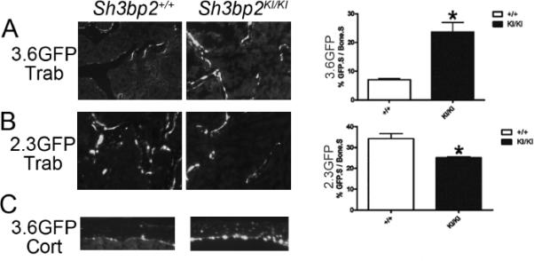
Fig. 3.

In vivo monitoring of cherubism Sh3bp2KI/KI osteoblast cultures using Col1a1 promoter-driven GFP. Sh3bp2+/+ and Sh3bp2KI/KI mice were crossed with transgenic mice overexpressing GFP driven by either 3.6-kb or 2.3-kb Col1a1 promoters. Frozen sections (5 μm) of femurs from 10-week-old mice showed increased numbers of 3.6-kb Col1a1 GFP positive cells (3.6GFP) in Sh3bp2KI/KI mice (A), but decreased numbers of 2.3-kb Col1a1 GFP positive cells (2.3GFP) (B). (Student's t-test; n=6 for pOBCol3.6GFPtpz; n=4 for pOBCol2.3GFPemd, *p<0.05). 3.6-kb Col1a1 promoter in osteocytes from femoral cortex of Sh3bp2KI/KI but not of Sh3bp2+/+ mice was active (C). Trab: trabecular bone. Cort: Cortical bone.
