Summary
Many cellular functions depend on rapid and localized actin polymerization/depolymerization. Yet, the de novo polymerization of actin in cells is kinetically unfavorable because of the instability of polymerization intermediates (small actin oligomers) and the actions of actin monomer binding proteins. Cells use filament nucleation and elongation factors to initiate and sustain polymerization. Structural biology is beginning to shed light on the diverse mechanisms by which these unrelated proteins initiate polymerization, undergo regulation, and mediate the transition of monomeric actin onto actin filaments. A prominent role is played by the W domain, which in some of these proteins occurs in tandem repeats that recruit multiple actin subunits. Pro-rich regions are also abundant and mediate the binding of profilin-actin complexes, which are the main source of polymerization competent actin in cells. Filament nucleation and elongation factors frequently interact with Rho family GTPases, which relay signals from membrane receptors to regulate actin cytoskeleton remodeling.
Keywords: Actin filament nucleation, elongation, Arp2/3 complex, Formin, Lmod, Spire, Cobl, VopL/VopF, Eva/VASP
Introduction
The dynamic remodeling of the actin cytoskeleton is essential for many cellular functions, including cell locomotion, cytokinesis and membrane trafficking. Additionally, many pathogens hijack the host cell actin cytoskeleton during infection. These processes involve rapid bursts of actin polymerization/depolymerization. But, somewhat paradoxically, the formation of new actin filaments in cells is kinetically unfavorable, because of the instability of polymerization intermediates (actin dimers, trimers and tetramers) and the actions of actin monomer binding proteins such as profilin and thymosin-β4 (Tβ4). This creates an opportunity for cells to actively regulate the de novo polymerization of actin by using actin filament nucleation and elongation factors.
The actin filament can be described as either a single left-handed short-pitch helix, with consecutive lateral subunits staggered with respect to one another by half a monomer length, or two right-handed long-pitch helices of head-to-tail bound actin subunits [1]. Filament nucleators work by different mechanisms, stabilizing small actin oligomers along either the long- or the short-pitch helices of the actin filament. With the exception of formins they all use the WASP-Homology 2 (WH2 or W) domain, a small and versatile actin-binding motif, for interaction with actin. In various proteins, including Spire, Cobl and VopL/VopF, the W domain occurs in tandem repeats that bind three to four actin subunits to form a nucleus. Structural considerations suggest that Nucleation Promoting Factors (NPFs), proteins that form a nucleus consisting of one to three actin subunits and the two actin-related proteins of Arp2/3 complex, can be viewed as a specialized form of tandem W domain nucleator. Formins are unique in that they use the formin-homology 2 (FH2) domain for interaction with actin and promote not only nucleation, but also processive barbed end elongation. In contrast, the elongation function among W-based nucleators has been outsourced to a dedicated family of proteins, Eva/VASP, which are related to NPFs.
Formins
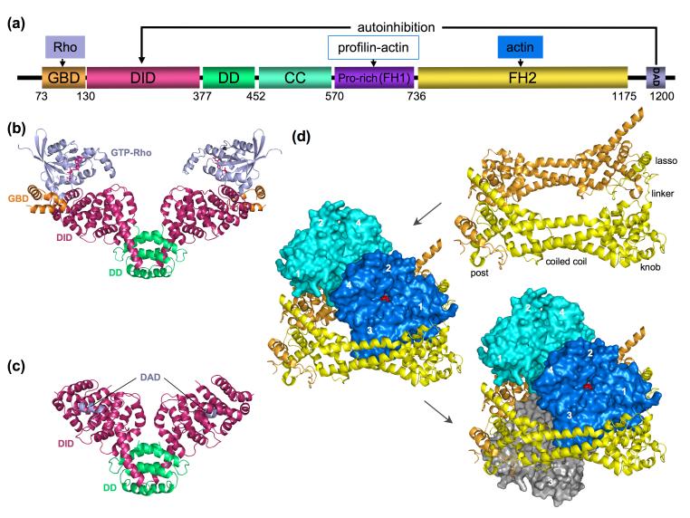
Among filament nucleation/elongation factors, formins are the best understood at the structural level [2-4]. In cellular processes ranging from cell locomotion and morphogenesis to cytokinesis, formins mediate the assembly of unbranched actin networks, such as filopodia, stress fibers and actin cables. Like most cytoskeletal proteins, formins are multidomain, multifunctional proteins. There is significant sequence variability within the formin family, but two regions of higher conservation known as the formin homology (FH) 1 and 2 domains were initially identified [5]. Biochemical and structural characterization have since led to a more accurate definition of domain boundaries and functions [3] (Figure 1a). The diaphanous-related formins (DRFs) are most extensively studied, including mDia 1, 2, and 3, DAAM, Bni1 and Bnr1. The N-terminal region of DRFs consists of four domains involved in regulation and dimerization: GTPase-binding domain (GBD), diaphanous inhibitory domain (DID), dimerization domain (DD) and coiled coil domain (CC). The middle region consists of a Pro-rich domain. The C-terminal region consists of the FH2 domain and diaphanous autoregulatory domain (DAD). In the resting state, DRFs exist in a folded autoinhibited conformation stabilized by internal DAD-DID interaction. DRFs function under the control of Rho-family GTPases that release DAD-DID autoinhibitory interactions [6]. Activation frees the FH2 domain, which is responsible for the two filament assembly functions, nucleation and elongation [7]. Recruitment of profilin-actin through the Pro-rich domain accelerates formin-mediated filament elongation [8].
Figure 1.
Domain diagram of DRFs and representative structures. (a) A prototypical DRF (domain boundaries correspond to mDia1) consists of the following domains: GBD (GTPase-binding domain), DID (diaphanous inhibitory domain), DD (dimerization domain), CC (coiled coil), FH1 and FH2 (formin homology 1 and 2 domains) and DAD (diaphanous autoregulatory domain). (b-c) Structures of N-terminal regulatory fragments with bound RhoC [9] and DAD [10]. Ribbon diagrams colored according to the domain diagram in part a. (d) Illustrations of the structure of the FH2-actin complex [12] showing, in a sequence, the crystallographic FH2 dimer (obtained after domain swapping the ‘lasso’ region from the lower subunit), the actin subunits bound to the dimer (blue and cyan), and a third actin from the complex below (gray). Each FH2 domain is structurally subdivide into lasso, linker, knob, coiled coil and post regions. This figure is inspired by similar figures in [3].
A series of overlapping structures of fragments of mouse mDia1 and yeast Bni1 produce a rather complete picture of the various domains, dimerization, authoinhibitory interactions, activation by Rho GTPases, and filament assembly mechanism (Figure 1b-d) [3]. A structure consisting of a GBD-DID-DD fragment of mDia1 in complex with GTP-RhoC shows that the switch I and II regions of the GTPase engage primarily the GBD [9] (Figure 1c). The GBD, thought to be unstructured on its own, adopts an all-helical conformation and together with the bound GTPase folds against the adjacent armadillo repeat DID domain. In another structure (Figure 1c), a DID-DD fragment of mDia1 was cocrystallized with a C-terminal DAD peptide, which interacts with the DID domain near the binding interface of the GBD and Rho [10]. Combined, the two structures suggest that there is only partial but sufficient overlap between the binding interfaces of the DAD and those of the GBD and Rho to explain the release of inhibition produced by the GTPase.
The FH2 dimer consists of two rod-shaped domains connected in head-to-tail fashion by highly flexible linkers [11] (Figure 1d). The flexibility of the connecting linkers and the overall ring-like shape of the structure suggested that the FH2 subunits could move semi-independently, ‘stair-stepping’ at the barbed end of the filament to allow for the incorporation of new actin subunits [11]. This model received additional support from the structure of a complex of the FH2 of Bni1 with actin [12]. The asymmetric unit of the crystal contains a single FH2 subunit with one actin bound. However, a C2 symmetry axis positions identical FH2-actin complexes at ±28 Å and rotated by 180° with respect to the reference complex. Consecutive FH2 subunits along the C2 axis form dimers, similar to that of the uncomplexed FH2 structure [11]. However, to make room for the bound actins, the separation between FH2 subunits is greater in the complex, allowed by flexibility of the linker region. The two actins bound to the crystallographic FH2 dimer adopt a pseudo short-pitch conformation (actin subunits of the short-pitch filament helix are rotated by 167° and translated by 27.5Å [1]). Although the short-pitch dimer of the FH2-actin structure is constrained by crystal symmetry, it is probably a good representation of the dimeric seed stabilized by formins during nucleation. The structure also provides support for the prevailing stair-stepping elongation model. Each FH2-actin dimer is in contact with a third actin subunit (gray subunit in Figure 1d), thought to represent a newly added and loosely connected actin subunit at the barbed end. The stair-stepping model predicts that the uppermost FH2 subunit moves to bind the newly added actin and stabilize its position at the barbed end of the filament. Other features of this model, such as potential rotation of formin at the barbed end during elongation and the correlation between nucleotide hydrolysis by actin and elongation, remain unresolved and are not discussed.
Tandem W domain-based filament nucleators
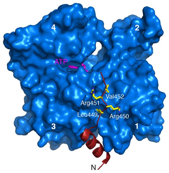
With the exception of formins, filament nucleators use the W domain for interaction with actin, although in Lmod and Arp2/3 complex other domains contribute as well. The W domain is short (17-27-aa) and poorly conserved [13]. Its N-terminal portion forms a helix that binds in the target-binding cleft between actin subdomains 1 and 3 [14,15] (Figure 2). The extended region after this helix has variable length and sequence, but comprises the conserved motif LKKT(V).
Figure 2.
Structure of the W-actin complex. The structure shown [15] is that of the W domain of the NPF protein WAVE2 (red) bound to actin (blue). Subdomains 1-4 of actin are labeled. The N-terminal helix of the W domain binds in the hydrophobic or target-binding cleft of actin, a common binding site for actin-binding proteins [14]. In WAVE2 the conserved LKKT(V) motif correspond to 449LRRV452 (side chains shown).
The W domain often occurs in tandem repeats, a common architecture among filament nucleators observed in Spire [16], Cobl [17], and VopL/VopF [18,19]. The actin monomers bound to the W domains of these proteins are thought to come together to form a filament-like polymerization nucleus (Figure 3). However, the nucleation mechanism of each protein is different, consistent with differences in their nucleation activities. Spire with four W domains has weak activity, whereas Cobl and VopL/VopF with three W domains are strong nucleators. Part of the explanation may lie in the variable linkers between W domains, in particular linker-2 between the second and third W domains, which dictate the relative arrangement of actin subunits in the polymerization nucleus. There is no high-resolution structure of an actin oligomer assembled by tandem W domains. A low-resolution Small Angle X-ray Scattering (SAXS) study [20] suggests that when the linkers are short, as in Spire, only actin subunits along the long-pitch helix of the filament can be connected. In contrast, the brain-enriched nucleator Cobl presents a long linker-2 whose length, but not its specific sequence, is crucial for Cobl’s strong nucleation activity [17]. A longer linker may allow successive W domains to connect actin subunits laterally, leading to the proposal that Cobl stabilizes a trimeric short-pitch nucleus [17]. Generally, strong nucleators stabilize short-pitch actin nuclei.
Figure 3.
Tandem W domain-based filament nucleators. Domain boundaries defined according to the sequences of Drosophila Spire (Q9U1K1), mouse Cobl (Q5NBX1) and Vibrio parahaemolyticus VopL (Q87GE5). Cartoons above the tandem W repeats depict the proposed nucleation mechanisms of Spire [16] and Cobl [17]. VopL’s nucleation mechanism is still unknown. Pro-rich sequences and potential dimerization domains are colored magenta and green, respectively.
Microbial pathogens often hijack the host cell cytoskeleton for infection. Vibrios are Gram-negative bacteria that cause wound infections, gastro-intestinal disease and diarrhea. Vibrio parahaemolyticus and Vibrio cholerae produce the related type III secretion system (T3SS) virulence factors VopL [18], and VopF [19]. VopL/VopF disrupt actin homeostasis, and appear to be required for infection. Both proteins present three W domains and are strong nucleators, suggesting that like Cobl they stabilize short-pitch polymerization nuclei. However, linker-2 in VopL/VopF is shorter than in Cobl, and because the length of this linker is critical for Cobl’s activity [17], the reasons for the strong nucleation activities of VopL/VopF are unknown.
Filament nucleation by NPFs-Arp2/3 complex
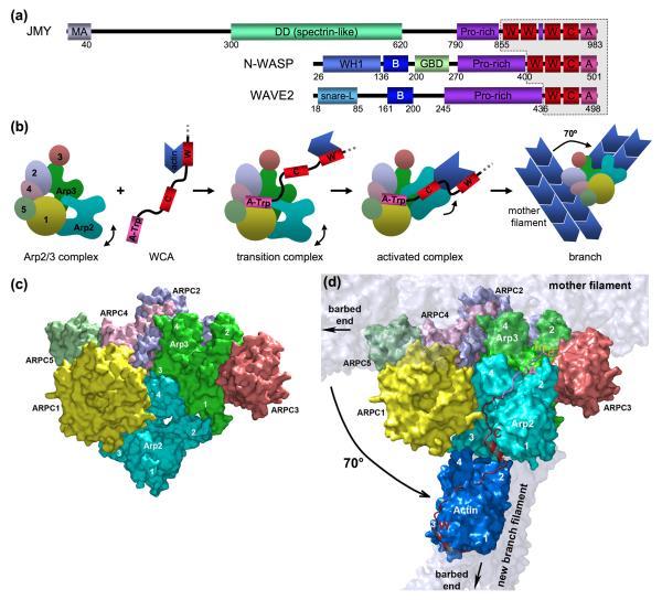
The W domain also participates in filament nucleation through NPFs of the Arp2/3 complex, which have between one and three W domains (Figure 4a). Arp2/3 complex consists of seven proteins, including two actin-related proteins, Arp2 and Arp3, and subunits ARPC1 to 5 (Figure 4b). By itself, Arp2/3 complex has low nucleation activity [21]. The discovery of ActA as a NPF at the surface of Listeria monocytogenes [22] and eukaryotic NPFs of the WASP/WAVE family [23-26] revealed the strong nucleation capacity of the combined NPFs-Arp2/3 complex.
Figure 4.
Actin filament nucleation by NPFs-Arp2/3 complex and NPFs. (a) Domain boundaries of three representative NPF proteins: mouse JMY (Q9QXM1), mouse N-WASP (Q91YD9) and human WAVE2 (Q9Y6W5). Note the resemblance with the filament nucleators shown in Figures 3, including the presence of Pro-rich regions and varying numbers of W domains (1 to 3). (b) The Arp2/3 complex consists of seven proteins, including the actin related proteins Arp2 and Arp3 and subunits ARPC1 to 5 (labeled 1 to 5). The WCA region of NPFs (contoured in part a) is the smallest fragment capable of catalyzing the formation of a polymerization nucleus, consisting of the two Arps and one to three actin subunits, and a conformational change in Arp2/3 complex that promotes monomer addition to the branch filament and binding of the nucleus to the side of a preexisting filament (mother filament). The branch grows from the barbed ends of the Arps at a 70° angle with respect to the mother filament. (c) Structure of inactive Arp2/3 complex [36,37]. Subdomains 1 and 2 of Arp2 are disordered in the structures, but were added here by analogy with actin. The Arp2/3 complex subunits are colored according to the diagram of part b. (d) SAXS-derived model of WCA-actin-Arp2/3 complex [43]. The orientation is the same as in part c. The mother and branch filaments are shown for reference, although this work did not address branch assembly. Note that Arp2 is proposed to move up compared to its location in part c. This study placed the first actin subunit of the branch at the barbed end of Arp2. The position of the W domain is known from the crystal structure of its complex with actin (ses Figure 2) and imposes constraints on the location of the C motif, which may bind in the hydrophobic cleft of Arp2. The position of the A motif (pink) is less well constrained, but this model is consisting with it binding at the interface between Arp3 and ARPC3, as suggested by biochemical studies [39-42].
Classical NPFs, including WASP/N-WASP/WAVE [23-26], WASH [27], WHAMM [28] and JMY [29], present a C-terminal WCA region (Figure 4a), which is the shortest fragment necessary for activation of nucleation with Arp2/3 complex [24]. This region consists of three discrete segments: W, C and A. The W domain(s), of which there can be between one and three, recruits the first actin subunit(s) of the new filament branch. The C (central or connecting) and A (acidic) motifs interact with subunits of Arp2/3 complex and stabilize the activated conformation by a still unknown mechanism. The actin monomer(s) bound to the W domain(s), together with Arp2 and Arp3, are thought to form a filament-like seed for nucleation of a filament branch that emerges at a 70° angle from the side of a preexisting filament (Figure 4b).
NPFs are themselves regulated by multiple factors, particularly by interaction with phospholipids and Rho-family GTPases at the membrane. WASP/N-WASP function under the control of Cdc42, whereas WAVE forms part of a multi-protein complex regulated by Rac [30-34]. From a structural point of view, the mechanism of regulation of WASP/N-WASP by Cdc42 is best understood and shares similarities with Rho-mediated regulation of DRFs. In the resting state WASP/N-WASP exist in a folded autoinhibited conformation stabilized by internal interaction of the WCA region with the N-terminal GBD, which blocks WCA interaction with Arp2/3 complex. NMR structures of the GBD in complex with a C-terminal WCA fragment [32] and Cdc42 [31] reveal that the two interactions are sterically incompatible, thus suggesting a mechanism of activation by steric competition of Cdc42 and WCA for a common binding interface. However, maximum activation of Arp2/3 complex results from co-stimulation by the binding of Cdc42 to the GBD and phospholipids to a basic patch positioned N-terminal to the GBD [35].
The crystal structure of Arp2/3 complex was first determined in the absence of nucleotide and NPF and revealed its inactive conformation [36], with Arp2 and Arp3 separated, the nucleotide cleft of Arp3 wide open and subdomains 1 and 2 of Arp2 disordered (Figure 4c). Nucleotide-bound structures were subsequently determined, but displayed essentially the same conformation, suggesting that nucleotide binding alone is insufficient to activate Arp2/3 complex [37]. Electron microscopy analysis of Arp2/3 complex in the branch provided evidence for a major conformational change, bringing Arp2 and Arp3 into a short-pitch dimer arrangement at the pointed end of the branch, and revealed structural changes within the mother filament itself [38].
None of the structures, however, resolves the interactions of NPFs with Arp2/3 complex. In biochemical studies WCA binds within short crosslinking distance to Arp2, ARPC1, Arp3 and ARPC3 [39-42]. In a recent study [43], the W domain of WCA was crosslinked to actin Cys-374 based on the structures of W-actin complexes [15]. Crosslinked WCA-actin forms a high-affinity 1:1 complex with Arp2/3 complex capped at the barbed end, and therefore suitable for structural investigation. SAXS analysis of this particle suggested that the first actin subunit binds at the barbed end of Arp2, constraining the binding site of the C motif to subunit Arp2, near the interface with ARPC1 (Figure 4d). Less can be said about the interactions of the A region, although it probably lies near the interface between Arp3 and ARPC3, consistent with the biochemical evidence [39-42]. This study offers testable hypotheses and a new way to address the problem of activation, but because of its limited resolution leaves unresolved the exact nature of the conformational change leading to activation and the precise role of WCA in this process.
NPFs-Arp2/3 complex can be viewed as a specialized form of tandem W domain nucleator [44]. In isolation, neither Arp2/3 complex nor the NPFs nucleate; they need each other for this activity. The NPFs, but not Arp2/3 complex, display similarities with other nucleators (Figures 3 and 4a), including the presence of Pro-rich regions and W repeats (1-3 in NPFs vs 3-4 in filament nucleators). Additionally, the C motif of NPFs is related to the W domain, and is proposed to bind Arp2 similarly to the W-actin interaction [15,43-46]. Arp2/3 complex itself can be considered as an actin dimer, with the five non-actin subunits serving primarily to dock the complex to the side of the mother filament. These considerations lead to a model of activation [44] in which the conserved tryptophan in the A region of NPFs links actin-loaded NPFs to the Arp2/3 complex (Figure 4b). After this initial encounter the actin subunit associated with the W domain binds at the barbed end of Arp2. Arp2, which is partially disordered in the inactive structure [36,37], is stabilized as a short-pitch dimer near Arp3 in the activated structure by additional interactions with the C motif and the first actin subunit of the branch. Arp2 is predicted to move mostly alone during activation, although minor changes throughout the complex are likely. Arp2/3 complex activation and branching (side binding to pre-existing filaments) may occur nearly simultaneously. NPFs are finally ejected from the branch, probably due to steric hindrance with actin subunits that begin joining the branch and with the mother filament.
Ena/VASP proteins as dedicated elongation factors among W-based filament nucleators
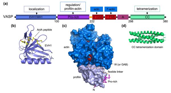
While formins promote both nucleation and elongation and use the FH2 domain for both activities, none of the W-based nucleators seems to promote elongation. This function appears to have been outsourced to a dedicated family of W-containing proteins, Ena/VASP, which on the other hand play no role on nucleation [47]. The domain organization of Ena/VASP is somewhat similar to that of WASP/N-WASP. Both contain N-terminal EVH1 (or WH1) domains, central Pro-rich regions and W-related sequences, known as the G- and F-actin binding (GAB and FAB) domains in Ena/VASP (Figure 5a). There is also an acidic patch in Ena/VASP following the FAB domain, albeit less acidic than in NPFs and lacking the important tryptophan. Finally, at the C-terminus, Ena/VASP has a coiled-coil tetramerization domain that is important for its filament bundling activity [48]. Although the evidence is less abundant than for formins, Ena/VASP has been shown to promote processive barbed end elongation and to compete with capping proteins at filament barbed ends [49-51]. Analogous to the stimulation of formin-mediated elongation produced by profilin [8], multiple studies have indicated that profilin-actin stimulates Ena/VASP-dependent Listeria motility [52-56].
Figure 5.
Domain diagram of VASP and representative structures. (a) Domain boundaries correspond to human VASP (P50552). Notice the resemblance with N-WASP (Figure 4a). (b) Structure of the EVH1 domain of Mena in complex with a Listeria ActA peptide [57]. (c) Structure of the ternary complex of profilin-actin with a VASP fragment comprising the last Pro-rich profilin-binding site and GAB domain [46]. The Gly-rich linker between these two domains was not visualized (discontinuous line). (d) Structure of the tetramerization domain, revealing a right-handed coiled-coil fold [59].
The EVH1 domain targets Ena/VASP to focal adhesions, filopodia and lamellipodia by binding to proteins that contain the consensus sequence FPPPP. Crystal structures of the EVH1 domain have been determined for various members of the family, including the complex with a Listeria ActA peptide shown in figure 5b [57], and reveal a fold related to the pleckstrin homology domain. The ternary structure of profilin-actin with a VASP fragment comprising the last Pro-rich segment and GAB domain (Figure 5c) suggests that profilin-actin complexes recruited to the Pro-rich region can be delivered directly to the barbed end of the elongating filament [46]. This structure also demonstrated that the GAB domain binds actin similarly to the W domain, and can bind actin simultaneously with profilin. What is more, the binding affinities of the Pro-rich and GAB domains are higher for profilin-actin than those of their separate interactions with profilin or actin [46,58]. Tetramerization could play an important role in Ena/VASP’s elongation mechanism, by sequentially allowing each subunit of the tetramer to release and advance during monomer addition to the barbed end while the other subunits remain attached to the growing filament. The structure of the tetramerization domain (Figure 5c) revealed an unusual, highly thermostable, right-handed coiled-coil fold [59].
Conclusions
The structural biology of actin filament nucleation and elongation has been a hot topic of research in recent years, due to the pivotal role these processes play in numerous cellular functions. The proteins involved are numerous, work by different mechanisms, and function under the control of diverse regulatory pathways. Significant progress has been made in our understanding of formin structure-function. The determination of atomic structures of Arp2/3 complex, W-actin complexes, and regulatory complexes with GTPases are also important breakthroughs. However, we still face a substantial gap of understanding of some of the most basic principles of filament nucleation and elongation, including the lack of high-resolution structures of activated Arp2/3 complex and tandem W nucleators in complex with actin. Even less is known about the recently discovered W-containing and muscle-enriched nucleator Lmod [60]. Most of the complexes are too large for NMR applications. Predictable, a major role will be played by the combined application of resolution-overlapping methods, including X-ray crystallography, electron microscopy and X-ray and neutron scattering. The intrinsically dynamic nature of filament nuclei and actin’s natural tendency to polymerize are two formidable obstacles hindering progress. Solving these problems will additionally require the ingenious application of protein engineering and biophysical methods, to produce monodisperse particles suitable for structural analyses.
Acknowledgements
Supported by NIH grants GM073791.
Footnotes
Publisher's Disclaimer: This is a PDF file of an unedited manuscript that has been accepted for publication. As a service to our customers we are providing this early version of the manuscript. The manuscript will undergo copyediting, typesetting, and review of the resulting proof before it is published in its final citable form. Please note that during the production process errors may be discovered which could affect the content, and all legal disclaimers that apply to the journal pertain.
References and recommended reading
Papers of particular interest have been highlighted as:
* of special interest
** of outstanding interest
- 1.Holmes KC. Structural biology: actin in a twist. Nature. 2009;457:389–390. doi: 10.1038/457389a. [DOI] [PubMed] [Google Scholar]
- 2.Paul AS, Pollard TD. Review of the mechanism of processive actin filament elongation by formins. Cell Motil Cytoskeleton. 2009;66:606–617. doi: 10.1002/cm.20379. [DOI] [PMC free article] [PubMed] [Google Scholar]
- 3.Goode BL, Eck MJ. Mechanism and function of formins in the control of actin assembly. Annu Rev Biochem. 2007;76:593–627. doi: 10.1146/annurev.biochem.75.103004.142647. [DOI] [PubMed] [Google Scholar]
- 4.Higgs HN. Formin proteins: a domain-based approach. Trends Biochem Sci. 2005;30:342–353. doi: 10.1016/j.tibs.2005.04.014. [DOI] [PubMed] [Google Scholar]
- 5.Castrillon DH, Wasserman SA. Diaphanous is required for cytokinesis in Drosophila and shares domains of similarity with the products of the limb deformity gene. Development. 1994;120:3367–3377. doi: 10.1242/dev.120.12.3367. [DOI] [PubMed] [Google Scholar]
- 6.Watanabe N, Kato T, Fujita A, Ishizaki T, Narumiya S. Cooperation between mDia1 and ROCK in Rho-induced actin reorganization. Nat Cell Biol. 1999;1:136–143. doi: 10.1038/11056. [DOI] [PubMed] [Google Scholar]
- 7.Pruyne D, Evangelista M, Yang C, Bi E, Zigmond S, Bretscher A, Boone C. Role of formins in actin assembly: nucleation and barbed-end association. Science. 2002;297:612–615. doi: 10.1126/science.1072309. Demonstration that formins function as filament nucleators and remain associated with barbed ends
- 8.Kovar DR, Harris ES, Mahaffy R, Higgs HN, Pollard TD. Control of the assembly of ATP- and ADP-actin by formins and profilin. Cell. 2006;124:423–435. doi: 10.1016/j.cell.2005.11.038. Direct observation of formin-mediated and profilin-stimulated processive filament elongation
- 9.Rose R, Weyand M, Lammers M, Ishizaki T, Ahmadian MR, Wittinghofer A. Structural and mechanistic insights into the interaction between Rho and mammalian Dia. Nature. 2005;435:513–518. doi: 10.1038/nature03604. First structure revealing the mechanism of formin activation by RhoC GTPase
- 10.Lammers M, Rose R, Scrima A, Wittinghofer A. The regulation of mDia1 by autoinhibition and its release by Rho*GTP. Embo J. 2005;24:4176–4187. doi: 10.1038/sj.emboj.7600879. First structure revealing the mechanism of formin auto-inhibition by internal DAD-DID interaction
- 11.Xu Y, Moseley JB, Sagot I, Poy F, Pellman D, Goode BL, Eck MJ. Crystal structures of a Formin Homology-2 domain reveal a tethered dimer architecture. Cell. 2004;116:711–723. doi: 10.1016/s0092-8674(04)00210-7. First structure of a formin FH2 domain
- 12.Otomo T, Tomchick DR, Otomo C, Panchal SC, Machius M, Rosen MK. Structural basis of actin filament nucleation and processive capping by a formin homology 2 domain. Nature. 2005;433:488–494. doi: 10.1038/nature03251. Structure of a formin-actin complex, providing structural insights into the mechanisms of nucleation and elongation
- 13.Dominguez R. The beta-thymosin/WH2 fold: multifunctionality and structure. Ann N Y Acad Sci. 2007;1112:86–94. doi: 10.1196/annals.1415.011. [DOI] [PubMed] [Google Scholar]
- 14.Dominguez R. Actin-binding proteins--a unifying hypothesis. Trends Biochem Sci. 2004;29:572–578. doi: 10.1016/j.tibs.2004.09.004. [DOI] [PubMed] [Google Scholar]
- 15.Chereau D, Kerff F, Graceffa P, Grabarek Z, Langsetmo K, Dominguez R. Actin-bound structures of Wiskott-Aldrich syndrome protein (WASP)-homology domain 2 and the implications for filament assembly. Proc Natl Acad Sci U S A. 2005;102:16644–16649. doi: 10.1073/pnas.0507021102. Structures of W-actin complexes, providing structural insights into the widespread mechanism of W-based actin nucleation
- 16.Quinlan ME, Heuser JE, Kerkhoff E, Mullins RD. Drosophila Spire is an actin nucleation factor. Nature. 2005;433:382–388. doi: 10.1038/nature03241. First tandem W-based nucleator to be discovered
- 17.Ahuja R, Pinyol R, Reichenbach N, Custer L, Klingensmith J, Kessels MM, Qualmann B. Cordon-Bleu Is an Actin Nucleation Factor and Controls Neuronal Morphology. Cell. 2007;131:337–350. doi: 10.1016/j.cell.2007.08.030. Discovery of a powerful tandem W-based nucleator, containing just three W domains and enriched in brain
- 18.Liverman AD, Cheng HC, Trosky JE, Leung DW, Yarbrough ML, Burdette DL, Rosen MK, Orth K. Arp2/3-independent assembly of actin by Vibrio type III effector VopL. Proc Natl Acad Sci U S A. 2007;104:17117–17122. doi: 10.1073/pnas.0703196104. Discovery of a powerful tandem W-based nucleator from a bacterial pathogen
- 19.Tam VC, Serruto D, Dziejman M, Brieher W, Mekalanos JJ. A Type III Secretion System in Vibrio cholerae Translocates a Formin/Spire Hybrid-like Actin Nucleator to Promote Intestinal Colonization. Cell Host and Microbe. 2007;1:95–107. doi: 10.1016/j.chom.2007.03.005. Discovery of a powerful tandem W-based nucleator from a bacterial pathogen
- 20.Rebowski G, Boczkowska M, Hayes DB, Guo L, Irving TC, Dominguez R. X-ray scattering study of actin polymerization nuclei assembled by tandem W domains. Proc Natl Acad Sci U S A. 2008;105:10785–10790. doi: 10.1073/pnas.0801650105. Low-resolution structure of engineered actin polymerization seeds assembled by tandem W domains
- 21.Mullins RD, Heuser JA, Pollard TD. The interaction of Arp2/3 complex with actin: nucleation, high affinity pointed end capping, and formation of branching networks of filaments. Proc Natl Acad Sci U S A. 1998;95:6181–6186. doi: 10.1073/pnas.95.11.6181. [DOI] [PMC free article] [PubMed] [Google Scholar]
- 22.Welch MD, Rosenblatt J, Skoble J, Portnoy DA, Mitchison TJ. Interaction of human Arp2/3 complex and the Listeria monocytogenes ActA protein in actin filament nucleation. Science. 1998;281:105–108. doi: 10.1126/science.281.5373.105. [DOI] [PubMed] [Google Scholar]
- 23.Machesky LM, Insall RH. Scar1 and the related Wiskott-Aldrich syndrome protein, WASP, regulate the actin cytoskeleton through the Arp2/3 complex. Curr Biol. 1998;8:1347–1356. doi: 10.1016/s0960-9822(98)00015-3. [DOI] [PubMed] [Google Scholar]
- 24.Machesky LM, Mullins RD, Higgs HN, Kaiser DA, Blanchoin L, May RC, Hall ME, Pollard TD. Scar, a WASp-related protein, activates nucleation of actin filaments by the Arp2/3 complex. Proc Natl Acad Sci U S A. 1999;96:3739–3744. doi: 10.1073/pnas.96.7.3739. [DOI] [PMC free article] [PubMed] [Google Scholar]
- 25.Rohatgi R, Ma L, Miki H, Lopez M, Kirchhausen T, Takenawa T, Kirschner MW. The interaction between N-WASP and the Arp2/3 complex links Cdc42-dependent signals to actin assembly. Cell. 1999;97:221–231. doi: 10.1016/s0092-8674(00)80732-1. [DOI] [PubMed] [Google Scholar]
- 26.Winter D, Lechler T, Li R. Activation of the yeast Arp2/3 complex by Bee1p, a WASP-family protein. Curr Biol. 1999;9:501–504. doi: 10.1016/s0960-9822(99)80218-8. [DOI] [PubMed] [Google Scholar]
- 27.Linardopoulou EV, Parghi SS, Friedman C, Osborn GE, Parkhurst SM, Trask BJ. Human subtelomeric WASH genes encode a new subclass of the WASP family. PLoS Genet. 2007;3:e237. doi: 10.1371/journal.pgen.0030237. [DOI] [PMC free article] [PubMed] [Google Scholar]
- 28.Campellone KG, Webb NJ, Znameroski EA, Welch MD. WHAMM is an Arp2/3 complex activator that binds microtubules and functions in ER to Golgi transport. Cell. 2008;134:148–161. doi: 10.1016/j.cell.2008.05.032. [DOI] [PMC free article] [PubMed] [Google Scholar]
- 29.Zuchero JB, Coutts AS, Quinlan ME, Thangue NB, Mullins RD. p53-cofactor JMY is a multifunctional actin nucleation factor. Nat Cell Biol. 2009;11:451–459. doi: 10.1038/ncb1852. [DOI] [PMC free article] [PubMed] [Google Scholar]
- 30.Ma L, Rohatgi R, Kirschner MW. The Arp2/3 complex mediates actin polymerization induced by the small GTP-binding protein Cdc42. Proc Natl Acad Sci U S A. 1998;95:15362–15367. doi: 10.1073/pnas.95.26.15362. [DOI] [PMC free article] [PubMed] [Google Scholar]
- 31.Abdul-Manan N, Aghazadeh B, Liu GA, Majumdar A, Ouerfelli O, Siminovitch KA, Rosen MK. Structure of Cdc42 in complex with the GTPase-binding domain of the ‘Wiskott-Aldrich syndrome’ protein. Nature. 1999;399:379–383. doi: 10.1038/20726. [DOI] [PubMed] [Google Scholar]
- 32.Kim AS, Kakalis LT, Abdul-Manan N, Liu GA, Rosen MK. Autoinhibition and activation mechanisms of the Wiskott-Aldrich syndrome protein. Nature. 2000;404:151–158. doi: 10.1038/35004513. Study providing structural insights into the mechanism of WASP regulation by GTPases
- 33.Eden S, Rohatgi R, Podtelejnikov AV, Mann M, Kirschner MW. Mechanism of regulation of WAVE1-induced actin nucleation by Rac1 and Nck. Nature. 2002;418:790–793. doi: 10.1038/nature00859. [DOI] [PubMed] [Google Scholar]
- 34.Bompard G, Caron E. Regulation of WASP/WAVE proteins: making a long story short. J Cell Biol. 2004;166:957–962. doi: 10.1083/jcb.200403127. [DOI] [PMC free article] [PubMed] [Google Scholar]
- 35.Prehoda KE, Scott JA, Mullins RD, Lim WA. Integration of multiple signals through cooperative regulation of the N-WASP-Arp2/3 complex. Science. 2000;290:801–806. doi: 10.1126/science.290.5492.801. [DOI] [PubMed] [Google Scholar]
- 36.Robinson RC, Turbedsky K, Kaiser DA, Marchand JB, Higgs HN, Choe S, Pollard TD. Crystal structure of Arp2/3 complex. Science. 2001;294:1679–1684. doi: 10.1126/science.1066333. First structure of the Arp2/3 complex, revealing its inactive conformation
- 37.Nolen BJ, Pollard TD. Insights into the influence of nucleotides on actin family proteins from seven structures of Arp2/3 complex. Mol Cell. 2007;26:449–457. doi: 10.1016/j.molcel.2007.04.017. [DOI] [PMC free article] [PubMed] [Google Scholar]
- 38.Rouiller I, Xu XP, Amann KJ, Egile C, Nickell S, Nicastro D, Li R, Pollard TD, Volkmann N, Hanein D. The structural basis of actin filament branching by the Arp2/3 complex. J Cell Biol. 2008;180:887–895. doi: 10.1083/jcb.200709092. EM structure of Arp2/3 complex in the branch, revealing changes within the Arp2/3 complex and the mother filament
- 39.Kelly AE, Kranitz H, Dotsch V, Mullins RD. Actin binding to the central domain of WASP/Scar proteins plays a critical role in the activation of the Arp2/3 complex. J Biol Chem. 2006;281:10589–10597. doi: 10.1074/jbc.M507470200. [DOI] [PMC free article] [PubMed] [Google Scholar]
- 40.Zalevsky J, Grigorova I, Mullins RD. Activation of the Arp2/3 complex by the Listeria acta protein. Acta binds two actin monomers and three subunits of the Arp2/3 complex. J Biol Chem. 2001;276:3468–3475. doi: 10.1074/jbc.M006407200. [DOI] [PubMed] [Google Scholar]
- 41.Weaver AM, Heuser JE, Karginov AV, Lee WL, Parsons JT, Cooper JA. Interaction of cortactin and N-WASp with Arp2/3 complex. Curr Biol. 2002;12:1270–1278. doi: 10.1016/s0960-9822(02)01035-7. [DOI] [PubMed] [Google Scholar]
- 42.Kreishman-Deitrick M, Goley ED, Burdine L, Denison C, Egile C, Li R, Murali N, Kodadek TJ, Welch MD, Rosen MK. NMR analyses of the activation of the Arp2/3 complex by neuronal Wiskott-Aldrich syndrome protein. Biochemistry. 2005;44:15247–15256. doi: 10.1021/bi051065n. [DOI] [PubMed] [Google Scholar]
- 43.Boczkowska M, Rebowski G, Petoukhov MV, Hayes DB, Svergun DI, Dominguez R. X-ray scattering study of activated Arp2/3 complex with bound actin-WCA. Structure. 2008;16:695–704. doi: 10.1016/j.str.2008.02.013. Low-resolution structure of Arp2/3 complex with bound actin and the WCA region of N-WASP, placing the first actin of the branch at the barbed end of Arp2
- 44.Dominguez R. Actin filament nucleation and elongation factors - structure-function relationships. Crit Rev Biochem Mol Biol. 2009;44:351–366. doi: 10.3109/10409230903277340. [DOI] [PMC free article] [PubMed] [Google Scholar]
- 45.Aguda AH, Burtnick LD, Robinson RC. The state of the filament. EMBO Rep. 2005;6:220–226. doi: 10.1038/sj.embor.7400363. [DOI] [PMC free article] [PubMed] [Google Scholar]
- 46.Ferron F, Rebowski G, Lee SH, Dominguez R. Structural basis for the recruitment of profilin-actin complexes during filament elongation by Ena/VASP. Embo J. 2007;26:4597–4606. doi: 10.1038/sj.emboj.7601874. Structure of ternary complex of profilin-actin with a Pro-rich-GAB fragment of VASP, suggesting a mechanism for the transfer of profilin-actin onto the barbed end of elongating filaments
- 47.Drees F, Gertler FB. Ena/VASP: proteins at the tip of the nervous system. Curr Opin Neurobiol. 2008;18:53–59. doi: 10.1016/j.conb.2008.05.007. [DOI] [PMC free article] [PubMed] [Google Scholar]
- 48.Bachmann C, Fischer L, Walter U, Reinhard M. The EVH2 domain of the vasodilator-stimulated phosphoprotein mediates tetramerization, F-actin binding, and actin bundle formation. J Biol Chem. 1999;274:23549–23557. doi: 10.1074/jbc.274.33.23549. [DOI] [PubMed] [Google Scholar]
- 49.Breitsprecher D, Kiesewetter AK, Linkner J, Urbanke C, Resch GP, Small JV, Faix J. Clustering of VASP actively drives processive, WH2 domain-mediated actin filament elongation. Embo J. 2008;27:2943–2954. doi: 10.1038/emboj.2008.211. Demonstration of processive filament elongation by VASP
- 50.Pasic L, Kotova T, Schafer DA. Ena/VASP proteins capture actin filament barbed ends. J Biol Chem. 2008;283:9814–9819. doi: 10.1074/jbc.M710475200. [DOI] [PMC free article] [PubMed] [Google Scholar]
- 51.Applewhite DA, Barzik M, Kojima SI, Svitkina TM, Gertler FB, Borisy GG. Ena/VASP Proteins Have an Anti-Capping Independent Function in Filopodia Formation. Mol Biol Cell. 2007 doi: 10.1091/mbc.E06-11-0990. [DOI] [PMC free article] [PubMed] [Google Scholar]
- 52.Kang F, Laine RO, Bubb MR, Southwick FS, Purich DL. Profilin interacts with the Gly-Pro-Pro-Pro-Pro-Pro sequences of vasodilator-stimulated phosphoprotein (VASP): implications for actin-based Listeria motility. Biochemistry. 1997;36:8384–8392. doi: 10.1021/bi970065n. [DOI] [PubMed] [Google Scholar]
- 53.Geese M, Schluter K, Rothkegel M, Jockusch BM, Wehland J, Sechi AS. Accumulation of profilin II at the surface of Listeria is concomitant with the onset of motility and correlates with bacterial speed. J Cell Sci. 2000;113(Pt 8):1415–1426. doi: 10.1242/jcs.113.8.1415. [DOI] [PubMed] [Google Scholar]
- 54.Dickinson RB, Southwick FS, Purich DL. A direct-transfer polymerization model explains how the multiple profilin-binding sites in the actoclampin motor promote rapid actin-based motility. Arch Biochem Biophys. 2002;406:296–301. doi: 10.1016/s0003-9861(02)00212-6. [DOI] [PubMed] [Google Scholar]
- 55.Grenklo S, Geese M, Lindberg U, Wehland J, Karlsson R, Sechi AS. A crucial role for profilin-actin in the intracellular motility of Listeria monocytogenes. EMBO Rep. 2003;4:523–529. doi: 10.1038/sj.embor.embor823. First demonstration of a profilin role in VASP-mediated elongation
- 56.Auerbuch V, Loureiro JJ, Gertler FB, Theriot JA, Portnoy DA. Ena/VASP proteins contribute to Listeria monocytogenes pathogenesis by controlling temporal and spatial persistence of bacterial actin-based motility. Mol Microbiol. 2003;49:1361–1375. doi: 10.1046/j.1365-2958.2003.03639.x. [DOI] [PubMed] [Google Scholar]
- 57.Prehoda KE, Lee DJ, Lim WA. Structure of the enabled/VASP homology 1 domain-peptide complex: a key component in the spatial control of actin assembly. Cell. 1999;97:471–480. doi: 10.1016/s0092-8674(00)80757-6. First structure of the EVH1 domain in complex with a target peptide
- 58.Chereau D, Dominguez R. Understanding the role of the G-actin-binding domain of Ena/VASP in actin assembly. J Struct Biol. 2006;155:195–201. doi: 10.1016/j.jsb.2006.01.012. [DOI] [PubMed] [Google Scholar]
- 59.Kuhnel K, Jarchau T, Wolf E, Schlichting I, Walter U, Wittinghofer A, Strelkov SV. The VASP tetramerization domain is a right-handed coiled coil based on a 15-residue repeat. Proc Natl Acad Sci U S A. 2004;101:17027–17032. doi: 10.1073/pnas.0403069101. [DOI] [PMC free article] [PubMed] [Google Scholar]
- 60.Chereau D, Boczkowska M, Skwarek-Maruszewska A, Fujiwara I, Hayes DB, Rebowski G, Lappalainen P, Pollard TD, Dominguez R. Leiomodin is an actin filament nucleator in muscle cells. Science. 2008;320:239–243. doi: 10.1126/science.1155313. Discovery of a filament nucleator in muscle cells, structurally unrelated to other known nucleators


