

Molecular imaging can potentially provide means for monitoring response to therapy in rheumatoid arthritis (RA) early in the course of disease [1].Quantitative measurements of RA disease activity made in the wrist by whole-body PET scanners, however, have inadequate accuracy because of limited spatial resolution [2]. A high-resolution PET/CT scanner for imaging extremities has been built at our institution [3]. In conjunction with a clinical MRI scanner, high-resolution PET/MR images can be obtained for the wrist. The CT image is used for PET/MR image coregistration.
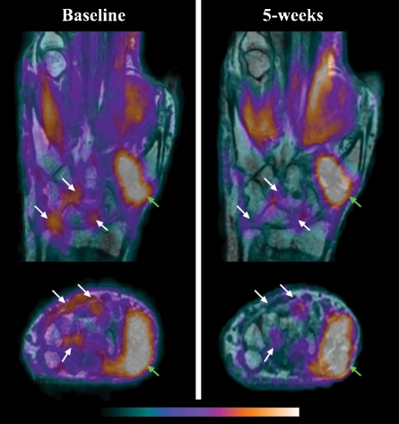
A 57-year-old female with established RA was stable until a recent clinical flare-up in the right wrist. Clinical examination revealed synovitis, swelling, and diminished range of motion. The patient also had a history of osteoarthritis (OA). An extremity 18F-FDG PET/CT scan immediately following MRI at baseline was performed on this patient. Tumor necrosis factor alpha (TNF-α) inhibitor (etanercept) therapy was then initiated as a part of the patient’s standard of care. The patient was re-scanned 5 weeks after starting treatment.
The figure shows high-resolution 18F-FDG PET images (pseudocolor) overlaid on pre-contrast MRI images (gray scale) at baseline (left column) and 5 weeks (right column). Significant reduction in PET signal (suggesting reduced inflammation) in the synovium and at sites of erosions (white arrows) is visible. The green arrow shows inflammation due to OA. Physician examination at 3 months confirmed that this patient responded positively to etanercept. This case illustrates the potential of high-resolution PET with MRI for quantitative visualization of early response to therapy in RA.
Acknowledgments
Open Access
This article is distributed under the terms of the Creative Commons Attribution Noncommercial License which permits any noncommercial use, distribution, and reproduction in any medium, provided the original author(s) and source are credited.
Footnotes
This work was funded by the NIH grants UL1-RR024146, R01CA129561, R01EB002138 and the UC Davis Imaging Research Center.
References
- 1.Brenner W. 18F-FDG PET in rheumatoid arthritis: there still is a long way to go. J Nucl Med. 2004;45(6):927–9. [PubMed] [Google Scholar]
- 2.Beckers C, Ribbens C, André B, Marcelis S, Kaye O, Mathy L, et al. Assessment of disease activity in rheumatoid arthritis with (18)F-FDG PET. J Nucl Med. 2004;45(6):956–64. [PubMed] [Google Scholar]
- 3.Bowen SL, Wu Y, Chaudhari AJ, Fu L, Packard NJ, Burkett GW, et al. Initial characterization of a dedicated breast PET/CT scanner during human imaging. J Nucl Med. 2009;50(9):1401–8. doi: 10.2967/jnumed.109.064428. [DOI] [PMC free article] [PubMed] [Google Scholar]