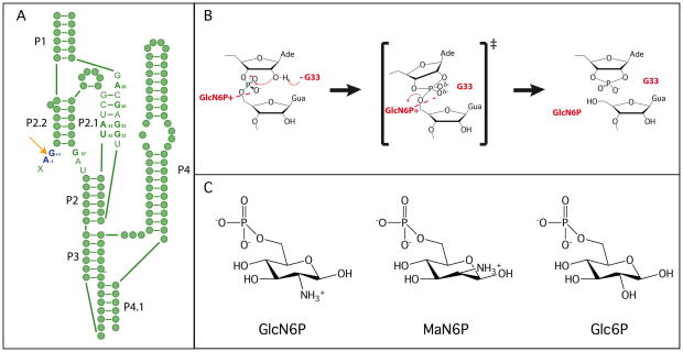
Figure 1.
Structure and mechanism of the glmS ribozyme. A. Secondary structure of the pre-cleaved glmS ribozyme. Green filled circles represent individual nucleotide and key active site nucleotides are indicated. The cleavage site is shown using an orange arrow, and the nucleotides flanking the scissile phosphate are shown in dark blue. Paired regions are numbered according to convention. B. The proposed catalytic mechanism of the glmS ribozyme. C. The three sugars used in the mechanistic and structural studies.

