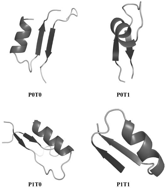
Figure 3. Representatives of the four different patterns of αβ2 units in proteins.
The P0T0, P0T1, P1T0, and P1T1 example units are extracted from proteins 1VQ1(A226-249), 2O1N(116-145), 1COZ(1-28), and 2P7H(A21-46) respectively. P indicates if the two strands are parallel or antiparallel to each other, while T relates to the relative position of the strands and the helix along the protein sequence. Figure drawn with Pymol.

