Abstract
The potential of the PRESAGE™/Optical-CT system as a comprehensive 3D dosimetry tool has been demonstrated. The current study focused on detailed characterization of robustness (intra-dosimeter uniformity and temporal stability) and reproducibility (inter-dosimeter reproducibility) of PRESAGE™ inserts compatible with the RPC H&N phantom. In addition, the accuracy and precision of PRESAGE dose measurement was also evaluated. Four identical PRESAGE™ dosimeters (10cm diameter and 7cm height cylinders) were irradiated with the same rotationally symmetric treatment plan using a Varian accelerator. The treatment plan was designed to rigorously evaluate robustness and reproducibility for multiple dose levels and in 3D. All dosimeters were scanned by optical-CT at daily intervals to study temporal stability. Dose comparisons were made between PRESAGE, ECLIPSE, and independent measurement with EBT film at a select depth. The use of improved optics and acquisition technique yielded substantially higher quality 3D dosimetry data from PRESAGE than has been achieved previously (noise reduced to ~1%, accuracy to within 3%). Data analysis showed excellent intra-dosimeter uniformity, temporal stability and inter-dosimeter reproducibility of relative radiochromic response. In general, the PRESAGE™ dose-distribution was found to agree better with EBT (~99% pass rate) than with ECLIPSE calculations (~92% pass rate) especially in penumbral regions for a 3% dose-difference and 3 mm distance-to-agreement evaluation criteria. The results demonstrate excellent robustness and reproducibility of the PRESAGE™ for relative 3D-dosimetry and represent a significant step towards incorporation in the RadOnc-clinic (e.g. integration with RPC phantom).
1. Introduction
PRESAGE™ is a relatively novel radiochromic 3D dosimeter, which has many favorable characteristics for dose readout using optical-CT1,2,3. Most studies have focused on comparing planned dose distribution with a one-time measurement of dose2,3. Comprehensive 3D characterization of the robustness and reproducibility of PRESAGE™ is still pending. In this study, experiments were specifically conducted to evaluate robustness (intra-dosimeter uniformity and temporal stability of response) and reproducibility (inter-dosimeter) of radiochromic response for multiple dose levels and in 3D. Some robustness and reproducibility characteristics were evaluated previously but were limited to single profile measurements3. Robustness and reproducibility were evaluated by irradiating multiple 3D dosimeters with an identical treatment plan, tracking the readout at periodic time intervals and inter-comparison of the readouts. The accuracy and precision of dosimetry was also characterized by comparison with treatment planning system (TPS) calculations and with EBT film measurements in the central plane. The treatment plan was carefully designed to facilitate rigorous investigation of robustness, reproducibility, precision and accuracy for multiple dose levels and in 3D (see methods). The PRESAGE dosimeters were designed to fit as inserts in the RPC H&N phantom to test the viability of the PRESAGE/optical-CT dosimetry system as a clinical tool (e.g. integration with the RPC IMRT credentialing test)
2. Materials and Methods
2.1. Experiment to evaluate robustness, reproducibility, accuracy and precision
Four identical PRESAGE™ 3D dosimeters were irradiated with an identical treatment plan. The dosimeters (10 cm diameter) were from the same batch of pre-mold mixture. The PRESAGE inserts had an effective Z number of 8.3, a physical density of 1.07g/cm3, and a CT number of ~180.
An x-ray CT scan was acquired and imported into an Eclipse workstation for planning. For the CT scan, the PRESAGE™ inserts were placed with their flat circular face on the treatment couch such that circular sections were available in the frontal plane of reconstruction (fig 1). The gantry was positioned at 180° and nine rotationally symmetric beams (collimator angular increments of 40° starting at 0°) were placed. The field size for all beams was 1×3 cm2. A dose of 4Gy (100%) was prescribed at the center of the first beam (0° collimator) and at a depth of 2 cm along the central axis of the PRESAGE™ insert. The second (40° collimator) and third beams (80° collimator) were weighted to 72% and 32% of the first beam respectively. The remaining beams were a repetition of the first three beams. The overall dose distribution in any axial slice was highly modulated and consisted of three identical sectors of dose distribution i.e. rotationally symmetric. Each sector consisted of dose delivered by a high dose (100%), a medium dose (72%) and a low dose (32%) beam. Along the long axis of PRESAGE inserts, there was only a slight variation of dose distribution. The significance of this treatment plan is that: i) it facilitated rigorous investigation of intra dosimeter uniformity of radiochromic response for multiple dose levels because of rotational symmetry of distribution, ii) it facilitated rigorous investigation of temporal variation and inter-dosimeter reproducibility for multiple dose levels and in 3D and iii) it enabled accurate EBT film dosimetry (insensitive to slight errors in film placement) for independent comparison in the axial plane. All PRESAGE inserts were irradiated with the same treatment plan but on different days. Accuracy and symmetry of delivery was ascertained by a quality assurance test whereby the collimator jaws were retracted, the cross-hair was aligned with the geometrical center of the circular face of dosimeters and the collimator was rotated to test the alignment of cross-hair with the geometrical center at all angular increments.
Fig. 1. Rotationally symmetric treatment plan.
A) Pictorial representation of planning and delivery. B) Frontal view from CT scan with field overlay showing placement of the nine fields in a rotationally symmetric pattern. C) Rotationally symmetric dose distribution.
The OCTOPUS™ 5X optical-CT scanner (MGS Research Inc, Madison, CT) was used for dose readout from PRESAGE™ inserts. The OCTOPUS™ 5X optical-CT scanner is an upgrade over previously used apparatus2. The upgrade facilitated ~5X faster scanning because of changes in motor configurations. Each 3D scan consisted of 15 slices with inter-slice spacing of 5 mm. Each slice comprised 600 linear projection scans with rotational increments of 0.6° commensurate with the Nyquist sampling criteria. The time required to scan each slice was ~7–8 minutes. Every projection had a matrix size of 168 mm (168 pixels with 1 mm spacing). An in house MATLAB-based (Mathworks, Natic, MA) code was used to reconstruct the 3D distribution of radiochromic response (optical density; units: /meter) with a resolution of 1 mm × 1 mm × 5 mm. The PRESAGE inserts were scanned at periodic time intervals of 24 hrs post irradiation to evaluate temporal variation. The relative dose distribution was proportional to the change in OD and is called PRESAGE dose. After optical-CT scanning, one of the dosimeters was cut centrally and an EBT film was inserted at the cut-plane for independent verification of dose distribution. A calibration curve was acquired on the day of irradiations to enable conversion to dose distribution (EBT dose)2. The EPSON® Perfection 4990 flatbed scanner was used for scanning EBT films and its use has been described previously.
2.2. Data analysis
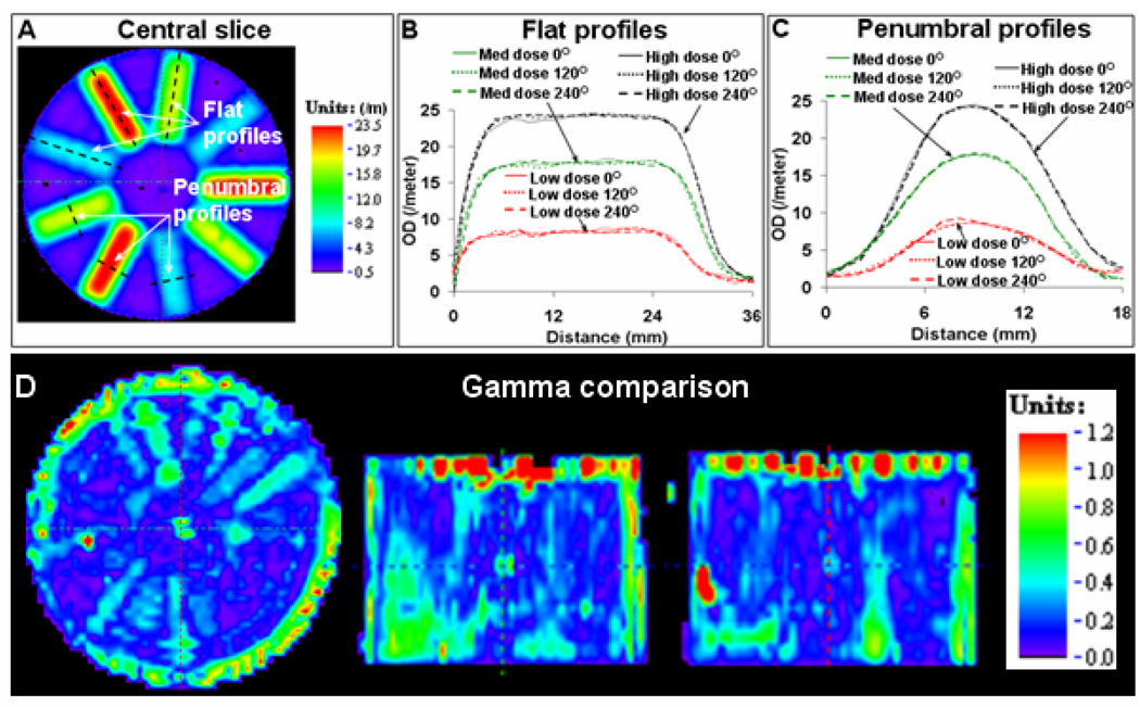
Data analysis was done in DoseQA (www.3cognition.com) or using in house routines in MATLAB. The regions receiving identical dose should elicit identical radiochromic response (change in optical density (OD)). This was the basis for the evaluation of intra-dosimeter uniformity of response and was facilitated by the rotationally symmetric treatment plan and delivery. A 3D PRESAGE dose was compared with itself but rotated by 120° and 240°. Dose profiles and gamma maps (2% dose difference and 1 mm distance-to-agreement (DTA) criteria) were used in comparative analysis. The 2% dose difference (accounting for highly modulated dose distribution) and 1 mm DTA criteria (accounting for setup error) were very stringent as compared to conventional clinical criteria.
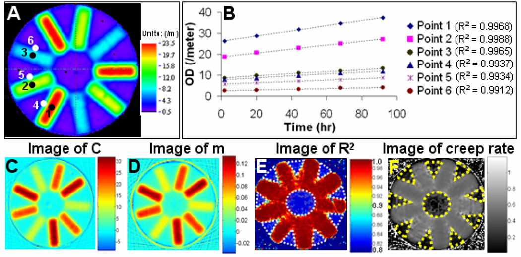
The 3D radiochromic response of a dosimeter was compared with 3D response at 24 hour intervals post-irradiation for upto 90 hrs to evaluate temporal stability. The radiochromic response showed a consistent creep (increase of OD) with time and this creep was evaluated for linearity with time. Each point in the 3D distribution was fit to a linear parametric model represented by; y = m×t + C; using linear least squared regression. Here, m (rate of net OD change or creep rate; units: /meter/hr) and C (initial OD; units: /meter) are the parameters of the model; y (estimate of net OD at any time t; units: /meter) is the dependent variable and t (time; units: hr) is the independent variable. The R2 value for regression at each data point was used to evaluate linearity of fit. The creep-rate (% change of OD / hr) is a measure of consistent aging of the radiochromic response at all dose levels and was used to evaluate linearity of temporal variation of OD.
Inter-dosimeter reproducibility was evaluated by comparing 3D dose distributions from the four different dosimeters. The evaluation consisted of profile comparisons and gamma evaluations. A criterion of 2% dose-difference (accounting for highly modulated distribution) and 2 mm distance-to-agreement (accounting for inter-dosimeter setup error) was used in gamma evaluations.
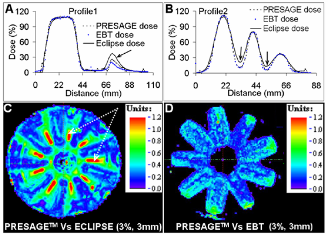
The accuracy and precision of PRESAGE dose was evaluated by comparison with independent EBT dose measurement in the central plane and with 3D calculations from the Eclipse treatment planning system (henceforth termed Eclipse dose). Dose profile comparisons and gamma evaluations were used. Gamma criteria of 3% dose-difference and 3 mm DTA was used in dose comparisons between independent datasets.
3. Results and Discussion
Improved optics and data-acquisition technique facilitated reconstruction of dose-distribution with reduced noise (1%–2%) and reduced edge artefact (~3mm from edge). Fig 2 shows dose distribution in the central slice and profiles along dotted lines which confirm reduced noise. Such high quality data facilitated rigorous evaluation of robustness and reproducibility characteristics of PRESAGE.
Fig. 2. PRESAGE dose in central plane.
A) Rotationally symmetric dose-distribution in central plane. B & C) Dose profiles along dotted lines in A show reduced noise and edge-artefact.
Evaluation of intra-dosimeter uniformity is shown in fig 3. The 3D readout from a PRESAGE dosimeter was compared with the same readouts, but rotated by 120° and 240° respectively. This was possible because of the rotationally symmetric treatment and delivery. Flat and penumbral dose profiles showed close agreement for all dose levels. These profiles were chosen because any differences in distribution are likely to show up because of steep dose gradients. Gamma comparisons showed excellent (>98% pass rate) agreement in 3D confirming excellent intra dosimeter uniformity of radiochromic response in 3D.
Fig. 3. Intra-dosimeter uniformity. PRESAGE distribution was registered with the same distribution rotated by 120° and 240°. Such comparison was possible because of radially symmetric distribution.
A) Central slice radiochromic response showing the position of profiles. B&C) Dose profiles along dotted lines in A show encouraging intra-dosimeter uniformity. D) Gamma comparison (2% dose-difference and 1 mm DTA) shows encouraging agreement in 3D.
Temporal variation of radiochromic response was evaluated by periodic tracking for ~90 hrs post-irradiation and results are shown in fig 4. Results showed that the dosimeters turned darker with time i.e. they showed temporal creep (increase in OD) post irradiation (fig 4b). The creep was linear with time as seen by the excellent fit (R2 > 0.98) to the linear regression model for all data points in the central slice. The images in fig 4 are representative for all slices. The creep rate was calculated from the parameters of the model and was estimated to be ~ 0.54% per hour. The results demonstrate excellent relative temporal stability.
Fig. 4. Temporal stability of radiochromic response.
A) Central slice radiochromic response. B) Temporal variation for six representative points which are shown in A, C, D, E & F) The linear regression model for each point in central slice. C&D are images of parameters C and m. E is an image of the R2 value of linear fit. F is an image of the creep rate calculated from the parameters of the fit.
Inter dosimeter reproducibility was evaluated by inter-comparison of PRESAGE dose from multiple dosimeters and was found to be excellent as shown in fig 5. The relative dose distributions showed excellent agreement (>98% gamma pass rate) to within 2% dose difference and within 2 mm DTA.
Fig. 5. Inter-dosimeter reproducibility of PRESAGE™.
Measurements from 4 different dosimeters irradiated with the same treatment plan showed excellent agreement to within 2% dose-difference and 2mm distance to agreement as seen in the profile plots of A and B and representative 3D gamma comparison map between dose readout from two different dosimeters.
Comparison of PRESAGE dose with EBT dose and Eclipse dose in the central slice is shown in fig 6. PRESAGE dose clearly agreed better with EBT dose than with Eclipse dose, particularly in penumbral regions. Excellent agreement of PRESAGE dose and EBT dose (3% dose-difference and 3 mm DTA) confirmed that PRESAGE was an accurate relative dosimeter.
Fig. 6. Accuracy of PRESAGE dose.
A&B) Profiles of PRESAGE, EBT and Eclipse dose. C) Gamma comparison between PRESAGE and Eclipse dose. D) Gamma comparison between PRESAGE dose and EBT dose. In general, PRESAGE dose agreed better with EBT dose than with Eclipse dose, particularly in penumbrae.
4. Conclusions
This study presents the first rigorous investigations into the robustness, reproducibility, precision and accuracy of 3D dosimetry in PRESAGE™ inserts compatible with the RPC H&N IMRT credentialing phantom. Improvements in optics and acquisition technique produced very high quality (noise ~ 2%) 3D data even at low dose levels. Results indicated that the PRESAGE™ inserts were extremely robust for relative 3D dosimetry as indicated by excellent intra-dosimeter consistency (within 2%) of radiochromic response and stability of relative dose distribution for > 90 hrs post-irradiation. Reproducibility of response for different 3D inserts was also evaluated and was highly encouraging (within 2%) at all dose levels. Accuracy of relative dose measurement was evaluated by comparison with independent EBT film measurement and was within 2% even along the penumbra and at low dose levels. The excellent quality of 3D dose measurement that was achieved with the PRESAGE™/OCTOPUS™ dosimetry system (comparable to EBT film) confirms its viability as a clinical 3D dosimetry tool (e.g. integration with RPC IMRT credentialing).
References
- 1.Adamovics J, Maryanski MJ. Characterisation of PRESAGE: A new 3-D radiochromic solid polymer dosemeter for ionizing radiation. Radiat Prot Dosimetry. 2006;120:107–112. doi: 10.1093/rpd/nci555. [DOI] [PubMed] [Google Scholar]
- 2.Guo P, Adamovics J, Oldham M. A practical three-dimensional dosimetry system for radiation therapy. Med Phys. 2006;33:3962–3972. doi: 10.1118/1.2349686. [DOI] [PMC free article] [PubMed] [Google Scholar]
- 3.Guo PY, Adamovics JA, Oldham M. Characterization of a new radiochromic three-dimensional dosimeter. Med Phys. 2006;33:1338–1345. doi: 10.1118/1.2192888. [DOI] [PMC free article] [PubMed] [Google Scholar]


